Original Article
Combined vascular resection and reconstruction
for advanced hilar cholangiocarcinoma
Zhimin Yu1*, Qing Sun2*, Yue Zhu3, Jie Wang1, Junyao Xu1
Guandong Provincial Key Laboratory of Malignant Tumor Epigenetics and Gene Regulation, Departments of 1Hepatobiliary Surgery, 2Pathology, 3Vascular and Thyroidal Surgery, Sun Yat-Sen Memorial Hospital, Sun Yat-Sen University, Guangzhou 510120, P. R. China. *Co-first authors.
Received September 20, 2016; Accepted November 22, 2016; Epub February 15, 2017; Published February 28, 2017
Abstract: Background: Hilar cholangiocarcinoma with vascular invasion was previously regarded as locally ad-vanced disease and a contraindication to curative resection. The significance of combined vascular resection and reconstruction has remained ambiguous and controversial. This study aimed to reveal the role and efficacy of concomitant vascular resection and reconstruction on survival of patients with hilar cholangiocarcinoma. Materials and Methods: From January 2006 to December 2014, totally 19 out of 95 hilar cholangiocarcinoma patients under -going curative resection were performed with combined vascular resection and reconstruction in a single institute including right hepatic artery alone (n=6), portal vein plus right hepatic artery (n=3) and portal vein alone (n=10), the remaining 67 patients underwent without vascular resection. The clinicopathologic features and survival out -comes of candidates were analyzed retrospectively. Results: The one, three and five-years survival rates in vascular reconstruction group were 78.8%, 21.3%, 0% respectively, comparing with that of non-vascular resection group 79.3%, 31.8%, 12.3% respectively (P=0.416). Median survival time in vascular reconstruction group was 17 months with mortality of 5.26% (1/19), which was close to that of non-vascular resection group. Pathological examination confirmed 36.84% patients (7/19) were with microscopic invasion. Multivariate analysis showed that CA-199 above 200 U/ml (P=0.035) and pathological differentiation (P=0.015) were independent prognostic factors of adverse effect on postoperative survival. Conclusion: Combined vascular resection and reconstruction for advanced hilar cholangiocarcinoma could improve survival with acceptable efficacy and safety in selected patients.
Keywords: Hilar cholangiocarcinoma, vascular resection, hepatic artery resection, portal vein resection, survival
Introduction
Hilar cholangiocarcinoma (HCCA) with the
pro-pensity of extensiveness infiltration usually
invades adjacent to major hilar vasculatures including portal vein, hepatic artery [1, 2]. Moreover, vascular invasion was previously regarded as locally advanced disease and a
contraindication of curative resection due to the fact that it was hard to achieve negative
margin [3]. However, currently, curative resec-tion but no alternative treatment has been the
only potential curable approach for HCCA and no alternative therapies has offered survival
comparable with surgical resection [4, 5]. With
the advance of surgical strategies and surgical techniques, inspiring survival outcomes for
advanced HCCA patients has come to be
pos-sible after simultaneously with vascular resec
-tion and reconstruc-tion [6-9]. We have also
performed combined vascular resection and reconstruction for advanced HCCA patients in
the recent decade. This retrospective study was undertaken to elucidate whether combined va-
scular resection and reconstruction for HCCA was beneficial and evaluate the safety and effi
-cacy of this procedure for selected advanced
HCCA patients.
Materials and methods
Patients
This study was approved by the local institu-
tional review board of Sun Yat-Sen Memorial Hospital, Sun Yat-Sen University. The data of this study was extracted from patients medical
established by the department of
Hepatobil-iary, Memorial Hospital, Sun Yat-Sen University. Between January 2006 and December 2014, a
total of 142 HCCA patients underwent surgical resection, and R0 resection confirmed patho -logically was achieved in 95 patients that were
the subjects of this study. Based on whether
concomitant vascular reconstruction was
per-formed, 95 patients were stratified into two
groups: vascular reconstruction group (n=19), non-vascular resection group (n=76). The ves-sels involved in vascular reconstruction group
were as followed: right hepatic artery alone
(n=6), portal vein plus right hepatic artery (n=3)
and portal vein alone (n=10). The details of clin
-icopathologic and demographic features of
enrolled patients were depicted in Table 1.
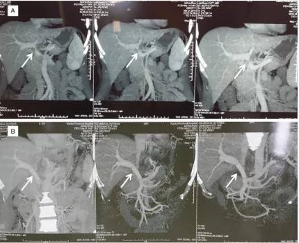
Preoperative assessment
To establish the nature, assess extent of tu-mor and identify vasculatures details as well
as estimate remnant liver volume, laboratory
examination and imaging workups of ultraso -nography, magnetic resonance cholangiopan-creatography (MRCP), multi-detector row
com-puted tomography (MDCT) were performed for
all candidates. Additionally, patients who were scheduled to accept major hepatectomy with serum total bilirubin over 200 umol/L were suggested with preoperative biliary drainage
prior to surgery. 26 out of 95 candidates under -went preoperative biliary drainage via percuta-neous transhepatic cholangial drainage (PTCD) or endoscopic nasal biliary drainages (ENBD)
including 8 patients of vascular reconstruction
group (PTCD=7, ENBD=1). In addition, no
preop-erative portal vein embolization (PVE) was indi -cated in any patients. Ultimately, general
condi-tion of all candidates met the requirements of Child-Pugh classification (grade A to B), which to certain extent warrants the safety of
surgery.
Operative procedures
Surgical procedures consisting of hemihepa -tectomy, extended hemihepa-tectomy, central hepatectomy, external bile duct resection with or without caudate lobectomy as well as com-bined invaded vessels resection and
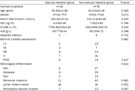
recon-struction were performed for patients individu -Table 1. Clinicopathologic and demographic features of 95 HCCA patients who underwent curative
resection
Vascular resection group Non-vascular resection group P-value
Number of patients n=19 n=76
Age (years) 55.40±11.88 61.03±10.48 0.045
Gender M=14, F=5 M=43, F=33 0.173
Serum total bililubin (umol/L) 242.8±137.14 213.11±143.95 0.420
CEA (ng/ml) 4.33±3.46 7.5±23.89 0.794
CA199 (U/ml) 7753.6±22914.28 1031.96±2081.51 0.103
ALB (g/L) 36.77±5.43 38.25±4.71 0.198
Hepatitis infection 3 9 0.701
Bismuth Corlette classification 0.691
I/II 2 15
IIIA 2 7
IIIB 8 23
IV 7 31
PTCD 8 18 0.107
Pathological differentiation 0.813
Well 6 27
Moderate 8 26
Poor 5 23
Perineural invasions 11 40 0.681
Lymph nodes invasion 16 31 0.001
[image:2.612.92.524.96.388.2]ally. Locoregional lymph nodes including nodes along the common hepatic artery, in the hepa-toduodenal ligament, and posterior
pancreati-coduodenal nodes were routinely dissected for
all patients, and aggressive lymph nodes dis-section was decided by the attending surgeons.
Upon completion of tumor resection, biliary
continuity was restored by Roux-en-Y
anasto-mosis. To further analyze the nature of tumor affecting the prognosis, tumor was classified as well differentiated, moderately differentiat
-ed, poorly differentiated adenocarcinoma after
resection according to the predominant
patho-up was performed with portal vein arterializa
-tion (PVA). Vascular patency after surgery
was assessed by Doppler ultrasonography or
MDCT. The details of surgical procedures for
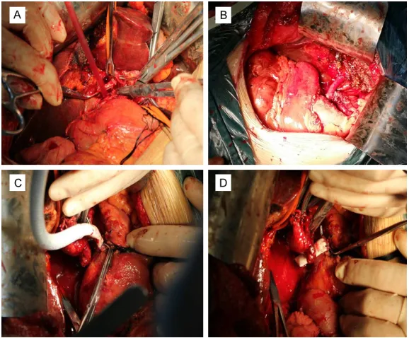
vascular resection and reconstruction were depicted in Table 2. A typical portal vein
recon-struction with vascular graft was presented in
Figures 1 and 2.
Morbidity and mortality
Mortality was defined as any postoperative
death occurring in-hospital stay. Major compli-Table 2. Surgical procedures of 19 HCCA patients with vascular recon
-struction
Details of vascular resection and reconstruction Number of patients
Bismuth Corlette classification 19
I 0
II 2
IIIA 2
IIIB 8
IV 7
Preoperative drainage 8
PTCD 7
ENBD 1
Surgical procedure
Left hepatectomy 14
With S1 lobectomy 8
Without S1 lobectomy 6
Extended left hepatecomy 3
With S1 lobectomy 2
Without S1 lobectomy 1
Right hepatectomy 1
With S1 lobectomy 0
Without S1 lobectomy 1
Extended right hepatectomy 1
Types of vessels invasion and reconstruction modes
Portal vein bifurcation only 10
End to end (E to E) 9
Vascular graft 1
Right hepatic artery only 6
End to end 5
Anastomosis with gastrointestial artery 1
Portal vein plus hepatic artery 3
Portal vein bifurcation (E to E) plus right hepatic artery (E to E) 2 Portal vein bifurcation (E to E) plus hepatic proper artery
(anastomosis gastrointestinal artery with right hepatic artery) 1
Portal vein bifurcation only 10
End to end (E to E ) 9
logic grading of
differ-entiation. Besides that, perineural invasions, ly- mph nodal metastases and microscopic vascular invasion that generated
from pathological results were also further
asse-ssed.
Approach to vascular resection and recon-struction
Preoperative MDCT or MRCP was routinely ap-
plied to assess infiltra
-tion of vessels. Surgical policies of portal vein or
hepatic artery resection and reconstruction were carried out only when vessels adhered to and
could not be freed from
tumor entity during
intra-operative skeletonization of hepatoduodenal liga -ment [10]. The portal vein was reconstructed by
model of end to end
anastomosis (n=12) or
vascular grafts (n=1)
be-tween the resected re- sidual trunk and corre-sponding branch, and the hepatic artery recon-struction relied on end to
end fashion (n=4) or
anastomosis with gastro-duodenal artery (n=2).
[image:3.612.93.398.97.560.2]cations were regarded as having a grade of III-IV complication according to the Clavien-Dindo Classification [11].
Statistics
Continuous variables were expressed as mean ± standard deviation. Categorical variables were expressed as numbers. Continuous or ca- tegorical variables comparison between the-
se two groups was performed by a Student’s t
test (two-tailed) or Mann-Whitney U tests and
χ2 test or Fisher’s exact test. Cumulative Sur-
vival time counted from the month of surgery was calculated with the Kaplan-Meier method
and difference in survival curves were com -pared with log-rank test, respectively. The Cox
proportional hazard model was used for multi
-variate analysis of survival after curative resec -tion basing on the interesting variables that
were statistically significant by univariate analy -sis. A P-value <0.05 was considered as
statisti-cally significance. Statistical analysis was
per-formed with SPSS software, version 17.0.
Results
Clinicopathologic and demographic features The current study population consisted of 57 male and 38 female patients with a mean age
minutes and that of hepatic artery reconstruc
-tion was 9.4 minutes. Vascular anastomosis
was constructed by continuous sutures with 7-0/8-0 prolene.
Morbidity and mortality
Postoperative complications occurred to 84.2%
patients (n=16) of 19 patients including 6 with
major complication. Bile leakage was the most
common complication (n=10, 52.6%), following
the ascites (n=3, 15.8%) and intra-abdominal
infection (n=3, 15.8%). One patient (5.26%)
encountered a second laparotomy due to in-
tra-abdominal bleeding within 7 days after sur -gery and one patient (5.26%) who underwent
portal vein reconstruction died of liver failure resulting in multiple organ failure within 2
weeks postoperatively. Neither arterial aneu-rysm nor vascular thrombi nor vascular occlu-sion nor vascular anastomosis bleeding was
identified in patients of vascular reconstruction group. The more details of morbidity and mor
-tality of each group was summarized in Table 3.
Survival
The 1-year, 3-year and 5-year survival rate were 78.8%, 21.3%, 0%, respectively in vascular reconstruction group with median survival time Figure 1. Portal vein resection and reconstruction with vascular graft. A:
Ready for resection; B: Removed the tumor mass with left hepatectomy; C: Vascular graft anastomosis with distal portal vein; D: Vascular graft anasto -mosis with proximal portal vein.
of 59.9 years (range, 28-82
years). According to the Bis-
muth-Corlette Classification,
9 patients were with type I, 8 with type II, 9 with type IIIA, 31 with type IIIB, 38 with type
IV. The clinicopathologic and demographic features show-ed no significant difference factors between two groups
in Table 1, except for age,
lymph node invasion.
Operation data
The mean operation time for
patients in vascular recon-struction group was 6.26±
1.08 h. The mean amount of
blood loss was 563.16±377.4
ml and blood transfusion was performed on 5 patients dur -ing operation with mean 3 U
red blood cell transfusion. The median time for portal
[image:4.612.89.378.71.310.2]of 17 months and 79.3%, 31.8%, 12.3%
res-pectively in non-vascular group with median
survival time of 22 months (P=0.416, Figure 3).
At the median follow up time of 32 months,
there are still 6 patients alive in vascular
recon-struction group. Moreover, 2 out of these 6
patients survived more than 30 months with
tumor free and one of them underwent right
hepatic artery plus portal vein reconstruction,
the other one was after hepatic artery recon -struction alone.
Analysis of prognostic factors
In this series, univariate analysis based on Kaplan-Meier method showed that
pathologi-cal differentiation, Lymph node invasion and CA-199 above 200 U/ml proved to be signifi
-cant factors (Figure 4). Simultaneously,
multi-variate Cox proportional hazards regression analysis also identified that CA-199 above 200
U/ml (P=0.035) and pathological differentia -tion (P=0.015) were independent prognostic
factors affecting postoperative survival (Table 4).
Discussion
Vascular invasion was a major obstacle to achieve radical resection for advanced HCCA previously. However, with the progress of
advanced hepatobiliarypancreatic surgeries, concomitant vascular resection and
recon-struction are currently recognized as a means to increase the rate of resectability with accept -able survival and mortality [12, 13]. The
[image:5.612.91.524.71.424.2]report-ed percentage of portal vein resection in HCCA varies from 9.8 to 37% and that from 1.7 to 18
% [14-16] of hepatic artery resection. The 5-year survival rate of combined portal vein reconstruction for advanced HCCA from lead -ing centers was close to 9.9%-25% with
mortal-ity of 16-17% [17-20], while few 5-year survivor
with hepatic artery reconstruction was
report-ed with higher mortality of 33-55% [1].
Nagigo et al [9] reported that the overall R0 resection rate was 66% with 1-, 3-, and 5-year
survival rates of 78.9%, 36.3%, 30.3% respec
-tively and mortality of 2% for 50 patients of
advanced HCCA who underwent simultaneous portal vein and hepatic artery reconstruction.
In the study of Miyazki et al [1], the 1-, 3-, 5-year
bifurcation [1, 6, 7, 9, 13], which was main rea
-son why most surgeons favor right hepatecto
-my for patients with type IV lesion. For one
thing, preserving the right hepatic artery and right portal vein could be an oncological
prob-lem with left or extended left hepatectomy,
which could cause tumor cell dissemination
[22]. For another, the left hepatic artery runs through the leftmost portion of the hepatoduo
-denal ligament and can be left undisturbed dur -ing right-sided hepatectomy [6]. Furthermore, it
is the fact that the right liver accounts for 60-70% of the total liver volume. Therefore, to
[image:6.612.94.380.84.527.2]certain extent, right hemihepatectomy or extended right hemihepatectomy carries more Table 3. Complications of 95 HCCA patients with curative resection
No. of patients in vascular reconstruction
group n=19
No. of patients in non-vascular resection group
n=76 Morbiditya
Grade IVa
Hepatic encephalopathy 0 1 (1.3%) Hepatic or renal insufficiency 0 4 (5.3%)
ARDS 0 2 (2.6%)
Grade IIIb
Intra-abdominal abscess 0 0
Liver abscess 0 0
Bilioenteric anastomosis bleeding 0 2 (2.6%) Intra-abdominal bleeding 1 (5.2%) 2 (2.6%) Grade IIIa
Intra-abdominal abscess 0 2 (2.6%) Gastrointestinal bleeding 1 (5.2%) 8 (10.5%)
Pleural effusion 1 (5.2%) 12 (15.8%)
Ascites 3 (15.8%) 17 (22.4%)
Liver absecess 0 1 (1.3%)
Grade II
Bile leakage 10 (52.6%) 28 (36.8%)
Pneumonia 2 (10.4%) 13 (17.1%)
Pulmonary abscess 0 1 (1.3%)
Intra-abdominal infection 3 (15.8%) 18 (23.7%)
Sepsis 0 8 (10.5%)
Wound infection 1 (5.2%) 2 (2.6%)
Grade I 4 (21.0%) 10 (13.16%)
No. of complications 19 136
No. of Patients with complications 16 (84.21%) 45 (59.2%) No. of Patients with major complications 6 (31.58%) 23 (30.26%) Postoperative hospital stays (day) 19±16.4 16±25.7 Mortality
In-hospital death 1 (5.2%) 3 (3.95%)
a: according to the Clavein-Dindo classification.
survival rates in portal vein reconstruction group were 47%, 31%, and 25%,
respec-tively and that of 17%, 0%,
0%, respectively in hepatic artery reconstruction group. Additionally, Gerhards et al
[21] performed combined vascular resection for 12
HCCA patients including hepatic artery (n=2), portal vein (n=3) and portal vein plus hepatic artery (n=7),
but the mortality of portal
vein resection group was
3/10 and 5/9 of hepatic
artery resection group. Ba- sed on the previous litera-tures published, it seemed
to reveal that survival of
combined hepatic artery re-
section was inferior to that
in portal vein resection and may bring about higher mor-tality. The possible
explana-tion for this discrepancy was
that hepatic artery resec-tion were required
com-bined resection of affected
portal vein in most patients, which may obligate longer
periods of liver ischemia for
vascular reconstruction and result in more severe isch-emic damage to the
rem-nant liver after major hepa -tectomy [1].
Additionally, right hepatic artery involvement is more
frequent in HCCA patients
surgical risks on
postopera-tive liver failure, even death. On the contrary, 9 patients of type IV lesion (47.37%) with right hepatic artery infiltrati-on were performed with left
hemihepatectomy and right hepatic artery reconstruction in this series. As a result, all these 9 patients obtained R0
resection and none of pa-tients suffered from acute liver failure. From a surgeon’s
perspective, surgical policy
was flexible that may largely
depend on the predominance
of tumor location, especially for type IV lesion with contra -lateral vascular invasion.
[image:7.612.93.518.76.360.2]In the present study, 1-year and 3-year survival rates were 78.8%, 21.3% respectively as Figure 3. Cumulative Survival curves were calculated with Kaplan-Meier method. Survival of combined vascular resection and reconstruction group was comparable of that in non-vascular resection and reconstruction (P=0.416). Univariate analysis identified that lymph node invasion, preoperative CA-199≥200 U/ml, and pathologic differentia -tion were the significant factors affecting postoperative survival.
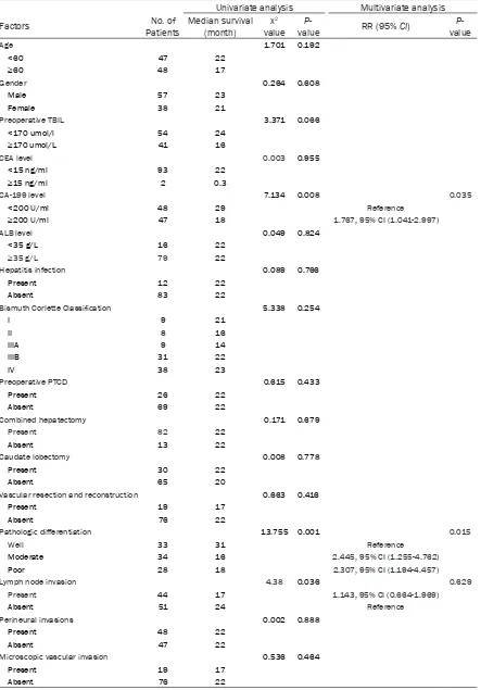
[image:7.612.88.378.432.674.2]Table 4. Univariate and Multivariate analysis of 95 HCCA patients with curative resection
Univariate analysis Multivariate analysis Factors Patients No. of Median survival (month) valuex2 value P- RR (95% CI) valueP
-Age 1.701 0.192
<60 47 22
≥60 48 17
Gender 0.264 0.608
Male 57 23
Female 38 21
Preoperative TBIL 3.371 0.066
<170 umol/l 54 24
≥170 umol/L 41 16
CEA level 0.003 0.955
<15 ng/ml 93 22
≥15 ng/ml 2 0.3
CA-199 level 7.134 0.008 0.035
<200 U/ml 48 29 Reference
≥200 U/ml 47 18 1.767, 95% CI (1.041-2.997)
ALB level 0.049 0.824
<35 g/L 16 22
≥35 g/L 79 22
Hepatitis infection 0.089 0.766
Present 12 22
Absent 83 22
Bismuth Corlette Classification 5.338 0.254
I 9 21
II 8 16
IIIA 9 14
IIIB 31 22
IV 38 23
Preoperative PTCD 0.615 0.433
Present 26 22
Absent 69 22
Combined hepatectomy 0.171 0.679
Present 82 22
Absent 13 22
Caudate lobectomy 0.008 0.778
Present 30 22
Absent 65 20
Vascular resection and reconstruction 0.663 0.416
Present 19 17
Absent 76 22
Pathologic differentiation 13.755 0.001 0.015
Well 33 31 Reference
Moderate 34 16 2.445, 95% CI (1.255-4.762)
Poor 28 18 2.307, 95% CI (1.194-4.457)
Lymph node invasion 4.38 0.036 0.629
Present 44 17 1.143, 95% CI (0.664-1.969)
Absent 51 24 Reference
Perineural invasions 0.002 0.888
Present 48 22
Absent 47 22
Microscopic vascular invasion 0.536 0.464
Present 19 17
well as the median survival time of 17 months
in vascular reconstruction group, which was
comparable of that in non-vascular resection
(P=0.416). Although no patient in vascular reconstruction group has survived over 5 years
till now, in comparison with median survival of 3 to 6 months of studies in terms of the natural history of advanced cholangiocarcinoma with -out any interventional procedures [23, 24], our
results demonstrated significant survival improvement and benefits. Additionally, 2
patient who underwent concomitant portal vein and hepatic artery reconstruction have
sur-vived for 40 months, 20 months respectively with tumor free and another one has survived for 30 months after combined hepatic artery reconstruction. Although the outcomes of our study was not favorable but acceptable, at
least, combined vascular resection and
recon-struction have offered selected patients an opportunity for long-term survival.
Nevertheless, the incidence of histologically
proven microscopic vascular invasion was
lower in recent series than that of macroscopic
vascular invasion [6, 9], a multivariate analysis
of a convictive study [6] revealed that macro
-scopic invasion of portal vein but micro-scopic
invasion had a negative impact on prognosis.
Approximately one third of resected portal vein specimens were not infiltrated microscopically, yet tumor infiltration adjacent to portal vein was
detected in most cases. Put another way, the resection margin would be positive without combined vascular resection. Undoubtedly, it
has been an approved evidence to emphasize the necessities and effects of combined vascu
-lar resection for advanced HCCA. Simi-larly, in
our series, pathologically microscopic vascular
invasion was found in only 7 (36.8%) resected
specimens in vascular reconstruction group (Figure 4). Multivariate analysis indicated that
pathological differentiation was an indepen
-dent prognostic factors for survival (P=0.015).
Obviously, whatever the feature of macroscopic vascular invasion or pathological differentia -tion seems to be associated with biological
behavior of HCCA. Therefore, further investiga
-tions are required to clarify the tumor nature that would cause far-reaching impact on sur
-vival for HCCA patients.
With no doubt, the toughest challenge for sur
-geons is how to identify vascular invasion and
to rebuild vascular patency when those with
severe tumor infiltration. Based on our limited
experience and reviews, for one thing, both of invasion of Glission sheath and detecting local
stenosis, distal pulses weaken or
disappear-ance of hepatic artery intraoperatively may sug -gest vascular invasion. For another, vascular resection without detection preoperatively by MDCT or MRCP should be still recommended
when vascular involvement was found on gross
inspection during operation. With regard to vas-cular reconstruction, we tend to adopt vasvas-cular
grafts or anastomosis with adjacent artery when suffering from the difficulty in hepatic
artery reconstruction. In this series, 2 patients
underwent anastomosis of right hepatic artery
and gastroduodenal artery, and another one
had vascular graft for portal vein reconstruc
-tion. Consequently, no liver failure occurred to each patient postoperatively. Additionally, PVA
is also proposed to be a salvage therapy to
maintain arterial inflow when hepatic artery
reconstruction is impossible [25]. Because all
of available reports were with small sample sizes, further investigations are required to measure its significance.
However, there were some drawbacks in this
retrospective study. First and foremost, the sur
-vival and mortality of hepatic artery reconstruc
-tion was different from that of portal vein recon -struction, but they were combined into a new
group due to small sample size, which may resulted in magnifying the survival benefits and
reducing the morbidity rate as well as making statistically unreasonable. Furthermore, in light
of the retrospective nature of most published
series including the current one, more valid data about combined vascular resection and
reconstruction should be provided by the future
multi-center prospective studies. Last but not
the least, owing to the randomization and con
-trol appearing hardly feasible, the actual bene
-fits of combined vascular resection and recon
-struction for advanced HCCA patients worth further exploring and clarifying.
In summary, combined vascular resection and
reconstruction can be performed with accept
-able prognosis and mortality for advanced
HCCA patients.
Acknowledgements
This work was supported by Grant 15 y kpy 20
Labo-ratory of Malignant Tumor Mechanism and Translational Medicine of Guangzhou Bureau of Science and Information Technology; Grant KLB 09001 from the Key Laboratory of
Mali-gnant Tumor Gene Regulation and Target
Therapy of Guangdong Higher Education
Insti-tutes.
Disclosure of conflict of interest
None.
Address correspondence to: Jie Wang and Junyao Xu, Department of Hepatobiliary Surgery, Sun Yat-Sen Memorial Hospital, Sun Yat-Yat-Sen University, #33 Yingfeng Road, Guangzhou 510120, P. R. China. Tel: 86-20-34071175; Fax: 86-20-81332853; E-mail: sumsjw@163.com (JW); Fax: 86-20-34091489; E-mail: xuyuny@mail.sysu.edu.cn (JYX)
References
[1] Miyazaki M, Kato A, Ito H, Kimura F, Shimizu H, Ohtsuka M, Yoshidome H, Yoshitomi H, Furu -kawa K and Nozawa S. Combined vascular re -section in operative re-section for hilar cholan -giocarcinoma: does it work or not? Surgery 2007; 141: 581-588.
[2] Hayashi S, Miyazaki M, Kondo Y and Nakajima N. Invasive growth patterns of hepatic hilar ductal carcinoma. A histologic analysis of 18 surgical cases. Cancer 1994; 73: 2922-2929. [3] Jarnagin WR, Fong Y, DeMatteo RP, Gonen M,
Burke EC, Bodniewicz BJ, Youssef BM, Klims -tra D and Blumgart LH. Staging, resectability, and outcome in 225 patients with hilar cholan-giocarcinoma. Ann Surg 2001; 234: 507-517; discussion 517-509.
[4] Ito F, Cho CS, Rikkers LF and Weber SM. Hilar cholangiocarcinoma: current management. Ann Surg 2009; 250: 210-218.
[5] Kondo S, Takada T, Miyazaki M, Miyakawa S, Tsukada K, Nagino M, Furuse J, Saito H, Tsuyu-guchi T, Yamamoto M, Kayahara M, Kimura F, Yoshitomi H, Nozawa S, Yoshida M, Wada K, Hirano S, Amano H, Miura F; Japanese Associ-ation of Biliary Surgery; Japanese Society of Hepato-Biliary-Pancreatic Surgery; Japan Soci-ety of Clinical Oncology. Guidelines for the management of biliary tract and ampullary car -cinomas: surgical treatment. J Hepatobiliary Pancreat Surg 2008; 15: 41-54.
[6] Ebata T, Nagino M, Kamiya J, Uesaka K, Naga-saka T and Nimura Y. Hepatectomy with portal vein resection for hilar cholangiocarcinoma: audit of 52 consecutive cases. Ann Surg 2003; 238: 720-727.
[7] Abbas S and Sandroussi C. Systematic review and meta-analysis of the role of vascular re -section in the treatment of hilar cholangiocar -cinoma. HPB (Oxford) 2013; 15: 492-503. [8] de Jong MC, Marques H, Clary BM, Bauer TW,
Marsh JW, Ribero D, Majno P, Hatzaras I, Wal -ters DM, Barbas AS, Mega R, Schulick RD, Choti MA, Geller DA, Barroso E, Mentha G, Ca-pussotti L and Pawlik TM. The impact of por-tal vein resection on outcomes for hilar cholan -giocarcinoma: a multi-institutional analysis of 305 cases. Cancer 2012; 118: 4737-4747. [9] Nagino M, Nimura Y, Nishio H, Ebata T, Igami T,
Matsushita M, Nishikimi N and Kamei Y. Hepa-tectomy with simultaneous resection of the portal vein and hepatic artery for advanced perihilar cholangiocarcinoma: an audit of 50 consecutive cases. Ann Surg 2010; 252: 115-123.
[10] Serrablo A and Tejedor L. Outcome of surgical resection in klatskin tumors. World J Gastroin-test Oncol 2013; 5: 147-158.
[11] Dindo D, Demartines N and Clavien PA. Clas-sification of surgical complications. Ann Surg 2004; 240: 205-213.
[12] Kondo S, Katoh H, Hirano S, Ambo Y, Tanaka E and Okushiba S. Portal vein resection and re -construction prior to hepatic dissection during right hepatectomy and caudate lobectomy for hepatobiliary cancer. Br J Surg 2003; 90: 694-697.
[13] Sakamoto Y, Sano T, Shimada K, Kosuge T, Kimata Y, Sakuraba M, Yamamoto J and Ojima H. Clinical significance of reconstruction of the right hepatic artery for biliary malignancy. Lan -genbecks Arch Surg 2006; 391: 203-208. [14] Nuzzo G, Giuliante F, Ardito F, Giovannini I, Ald
-righetti L, Belli G, Bresadola F, Calise F, Dalla Valle R, D’Amico DF, Gennari L, Giulini SM, Guglielmi A, Jovine E, Pellicci R, Pernthaler H, Pinna AD, Puleo S, Torzilli G, Capussotti L; Ital -ian Chapter of the International Hepato-Pan -creato-Biliary Association, Cillo U, Ercolani G, Ferrucci M, Mastrangelo L, Portolani N, Puli-tanò C, Ribero D, Ruzzenente A, Scuderi V, Fed -erico B. Improvement in perioperative and long-term outcome after surgical treatment of hilar cholangiocarcinoma: results of an Italian multicenter analysis of 440 patients. Arch Surg 2012; 147: 26-34.
[15] Igami T, Nishio H, Ebata T, Yokoyama Y, Suga-wara G, Nimura Y and Nagino M. Surgical treat-ment of hilar cholangiocarcinoma in the “new era”: the Nagoya university experience. J Hepa-tobiliary Pancreat Sci 2010; 17: 449-454. [16] Lee SG, Song GW, Hwang S, Ha TY, Moon DB,
experience. J Hepatobiliary Pancreat Sci 2010; 17: 476-489.
[17] Klempnauer J, Ridder GJ, von Wasielewski R, Werner M, Weimann A and Pichlmayr R. Resec-tional surgery of hilar cholangiocarcinoma: a multivariate analysis of prognostic factors. J Clin Oncol 1997; 15: 947-954.
[18] Miyazaki M, Ito H, Nakagawa K, Ambiru S, Shi -mizu H, Okaya T, Shinmura K and Nakajima N. Parenchyma-preserving hepatectomy in the surgical treatment of hilar cholangiocarcino -ma. J Am Coll Surg 1999; 189: 575-583. [19] Neuhaus P, Jonas S, Bechstein WO, Lohmann
R, Radke C, Kling N, Wex C, Lobeck H and Hin-tze R. Extended resections for hilar cholangio -carcinoma. Ann Surg 1999; 230: 808-818; discussion 819.
[20] Lee SG, Lee YJ, Park KM, Hwang S and Min PC. One hundred and eleven liver resections for hilar bile duct cancer. J Hepatobiliary Pancreat Surg 2000; 7: 135-141.
[21] Gerhards MF, van Gulik TM, de Wit LT, Obertop H and Gouma DJ. Evaluation of morbidity and mortality after resection for hilar cholangiocar -cinoma-a single center experience. Surgery 2000; 127: 395-404.
[22] Neuhaus P, Jonas S, Settmacher U, Thelen A, Benckert C, Lopez-Hanninen E and Hintze RE. Surgical management of proximal bile duct cancer: extended right lobe resection increas-es rincreas-esectability and radicality. Langenbecks Arch Surg 2003; 388: 194-200.
[23] Farley DR, Weaver AL and Nagorney DM. “Nat -ural history” of unresected cholangiocarcino -ma: patient outcome after noncurative inter -vention. Mayo Clin Proc 1995; 70: 425-429. [24] Park J, Kim MH, Kim KP, Park do H, Moon SH,
Song TJ, Eum J, Lee SS, Seo DW and Lee SK. Natural history and prognostic factors of ad -vanced cholangiocarcinoma without surgery, chemotherapy, or radiotherapy: a large-scale observational study. Gut Liver 2009; 3: 298-305.