Original Article
Cornuside ameliorates airway inflammation via Toll-like
receptor 4 and Notch1 in asthmatic mice induced
by lipopolysaccharide and ovalbumin
Liangchang Li1*, Guangyu Jin2*, Yan Jin3, Jingzhi Jiang1, JinshiYang3, Yunho Choi4, Zhewu Jin1, Mingyu Zheng3, Guanghai Yan1
1Department of Anatomy, Histology and Embryology, School of Basic Medical Sciences, Yanbian University, Yanji, P. R. China; 2Yanbian University Hospital, Medicine College, Yanbian University, Yanji, P. R. China; 3College of Pharmacy, Yanbian University, Yanji, P. R. China; 4Department of Anatomy, Medical School, Institute for Medical Sciences, Chonbuk National University, Jeonju, Republic of Korea. *Equal contributors.
Received February 2, 2016; Accepted July 10, 2016; Epub August 15, 2016; Published August 30, 2016
Abstract: To determine the potential role of cornuside in the regulation of allergic airway inflammation in lipopoly-saccharide (LPS) and ovalbumin (OVA)-induced asthma. BALB/c mouse model of LPS and OVA-induced asthma was used to evaluate the effect of cornuside on Toll-like receptor 4 (TLR4) and Nocth1 signaling pathway. Hematoxylin and eosin staining was performed for histological studies of murine lung tissues. Enzyme-linked immunosorbent as-say was employed to measure protein levels in serum, while Western blotting was carried out for the determination of protein expression in tissues. Airway hyperresponsiveness was measured 2 days after the last OVA challenge. After induction by LPS and OVA, the mice had increased numbers of inflammatory cells, increased levels of IL-4, IL-5 and IL-13 in bronchoalveolar lavage fluids (BAL) and lung tissues, increased total and OVA-specific IgE levels in serum and increased expression of TLR4 and Nocth1 and phosphorylation of STAT3 in lung tissues. Administration of cornuside markedly reduced airway inflammatory cell recruitment and peribronchiolar inflammation, decreased the production of various cytokines in BAL fluids and lung tissues, and lowered the levels of total and OVA-specific IgE in serum. In addition, the increased expression of TLR4 and Nocth1 and phosphorylation of STAT3 after LPS and OVA inhalation was inhibited by the administration of cornuside. The present study demonstrates that the signaling pathway between TLR4 and Notch1 may be able to coordinate with STAT3 and cornuside regulates TLR4 and Nocth1 signaling pathway to attenuate allergic airway inflammation in LPS and OVA-induced asthma. These findings provide a crucial molecular mechanism for the potential use of cornuside to prevent and/or treat asthma and other airway inflammatory disorders.
Keywords: Cornuside, asthma, TLR4, Notch1, STAT3, airway inflammation
Introduction
Asthma remains a major cause of morbidity in developed nations and is a leading cause of hospitalization [1]. The characteristic fea-tures of asthma are airway inflammation, epi-thelial damage, reversible airflow obstruction, airway hyperreactivity, and airway remodeling [2]. It is well recognized that respiratory in- fection mediates allergic airway inflammation [3]. However, genetic and environmental fac-tors that contribute to progression of asthma remain poorly understood. Pattern recognition receptors (PRRs) is an umbrella term for sever-al receptor families that have specific abilities to recognize various microbes, and to initiate
Several studies have shown that TLR4 activa-tion by LPS promotes inflammatory mecha-nisms including nuclear factor (NF)-κB and Janus-activated kinase (JAK)/signal transduc-ers and activators of transcription (STAT) path-ways [10]. STAT3 is a well-known transcription factor that regulates a variety of cellular pro-cesses, including cell proliferation and survival, oncogenesis and cancer metastasis. STAT3 also has a close interaction with Notch recep-tors in various physiological and pathological conditions, including proliferation, differentia-tion, and apoptosis [12]. Upon ligand-depen-dent activation, Notch is cleaved and releases the Notch intracellular domain, which partici-pates in a transcriptional complex in the nucle-us to regulate Notch-dependent gene expres-sion [13]. Blockade of Notch has been reported to inhibit T lymphocyte activation and airway inflammation in asthma [14, 15]. TLR4 and Notch signaling pathways are all involved in asthma, but these cell signaling pathways are not completely independent of each other. For example, cross-talks take place between differ-ent signaling pathways. We hypothesize that signaling pathways between TLR4 and Notch coordinate with each other in regulating airway inflammation in asthma.
Cornuside is a kind of secoiridoid glucoside iso-lated from the fruit of Cornus officinalis Sieb. et Zucc., which is a traditional oriental medicine for treating inflammatory diseases and invigo-rating blood circulation. A crude extract from the fruits of Cornus officinalis has been found to have pharmacological actions such as anti-neoplasmic, anti-inflammatory, hepatoprotec-tive, and antidiabetic effects [16-18]. Recent reports have demonstrated that cornuside could suppress the expression of cytokine-induced pro-inflammatory and adhesion mole-cules in human endothelial cells, and could pro-tect cultured rat cortical cells against damages induced by oxygen-glucose deprivation [19, 20]. Furthermore, cornuside has been demon-strated to inhibit LPS-induced nitric oxide (NO) production in cultured mouse macrophages [21]. Our previous studies have revealed that cornuside inhibits acute hepatic injury induced by carbon tetrachloride, and suppresses LPS-induced inflammatory mediators by inhibiting NF-κB activation in RAW 264.7 macrophages [22, 23]. However, a direct relationship between
cornuside and airway inflammation has not yet been understood in murine model of asthma. In the present study, we determine the effect of cornuside on airway inflammation in animal model of asthma. In addition, we examine whether TLR4 and Notch signaling pathways are involved in the pathophysiological process of asthma.
Materials and methods
Animals
LPS exposure
In brief, the mice were exposed to 0.1 μg LPS (from Escherichia coli 0111:B4; Sigma-Aldrich, St. Louis, MO, USA) on day 2 of sensitization an hour before antigen administration and on each day of OVA-aerosol challenge in 100 μL sterile saline [24]. Mice in control group were given LPS-free saline.
Harvest of bronchoalveolar lavage (BAL) fluid
and cytospin preparations
Immediately following assessment of airway responsiveness, the mice were anesthetized by intraperitoneal injection of pentobarbital (50 mg/kg body weight; Wuhan Boster Biological Technology, Ltd., Wuhan, China) and the tra- cheas were cannulated while gently massag- ing the thorax. The lungs were lavaged with 0.7 ml of PBS. BAL fluid samples were collected and the number of total cells in a 0.05 mL ali-quot was counted using a hemocytometer. The remaining samples were centrifuged at 200 g for 10 min at 4°C (model 5424R,
Eppendorf-4% paraformaldehyde, and embedded in paraf-fin. Specimens were cut into 4 μm sections by Leica model 2165 rotary microtome (Leica, Nussloch, Germany). The microsections were stained with hematoxylin and eosin (Richard-Allan Scientific, Kalamazoo, MI, USA) and exam-ined under the microscope (CX31 microscope; Olympus Corporation, Tokyo, Japan) with a magnification of × 100.
Enzyme-linked immunosorbent assay (ELISA)
IL-4, IL-5, and IL-13 levels in BAL and IgE in serum were determined using mouse ELISA kits for IL-4, IL-5, IL-13 and IgE (R&D Systems, Minneapolis, MN, USA) according to the manu-facturer’s protocols.
[image:3.612.93.371.72.301.2]To measure total IgE and OVA-specific IgE, 96- well microtiter plates were coated overnight with isotype-specific coating (total IgE) and 10 mg/mL OVA in PBS-Tween 20 (OVA-specific IgE). After washing and blocking of plate, sam-ples were incubated for 2 hours. Subsequent- ly, 96-well plates were washed, and HRP-con- Figure 1. Effect of cornuside on total and differential cellular components
of BAL in LPS and OVA-induced asthmatic mice. Numbers of counted cells of each type in BAL. LPS and OVA-induced asthmatic mice were orally given cornuside or DXM (positive control) daily on days 13-19 for seven consecu-tive days after the first sensitization. The effects of cornuside and DXM on LPS and OVA-induced changes in cell counts in BAL fluid were analyzed 24 h after the last OVA challenge. Results of five independent experiments (7 mice/group) were expressed as means ± standard errors of the mean. #, P < 0.05 compared with control group; *, P < 0.05 compared with LPS and OVA-induced group.
Netheler, Hamburg, Germany) and the supernatants were stored at -70°C for the as- say of IL-4, IL-5 and IL-13 levels. The cell pellets were resuspended in PBS and cy- tospin preparations (Cytos- pin 3, Shandon Life Scien- ces, Astmor, UK) of BAL cells were stained with Diff-Quik solution (International Rea- gents, Kobe, Japan). Two inde-pendent, blinded investiga-tors counted the cells using a microscope (CX31 micro-scope; Olympus Corporation, Tokyo, Japan). Approximately 400 cells were counted in each of four different rand- om locations. Inter-investiga- tor variation was < 5%. The mean number from the two investigators was used to estimate cell differentials. Hematoxylin-eosin (HE) stain-ing
jugated goat anti-mouse total IgE and OVA spe-cific IgE were added. After washing four times, 200 μL of o-phenylenediamine dihydrochloride (Sigma-Aldrich, St. Louis, MO, USA) was added to each well. The plate was incubated for 10 min in the dark and then the absorbance was determined at 450 nm using a Bio-Rad 680 microplate reader (Bio-Rad, Hercules, CA, USA). Total IgE and OVA-specific IgE concentrations were calculated from a standard curve gener-ated using 250 ng/mL recombinant IgE.
Western blotting
Lung tissues were homogenized in the pres-ence of protease inhibitors and protein concen-trations were determined using the Bradford reagent (Bio-Rad, Hercules, CA, USA). Protein sample (30 μg) from the lung homogenates was loaded per lane on 12% sodium dodecyl sulfate polyacrylamide gel electropheresis gel. Electrophoresis was then performed. The pro-teins were then transferred to nitrocellulose membranes. Western blotting analysis was performed using polyclonal antibodies against IL-5, TLR4, STAT3, p-STAT3, notch1, β-actin (Santa Cruz, Santa Cruz, CA, USA), IL-13 (R&D
Systems, Minneapolis, MN, USA) and IL-4 (Se- rotec, Oxford, UK). The binding of all the anti-bodies was detected using an electrochemi- luminescence detection system (Amersham, Arlington Heights, IL, USA) according to the manufacturer’s instructions.
Assessment of AHR
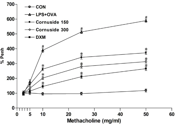
[image:4.612.92.524.71.288.2]Penh over the baseline, following challenges performed with each concentration of Mch, where the baseline Penh (after PBS challenge) was expressed as 100%.
tures of asthma, HE staining was used. His- tological analyses revealed that mice with as- thma induced by LPS and OVA had more wide-spread perivascular and peribronchiolar inflam-Figure 3. Effect of cornuside on Th2 cytokines in BAL fluids and lung tissues
of LPS and OVA-induced asthmatic mice. A. Protein expressions of IL-4, IL-5 and IL-13 in lung tissues after 24 h measured by Western blotting. B. The levels of IL-4, IL-5 and IL-13 in BAL fluids quantified by ELISA. BAL fluids were collected 24 h after the last challenge. Results from five independent experi-ments with 7 mice/group were given as means ± SEM. #, P < 0.05 compared with control; *, P < 0.05 compared with LPS and OVA -induced group. CON, saline-treated mice; LPS+OVA, LPS and OVA-induced asthmatic mice; Cornu-side 150, LPS and OVA-induced asthmatic mice treated with 150 mg/kg cor-nuside; Cornuside 300, LPS and OVA-induced asthmatic mice treated with 300 mg/kg cornuside; DXM, LPS and OVA-induced asthmatic mice treated with 1 mg/kg DXM.
Statistical analysis
Data were expressed as me- ans ± SEM. Statistical eva- luation of the data was per-formed using ANOVA, follow- ed by Dunnett’s post-hoc test by SPSS 18.0 statistical software (IBM, Armonk, NY, USA). Results with P < 0.05 were considered statistically significant.
Results
Cornuside attenuates cellular
changes in BAL fluids in asth -matic mice induced by LPS plus OVA
To study the effect of cornus-ide on cells in BLA fluids in asthmatic mice induced by LPS and OVA, samples were collected and the number of total cells was counted using a hemocytometer. Total num-bers of eosinophils, lympho-cytes, and neutrophils in BAL fluids were significantly incre- ased at 24 h after LPS and OVA inhalation compared with those after saline inhalation. By contrast, the numbers of these cells were significant- ly reduced by the adminis- tration of cornuside or refer-ence drug DXM (Figure 1). The result suggests that cornus-ide attenuates cellular chang-es in BAL fluids in asthmatic mice induced by LPS and OVA. Treatment with cornuside
inhibits the infiltration of inflammatory cells and at
-tenuates airway inflammation
induced by LPS and OVA
[image:5.612.93.375.68.514.2]fea-matory cell infiltration compared with control. However, mice treated with cornuside or DXM showed significantly reduced degrees of inflam-matory cell infiltration in peribronchiolar and perivascular regions (Figure 2). These results indicate that treatment with cornuside inhibits
Cornuside lowers the increased total IgE and
OVA-specific IgE levels in the serum of mice
with asthma induced by LPS and OVA
[image:6.612.90.372.67.519.2]To determine the levels of total IgE and OVA-specific IgE, ELISA was performed at 24 h after Figure 4. Effect of cornuside on (A) total IgE and (B) OVA-specific IgE levels in
serum of OVA-induced asthma. Serum was collected 24 h after the last chal-lenge. The level of IgE was quantified by ELISA. Results from five independent experiments with 7 mice/group were given as means ± SEM. #, P < 0.05 compared with control; *, P < 0.05 compared with LPS and OVA-induced group. CON, saline-treated mice; LPS+OVA, LPS and OVA-induced asthmatic mice; Cornuside 150, LPS and OVA-induced asthmatic mice treated with 150 mg/kg cornuside; Cornuside 300, LPS and OVA-induced asthmatic mice treated with 300 mg/kg cornuside; DXM, LPS and OVA-induced asthmatic mice treated with 1 mg/kg DXM.
the infiltration of inflamma- tory cells and attenuates air-way inflammation induced by LPS and OVA.
Cornuside reduces the in-creased in IL-4, IL-5 and IL-13 protein levels in lung tissues
and BAL fluids of mice with
asthma induced by LPS and OVA
the final OVA challenge. Total IgE and
[image:7.612.91.372.68.521.2]OVA-specific IgE levels in serum were increased sig- To investigate the effect of cornuside on the development of AHR in mice, we determined Figure 5. Effect of cornuside on the expression of TLR4 and Nocth1 and the
phosphorylation of STAT3 in lung tissues of LPS and OVA-induced asthmatic mice. LPS and OVA-induced asthmatic mice were orally given cornuside or DXM daily on days 13-19 for seven consecutive days after the first sensitiza-tion. Lung homogenates were prepared 24 h after the last OVA challenge. A. The expression of TLR4 and Notch1 and the phosphorylation of STAT3 as-sessed by Western blotting. B. Quantification of Western blots by densitomet-ric analysis relative to β-actin or total STAT3. Results from five independent experiments with 7 mice/group were given as means ± SEM. #, P < 0.05 compared with control; *, P < 0.05 compared with LPS and OVA-induced group. CON, saline-treated mice; LPS+OVA, LPS and OVA-induced asthmatic mice; Cornuside 150, LPS and OVA-induced asthmatic mice treated with 150 mg/kg cornuside; Cornuside 300, LPS and OVA-induced asthmatic mice treated with 300 mg/kg cornuside; DXM, LPS and OVA-induced asthmatic mice treated with 1 mg/kg DXM.
nificantly after LPS and OVA inhalation compared with th- ose after saline inhalation (Figure 4). By contrast, the increased total IgE and OVA-specific IgE levels in serum were significantly reduced by the administration of cornus-ide or DXM (Figure 4). The result indicates that cornus-ide lowers the increased to- tal IgE and OVA-specific IgE levels in the serum of mice with asthma induced by LPS and OVA.
Cornuside inhibits the expres-sion of TLR4 and Nocth1 and the phosphorylation of STAT3 in lung tissues of asthmatic mice induced by LPS and OVA
To test the expression of TLR4 and Nocth1 and the phosphorylation of STAT3 in lung tissues, we used Wes- tern blotting. Western blots revealed that the levels of TLR4 and Notch1 and the phosphorylation of STAT3 in lung tissues were increased at 24 h after LPS and OVA inhalation compared with th- ose in control. Furthermore, the increased TLR4 and No- tch1 levels and STAT3 phos-phorylation at 24 h after LPS and OVA inhalation were de- creased by the administra- tion of cornuside or DXM (Figure 5). The result sug-gests that cornuside inhibits the expression of TLR4 and Nocth1 and the phosphoryla-tion of STAT3 in lung tissues of asthmatic mice induced by LPS and OVA.
airway responsiveness using Penh. The data showed that Penh was significantly increased in LPS and OVA-induced asthmatic mice in response to Mch inhalation compared with con-trol mice. Cornuside or DXM dramatically pre-vented AHR to inhaled Mch (Figure 6). The result indicates that cornuside reduces Mch-induced AHR in mice with asthma Mch-induced by LPS and OVA.
Discussion
Inflammatory and allergic asthma is character-ized by the infiltration of eosinophils, mast cells, and T lymphocytes into airway epithelia [26, 27]. The interactions between these cells and airway epithelial cells play important roles in the pathogenesis of asthma [28]. Specific cytokines such as IL-4, IL-5, and IL-13 cause several key features of allergic asthma [29]. LPS is a major component of the outer mem-brane of gram-negative bacteria and its expo-sure is a major risk factor for asthma [30]. There is apparent controversy about the role of LPS in asthma and allergic inflammation as evi-denced by epidemiological and experimental
with these biochemical findings, HE staining has shown that cornuside treatments amelio-rate airway inflammation.
[image:8.612.93.374.72.272.2]Airway inflammation is connected by com- plex signaling networks. Therefore, the mole- cular mechanisms of this disorder are poorly understood. There is abundant evidence that demonstrates that activation of TLR pathway conditions subsequent responses to sensitiza-tion with soluble antigens. In the absence of adjuvant, stimulation of TLR4 on airway struc-tural cells has been shown to be necessary for priming innate immune responses and for the development of airway disease in respon- se to inhaled house dust mite allergen [32]. Recent reports have also shown that TLR4 expression on stromal cells promotes Th2-biased allergic sensitization to OVA via the ways and the development of subsequent air-way disease [33], and exposure to LPS incre- ases the severity of asthma, which activates TLR4 signaling in the regulation of Th2-driven airway disease [34]. In the present study, LPS and OVA have stimulated TLR4 expression as- sociated with allergic inflammation in OVA-in- Figure 6. Effect of cornuside on LPS and OVA-induced AHR. All animals were
nebulized with various concentrations of methacholine (2.5, 5, 10, 25, and 50 mg/mL) as a bronchoconstrictor. Data were expressed as the percentage increase in Penh over the baseline, where the baseline Penh of the saline-treated control group was expressed as 100%. Results from five independent experiments with 7 mice/group were given as means ± SEM. #, P < 0.05 compared with control; *, P < 0.05 compared with LPS and OVA -induced group. CON, saline-treated mice; LPS+OVA, LPS and OVA-induced asthmatic mice; Cornuside 150, LPS and OVA-induced asthmatic mice treated with 150 mg/kg cornuside; Cornuside 300, LPS and OVA-induced asthmatic mice treated with 300 mg/kg cornuside; DXM, LPS and OVA-induced asthmatic mice treated with 1 mg/kg DXM.
duce asthmatic model mice. Cornuside has suppressed the increase of TLR4 enhanced by LPS and OVA. Activated TLR4 leads to the pro-motion of inflammatory mechanisms including several downstream pathways of mitogen-acti-vated protein kinase, NF-κB, and JAK/STAT [35]. STAT proteins, cytokine-inducible tran-scription factors, are crucial for cytokine signal-ing and acute phase responses [36]. However, their role in mediating allergic responses in asthma is not well defined. One study has shown that airway epithelial STAT3 is responsi-ble for allergic inflammation by modulating Th2 cell recruitment and effector function in a murine model of chronic asthma [37]. Likewise, inhibition of STAT3 ameliorates experimental asthma by modulating lung CD11c (+) dendritic cell phenotype and function [38]. Therefore, targeting of STAT3 may provide the basis for a novel therapy for asthmatic inflammation. The present study indicates that STAT3 activation may be linked to TLR4 signaling and inhibited in LPS and OVA-induced mice that are treated with cornuside.
Notch signaling pathway is involved in the control of cell identity, proliferation, differen- tiation and apoptosis in various animals [39]. Mammalian Notch receptors (Notch1-4), a fam-ily of transmembrane proteins, have tradition-ally been thought to play an important role in the regulation of cellular development, differ- entiation, and apoptosis [12, 40]. However, recent studies have demonstrated that inhi- bition of Notch activation dramatically decre- ases T cell proliferation in both CD4 and CD8 cells [41], and jagged is an allophycocyanin-derived signal for IL-4-independent Th2 differ-entiation, with Delta being an APC-derived Th1 differentiation signal [42]. However, after Notch1 is blocked by Notch1-specific small interfering RNA, IL-4 is decreased and IFN-γ is increased in activated lung T cells, suggesting that Notch1 signal plays a role in Th1/Th2 dif-ferentiation of asthma [43]. Notch pathway crosstalk with STAT3 is also implicated and activation of Notch signaling can be promoted by enhancing STAT3 phosphorylation [44, 45]. We hypothesize that Notch pathway may also be involved in LPS and OVA-induced asthma. Notably, our study has shown that TLR4 and Notch1, including STAT3, have high expression in LPS and OVA-induced asthmatic mice, which is decreased by cornuside. These results
sug-gest that TLR4 and Notch signaling pathways are all involved in asthma, and crosstalk occurs between the two cell signaling pathways. Furthermore, the signaling pathway between TLR4 and Notch may be able to coordinate with STAT3.
In summary, we have examined the effects of cornuside on airway inflammation in LPS and OVA-induced asthmatic mice. At the same time, signaling pathway crosstalk between TLR4 and Notch in this process is elucidated. After treat-ment with cornuside, expression of TLR4 and Notch1 and phosphorylation of STAT3 in LPS and OVA-induced asthmatic mice are reduced. In addition, the level of Th2 cytokine, airway inflammation, and airway AHR are also attenu-ated. It is suggested that the blocking effect of cornuside on LPS and OVA-induced airway inflammation is mediated in part by regulating TLR4 and Notch pathways. The present study also provides a crucial molecular basis for the preventive and/or therapeutic capability of cor-nuside, for allergic airway diseases.
Acknowledgements
This study was supported by the National Natural Science Foundation of China (Nos. 81260665, 81560679, and 81560004), the Project of Research & Innovation of Jilin Youth Leader and Team (No. 20140519013JH), and the Natural Science Research Foundation of Jilin Province for Sciences and Technology (grant no. 20160101210JC).
Disclosure of conflict of interest
None.
Address correspondence to: Guanghai Yan, Depart- ment of Anatomy, Histology and Embryology, Yan- bian University, No. 977 Gongyuan Road, Yanji 13- 3002, Jilin Province, P. R. China. Tel: +86-433-243- 5137; Fax: +86-433-2435136; E-mail: ghyan@ybu. edu.cn; Mingyu Zheng, College of Pharmacy, Yan- bian University, No. 977 Gongyuan Road, Yanji 13- 3002, Jilin Province, P. R. China. Tel: +86-433-243- 6203; Fax: +86-433-2436205; E-mail: myzheng@ ybu.edu.cn
References
stress influences bronchial asthma pathogen-esis by modulating nuclear factor κB activa-tion. J Allergy Clin Immunol 2013; 132: 1397-408.
[2] Bostantzoglou C, Delimpoura V, Samitas K, Zervas E, Kanniess F and Gaga M. Clinical asthma phenotypes in the real world: opportu-nities and challenges. Breathe (Sheff) 2015; 11: 186-93.
[3] Gern JE. Viral and bacterial infections in the development and progression of asthma. J Allergy Clin Immunol 2000; 105: S497-502. [4] Kumar H, Kawai T and Akira S. Pathogen
rec-ognition by the innate immune system. Int Rev Immunol 2011; 30: 16-34.
[5] Gon Y. Toll-like receptors and airway inflamma-tion. Allergol Int 2008; 57: 33-7.
[6] Takeda K, Kaisho T and Akira S. Toll-like recep-tors. Annu Rev Immunol 2003; 21: 335-76. [7] Medzhitov R. Toll-like receptors and innate
im-munity. Nat Rev Immunol 2001; 1: 135-45. [8] Dong L, Li H, Wang S and Li Y. Different doses
of lipopolysaccharides regulate the lung in-flammation of asthmatic mice via TLR4 path-way in alveolar macrophages. J Asthma 2009; 46: 229-33.
[9] Eisenbarth SC, Piggott DA, Huleatt JW, Visintin I, Herrick CA and Bottomly K. Lipopolysac- charide-enhanced, toll-like receptor 4-depen-dent T helper cell type 2 responses to inhaled antigen. J Exp Med 2002; 196: 1645-51. [10] Watanabe J, Miyazaki Y, Zimmerman GA, Al-
bertine KH and McIntyre TM. Endotoxin con-tamination of ovalbumin suppresses murine immunologic responses and development of airway hyper-reactivity. J Biol Chem 2003; 278: 42361-8.
[11] Whitehead GS, Thomas SY and Cook DN. Mo- dulation of distinct asthmatic phenotypes in mice by dose-dependent inhalation of micro-bial products. Environ Health Perspect 2014; 122: 34-42.
[12] Fortini ME. Notch signaling: the core pathway and its posttranslational regulation. Dev Cell 2009; 16: 633-47.
[13] Mazzone M, Selfors LM, Albeck J, Overholtzer M, Sale S, Carroll DL, Pandya D, Lu Y, Mills GB, Aster JC, Artavanis-Tsakonas S and Brugge JS. Dose-dependent induction of distinct pheno-typic responses to Notch pathway activation in mammary epithelial cells. Proc Natl Acad Sci U S A 2010; 107: 5012-7.
[14] Zhou M, Cui ZL, Guo XJ, Ren LP, Yang M, Fan ZW, Han RC and Xu WG. Blockade of Notch Signalling by gamma-Secretase Inhibitor in Lung T Cells of Asthmatic Mice Affects T Cell Differentiation and Pulmonary Inflammation. Inflammation 2015; 38: 1281-8.
[15] Zhang W, Nie Y, Chong L, Cai X, Zhang H, Lin B, Liang Y and Li C. PI3K and Notch signal
path-ways coordinately regulate the activation and proliferation of T lymphocytes in asthma. Life Sci 2013; 92: 890-5.
[16] Chu Q, Satoh K, Kanamoto T, Terakubo S, Nakashima H, Wang Q and Sakagami H. Antitumor potential of three herbal extracts against human oral squamous cell lines. Anticancer Res 2009; 29: 3211-9.
[17] Sung YH, Chang HK, Kim SE, Kim YM, Seo JH, Shin MC, Shin MS, Yi JW, Shin DH, Kim H and Kim CJ. Anti-inflammatory and analgesic ef-fects of the aqueous extract of corni fructus in murine RAW 264.7 macrophage cells. J Med Food 2009; 12: 788-95.
[18] Chen CC, Hsu CY, Chen CY and Liu HK. Fructus Corni suppresses hepatic gluconeogenesis related gene transcription, enhances gluco- se responsiveness of pancreatic beta-cells, and prevents toxin induced beta-cell death. J Ethnopharmacol 2008; 117: 483-90.
[19] Kang DG, Moon MK, Lee AS, Kwon TO, Kim JS and Lee HS. Cornuside suppresses cytokine-induced proinflammatory and adhesion mole-cules in the human umbilical vein endothelial cells. Biol Pharm Bull 2007; 30: 1796-9. [20] Jiang WL, Chen XG, Zhu HB, Hou J and Tian
JW. Cornuside attenuates apoptosis and ame-liorates mitochondrial energy metabolism in rat cortical neurons. Pharmacology 2009; 84: 162-70.
[21] Philippeaux MM, Bargetzi JP, Pache JC, Robert J, Spiliopoulos A and Mauel J. Culture and functional studies of mouse macrophages on native-like fibrillar type I collagen. Eur J Cell Biol 2009; 88: 243-56.
[22] Song SZ, Choi YH, Jin GY, Li GZ and Yan GH. Protective effect of cornuside against carbon tetrachloride-induced acute hepatic injury. Biosci Biotechnol Biochem 2011; 75: 656-61. [23] Choi YH, Jin GY, Li GZ and Yan GH. Cornu-
side suppresses lipopolysaccharide-induced inflammatory mediators by inhibiting nuclear factor-kappa B activation in RAW 264.7 ma- crophages. Biol Pharm Bull 2011; 34: 959-66. [24] Kumari A, Dash D and Singh R. Lipopolysac-
charide (LPS) exposure differently affects aller-gic asthma exacerbations and its ameliora- tion by intranasal curcumin in mice. Cytokine 2015; 76: 334-42.
[25] Choi IW, Kim DK, Ko HM and Lee HK. Ad- ministration of antisense phosphorothioate oligonucleotide to the p65 subunit of NF-kappaB inhibits established asthmatic reac-tion in mice. Int Immunopharmacol 2004; 4: 1817-28.
[26] Hogan SP. Recent advances in eosinophil biol-ogy. Int Arch Allergy Immunol 2007; 143 Suppl 1: 3-14.
[28] Kuipers H and Lambrecht BN. The interplay of dendritic cells, Th2 cells and regulatory T cells in asthma. Curr Opin Immunol 2004; 16: 702-8.
[29] Oliphant CJ, Barlow JL and McKenzie AN. Insights into the initiation of type 2 immune responses. Immunology 2011; 134: 378-85. [30] Thorne PS, Kulhankova K, Yin M, Cohn R,
Arbes SJ Jr and Zeldin DC. Endotoxin exposure is a risk factor for asthma: the national survey of endotoxin in United States housing. Am J Respir Crit Care Med 2005; 172: 1371-7. [31] Reed CE and Milton DK. Endotoxin-stimulated
innate immunity: A contributing factor for asth-ma. J Allergy Clin Immunol 2001; 108: 157-66. [32] Hammad H, Chieppa M, Perros F, Willart MA,
Germain RN and Lambrecht BN. House dust mite allergen induces asthma via Toll-like re-ceptor 4 triggering of airway structural cells. Nat Med 2009; 15: 410-6.
[33] Tan AM, Chen HC, Pochard P, Eisenbarth SC, Herrick CA and Bottomly HK. TLR4 signaling in stromal cells is critical for the initiation of al-lergic Th2 responses to inhaled antigen. J Immunol 2010; 184: 3535-44.
[34] Lampinen M, Carlson M, Hakansson LD and Venge P. Cytokine-regulated accumulation of eosinophils in inflammatory disease. Allergy 2004; 59: 793-805.
[35] Kacimi R, Giffard RG and Yenari MA. Endotoxin-activated microglia injure brain derived endo-thelial cells via NF-kappaB, JAK-STAT and JNK stress kinase pathways. J Inflamm (Lond) 2011; 8: 7.
[36] Aggarwal BB, Kunnumakkara AB, Harikumar KB, Gupta SR, Tharakan ST, Koca C, Dey S and Sung B. Signal transducer and activator of transcription-3, inflammation, and cancer: how intimate is the relationship? Ann N Y Acad Sci 2009; 1171: 59-76.
[37] Simeone-Penney MC, Severgnini M, Tu P, Homer RJ, Mariani TJ, Cohn L and Simon AR. Airway epithelial STAT3 is required for allergic inflammation in a murine model of asthma. J Immunol 2007; 178: 6191-9.
[38] Hausding M, Tepe M, Ubel C, Lehr HA, Rohrig B, Hohn Y, Pautz A, Eigenbrod T, Anke T, Kleinert H, Erkel G and Finotto S. Induction of tolerogenic lung CD4+ T cells by local treat-ment with a pSTAT-3 and pSTAT-5 inhibitor ameliorated experimental allergic asthma. Int Immunol 2011; 23: 1-15.
[39] Gazave E, Lapebie P, Richards GS, Brunet F, Ereskovsky AV, Degnan BM, Borchiellini C, Vervoort M and Renard E. Origin and evolu- tion of the Notch signalling pathway: an over-view from eukaryotic genomes. BMC Evol Biol 2009; 9: 249.
[40] Radtke F, Fasnacht N and Macdonald HR. Notch signaling in the immune system. Im- munity 2010; 32: 14-27.
[41] Palaga T, Miele L, Golde TE and Osborne BA. TCR-mediated Notch signaling regulates pro- liferation and IFN-gamma production in peri- pheral T cells. J Immunol 2003; 171: 3019-24. [42] Amsen D, Blander JM, Lee GR, Tanigaki K,
Honjo T and Flavell RA. Instruction of distinct CD4 T helper cell fates by different notch li-gands on antigen-presenting cells. Cell 2004; 117: 515-26.
[43] Guo XJ, Zhou M, Ren LP, Yang M, Huang SG and Xu WG. Small interfering RNA-mediated knockdown of Notch1 in lung. Chin Med J 2009; 122: 2647-51.
[44] Wu WK, Cho CH, Lee CW, Fan D, Wu K, Yu J and Sung JJ. Dysregulation of cellular signal- ing in gastric cancer. Can Let 2010; 295: 144-53.