Original Article
Wear particle-induced interleukin-34 expression
contributed to osteoclastogenesis and
involved in aseptic loosening
Naicheng Liu1, Gang Zhou2, Jingjing Gan2, Tongguo Shi2, Zhenzhen Wang2, Zhenheng Wang1, Jianning Zhao1
1Department of Orthopedics, Jinling Hospital, School of Medicine, Nanjing University, Nanjing, Jiangsu, China; 2State Key Laboratory of Pharmaceutical Biotechnology, School of Life Sciences, Nanjing University, Nanjing,
Jiangsu, China
Received December 21, 2015; Accepted April 3, 2016; Epub June 15, 2016; Published June 30, 2016
Abstract: Purpose: This study explored the expression of interleukin-34 (IL-34) on the peri-implant membranes of patients with aseptic loosening and whether its expression is induced by wear particles, and moreover whether IL-34 could contribute to osteoclastogenesis. Methods: IL-34 was detected in the periprosthetic membranes of pa-tients with aseptic loosening by real-time polymerase chain reaction (RT-PCR) assay and its expression was studied by qPCR and ELISA in human fibroblasts after stimulation by TiAl6V4 particles (TiPs), CoCrMo metal particles (CoPs), and treatment with inhibitors of intracellular pathways. Tartrate-resistant acid phosphatase (TRAP) staining was performed to determine the formation of osteoclasts after bone marrow macrophages treated with IL-34 or M-CSF in the presence of receptor activator of nuclear factor kappa-B ligand (RANKL). Results: IL-34 was expressed in peri-implant membranes of patients with aseptic loosening. The expression was stimulated by wear particles via the JNK and p44/42 MAPK signaling pathways in vitro. Furthermore, the administration of IL-34 and RANKL was able to in-crease osteoclastogenesis in vitro. Conclusions: Our results reveal that IL-34 is involved in the pathological process of particle-induced aseptic loosening and may be a potential therapeutic target for inhibiting osteoclastogenesis.
Keywords: Wear particles, IL-34, osteolysis, osteoclastogenesis, fibroblasts
Introduction
Aseptic loosening, which has been ascribed to periprosthetic osteolysis, is a serious long-term complication of total joint replacement. This disorder may lead to implant instability and sur-gical revision. The balance of bone formation and resorption is broken by the complex re- sponse to phagocytosis of wear debris, which leads to particle-induced osteolysis [1]. Alt- hough numerous studies have demonstrated that activated osteoclasts are responsible for aseptic loosening, the specific impacts of par-ticles on osteoclastogenesis have not been clearly investigated. The formation of osteo-clasts depends mainly on RANKL and on mac-rophage-colony stimulating factor (M-CSF), which is also known as CSF-1. It has been dem-onstrated that M-CSF binds to its receptor CSF1R on osteoclast progenitor cells in concert with RANKL binding to the RANK-receptor on
the same cell, thus leading to the differentia-tion and activadifferentia-tion of osteoclasts [2]. Thus, based on their role in osteoclastogenesis, it is clear that M-CSF and RANKL are involved in the pathogenesis of aseptic loosening. M-CSF can modulate inflammatory osteolysis and plays an important role in bone destruction [3]. The depletion of M-CSF attenuates the inflammato-ry response in rheumatoid arthritis animal models, whereas the administration of M-CSF aggravates inflammation and bone resorption [4, 5]. Hence, M-CSF inhibition has been con-sidered a potential means of treating inflamma-tory osteolysis. However, it has been observed that mice deficient in CSF1R exhibit more severe phenotypic characteristics than Csf1op/
complications of the anti-cytokine therapy [7], it is necessary to find another substitute cy- tokine.
Recently, a new cytokine interleukin-34 (IL-34) was identified as an alternative ligand for CS- F1R [8]. Previous studies have confirmed the expression of IL-34 in gingival fibroblasts, ost- eoblasts and the synovial tissue of patients with arthritis, and it can promote osteoclasto-genesis with the same efficiency as M-CSF [9-13]. As a downstream effector of TNF-α and IL-1β [10], IL-34 may contribute to inflammation and bone destruction in inflammatory bone dis-eases. However, the role of IL-34 in the patho-genesis of aseptic loosening has never been discussed yet. Accordingly, we hypothesized that IL-34 might play a key role in wear particle-induced osteoclastogenesis and be a part of the pathogenesis of aseptic loosening.
The aim of this study was to explore whether IL-34 is expressed by the periprosthetic mem-branes of patients with aseptic loosening and whether this expression is induced by wear par-ticles. Next, we sought to analyze whether IL-34 can be substituted for M-CSF in RANKL-induced osteoclastogenesis in vitro, and we studied the mechanism by which IL-34 can support osteoclastogenesis.
Material and methods
Reagents
DMEM and α-MEM were purchased from Hy- clone (Logan, UT, USA), and fetal bovine serum (FBS) was obtained from Gibco (Carlsbad, CA, USA). RIPA lysis buffer was purchased from Beyotimme (Nantong, China). JNK inhibitor and
p44/42 MAPK inhibitor were purchased from Sigma-Aldrich (St. Louis, MO, USA).
Specimens from patients
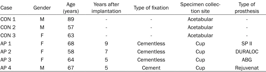
Periprosthetic interface membranes were ob- tained during revision operations of patients with aseptic loosening, after the absence of infection was confirmed. The control speci-mens were obtained from patients with femoral neck fracture during their primary total hip replacement. Clinical data on these patients are shown in Table 1.
Particle preparation
The TiAl6V4 and CoCrMo particles, which were kindly provided by Dr Zhenzhong Zhang (The College of Materials Science and Engineering of Nanjing University of Technology), had a mean particle diameter of 51.7 nm. Particles were suspended in phosphate buffered saline (PBS, pH 7.2-7.4) at a concentration of 50 mg/ ml as stock solutions. The particles were auto-claved for 15 min at 121°C and 15 psi for ster-ilization. For the in vitro experiments, particles were further diluted in cell culture medium to attain concentrations ranging from 10 to 500 µg/ml and ultrasonicated for 10 min before exposing them to cells. All particles were free of endotoxin, as determined using a quantitative Limulus Amebocyte Lysate (LAL) Assay (Charles River, Grand Island, UK) at a detection level of 0.25% EU/ml.
Fibroblast cultures
[image:2.612.92.519.85.202.2]Synovial fibroblasts (SF) were isolated from specimens of patients with femoral neck frac-tures without any evidence of synovial reaction. Table 1. Clinical data on patients
Case Gender (years)Age implantationYears after Type of fixation Specimen collec-tion site prosthesisType of
CON 1 M 89 - - Acetabular
-CON 2 M 57 - - Acetabular
-CON 3 F 63 - - Acetabular
-AP 1 F 68 9 Cementless Cup SP II
AP 2 F 58 7 Cementless Cup DURALOC
AP 3 F 64 5 Cementless Cup ABG
AP 4 M 67 5 Cement Cup Rejuvenat
Written consent was received before the isola-tion of fibroblasts. According to convenisola-tional protocols, cells were obtained by enzymatic digestion and cultured in DMEM medium sup-plemented with 10% FBS, penicillin (100 IU/ml) and streptomycin (100 µg/ml). The fibroblasts were then maintained at 37°C in a humidified atmosphere of 5% CO2 and 95% air. For in vitro experiments, cells were cultured in the absence (controls) or presence of wear particles with dif-ferent concentrations (0, 50, 100, 500 µg/ml) for 24 h. For time-course experiments, cells were incubated with particles (100 µg/ml) for different time periods (1 h, 3 h, 6 h, 12 h, 24 h).
RNA isolation and quantitative polymerase chain reaction (qPCR) assay
Total RNA was extracted using TRIzol reagent (Life Technologies, Grand Island, NY, USA), and 1 μg total RNA was used for first strand cDNA synthesisusing the PrimeScript RT Master Mix kit (TaKaRa Bio, Shiga, Japan). Real-time PCR was performed using LightCycler FastStart DNA Master SYBR Green I (Roche Diagnostics, Indianapolis, IN, USA) according to the manu-facturer’s protocol. Primers for β-actin were used as internal controls. The primer sequenc-es were as follows: Forward: 5’-GGACACACTTC- TGGGGACA-3’ and reverse: 5’-CCAAAGCCACG- TCAAGTAGG-3’ for mouse IL-34; and forward: 5’-GACCTCTATGCCAACACAGTGC-3’ and rever-
row and cultured in α-MEM medium supple-mented with 10% FBS, penicillin (100 IU/ml) and streptomycin (100 µg/ml) at 37°C and 5% CO2 until reaching 90% confluence. The cells were then seeded into 24-well plates in the presence of 30 ng/mL IL-34 (R&D Systems, Minneapolis, MN) and 20 ng/ml RANKL (R&D Systems, Minneapolis, MN) or 30 ng/mL M-CSF (R&D Systems, Minneapolis, MN) and 20 ng/ml RANKL. The culture media were replaced every 2 days until mature osteoclasts were formed. Then, the cells were analyzed for TRAP activity using a commercial kit (Jiancheng Biotech, Nanjing, China). TRAP-positive cells with more than three nuclei were considered osteoclasts and counted under a microscope.
Western blotting
[image:3.612.94.381.71.271.2]Cells prepared for western blotting were lysed in RIPA lysis buffer including a cocktail of pro-tein inhibitors on ice for 30 min. The lysates were centrifuged at 12,000 g for 10 min at 4°C; then, the supernatants were collected, and the protein concentrations were determined using a BCA protein assay kit (Biocolor Bioscience and Technology Co., Shanghai, China). Proteins (30 μg of each group) were run on 10% SDS-PAGE before being transferred to polyvinylidene fluoride membrane (Pall Co., East Hills, NY, USA), then incubated with the following primary antibodies: anti-Phospho-JNK (4668, 1:1000,
Figure 1. Expression of hIL-34 mRNA in periprosthetic membrane of patients with aseptic loosening. Relative levels of hIL-34 mRNA measured by real-time PCR in interface membranes of patients with aseptic loosening. CON, control; AP, aseptic loosening; n.d., not detected. Data are represented as the means ± S.D. for each group.
se: 5’-GTACTCCTGCTTGCT- GATCCAC-3’ for mouse β- actin.
ELISA assay for IL-34
The cytokines IL-34 were quantified using specific ELISA kits (Senbeijia Bio- tech, Nanjing, China). All pr- ocesses were conducted according to the manufac-turer’s instructions.
In vitro osteoclastogenesis assay
Cell Signaling Technology), anti-Phospho-ER- K1/2 (4370, 1:1000, Cell Signaling Technology) and the following secondary antibodies: horse-radish peroxidase (HRP)-conjugated anti-rabbit IgG (7074, 1:2000, Cell Signaling Technology). The protein bands were then detected, and the band density was analyzed using ImageJ 1.41 (National Institutes of Health, Bethesda, MD, USA).
Statistical analysis
The date was expressed as the mean values ± standard deviation (S.D.). Statistical analysis
was performed by one-way ANOVA test. The results were considered to show a significant difference at p values < 0.05.
Results
Expression of IL-34 in the peri-implant mem-brane of patients with aseptic loosening
[image:5.612.91.525.70.504.2]Previous studies have reported that IL-34 can promote osteoclastogenesis [15]. In this study, we were interested in whether IL-34 was involved in aseptic loosening. Specimens of periprosthetic membranes from patients with
aseptic loosening were obtained for real-time PCR. The data showed that IL-34 mRNA was expressed in periprosthetic membranes, but it could not be detected in the control tissue (Figure 1). The results suggested that IL-34 was differentially expressed by periprosthetic mem-branes and control tissues, which might be involved in the pathological process of aseptic loosening.
Upregulation of IL-34 by wear particles in hu-man fibroblasts
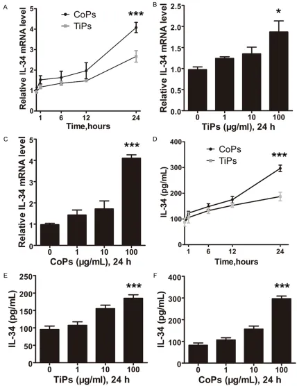
As fibroblasts compose nearly 70% of the cells in the peri-implant membrane and respond directly to wear particles [16], we next investi-gated whether wear particles could induce IL-34 expression in human fibroblasts. RT-PCR analyses showed that both TiPs and CoPs enhanced IL-34 mRNA expression in a dose- and time-dependent manner in fibroblasts (Figure 2A-C). To further confirm that IL-34 can be induced by wear particles, ELISA analyses were also performed. The expression of IL-34 was induced in a dose-dependent manner by TiPs with a 2-fold change at 100 µg/ml and by CoPs with a 3-fold change at 100 µg/ml (Figure 2D, 2E). In addition, the expression of IL-34 pro-tein was markedly elevated by wear debris in a time-dependent manner (Figure 2F).
Upregulation of osteoclastogenesis by IL-34
To further determine the role of IL-34 in osteo-clastogenesis, the combined effect of IL-34 and RANKL on the formation of osteoclasts was assessed in vitro using BMMs. As shown in Figure 3A-C, IL-34 cotreatment with RANKL efficiently increased the formation of osteo-clasts. The quantification of TRAP-positive osteoclasts revealed that the effect of IL-34 was comparable to that of M-CSF at the same concentration (Figure 3D). These data indicat-ed that IL-34 upregulatindicat-ed RANKL-inducindicat-ed ost- eoclastogenesis.
JNK and p44/42 MAPK mediated wear particle-induced IL-34 expression
It has been reported that mitogen-activated protein kinases (MAPKs) such as c-Jun N-ter- minal kinase (JNK) and p44/42 MAPK (also known as ERK-1/2) are important intracellular mediators of inflammatory cytokine-mediated IL-34 expression [17]. To determine the
mecha-nism through which wear particles induce IL-34 expression, the levels of phosphorylated JNK and ERK 1/2 in wear debris-stimulated cells were examined by western blot. Figure 4A-D showed that although wear debris upregulated the expression of IL-34, it also induced the phosphorylation of JNK and ERK 1/2. More importantly, the increased IL-34 expression was rescued by both JNK inhibitor and p44/42 MAPK inhibitor (Figure 4E, 4F). The results sug-gested that both the JNK and p44/42 MAPK pathways were involved in wear particle-induced IL-34 expression in fibroblasts. Discussion
Total joint arthroplasty is commonly used to treat end-stage joint diseases. However, peri-prosthetic bone resorption and subsequent aseptic loosening induced by wear particles remain the major causes of failed arthroplasty. Although numerous efforts have been made to improve the biomaterials and the implant design as well as the surgical techniques, it has remained impossible to eliminate particle gen-eration from bearing surfaces [1]. Hence, the inhibition of osteoclast formation is considered a potential target for therapy.
expression of IL-34 in fibroblasts can be upreg-ulated by wear particles. TiPs and CoPs have been proved to activate MAPK signaling path-ways [18]. We show in this article that the acti-vation of these pathways is required for the wear particle-induced expression of IL-34. However, we have to point out that this study has examined only the expression of IL-34 induced by wear particles in human Synovial fibroblasts from patients with femoral neck fractures, the same experiments should be car-ried out on cells from loosening patients in the future.
Previous studies showed that IL-34 not only plays a critical role in regulating osteoclasto-genesis but also improves the bone resorbing activity of osteoclasts both in vitro and in vivo
[15]. We also show in this paper that the admin-istration of IL-34 and RANKL increases the number of multinucleated osteoclasts. There- fore, together with the previous conclusions, our data suggest that IL-34 may play a predomi-nant role in particle-induced osteoclastoge- nesis.
In conclusion, we demonstrated in this study for the first time that the newly discovered cyto-kine IL-34 is expressed by the periprosthetic membranes of patients with aseptic loosening and that the implant-derived wear particles, TiPs and CoPs, enhance IL-34 expression in fibroblasts via JNK and p44/42 MAPK signaling pathways. Furthermore, the administration of IL-34 upregulated RANKL-induced osteoclasto-genesis. Our findings suggest that IL-34 may play a role in particle-induced osteolysis, and further explorations are needed to determine whether it is a potential therapeutic target for aseptic loosening.
Acknowledgements
This work was supported by the National Natural Science Foundation of China (815- 72111), the Department of orthopedics clinical research center of Jiangsu Province, China (BL2012002), the Graduate Education Inno- vation Project of Jiangsu Province (KYZZ15_ 0045), the Scientific Research Foundation of Graduate School of Nanjing University (20- 15CL12, 2015CL11) and the Scientific and Technological Project of Nanjing, China (20- 1402007).
Disclosure of conflict of interest
None.
Address correspondence to: Zhenheng Wang and Jianning Zhao, Department of Orthopedics, Jinling Hospital, School of Medicine, Nanjing University, Zhongshan Road, No. 305, Nanjing 210093, Jiang- su, China. E-mail: [email protected] (ZHW); [email protected] (JNZ)
Referrences
[1] Sundfeldt M, V Carlsson L, B Johansson C, Thomsen P and Gretzer C. Aseptic loosening, not only a question of wear: a review of differ-ent theories. Acta orthopaedica 2006; 77: 177-197.
[2] Ingham E and Fisher J. The role of macro-phages in osteolysis of total joint replacement. Biomaterials 2005; 26: 1271-1286.
[3] Xu JW, Konttinen YT, Waris V, Pätiälä H, Sorsa T and Santavirta S. Macrophage-colony stimu-lating factor (M-CSF) is increased in the synovi-al-like membrane of the periprosthetic tissues in the aseptic loosening of total hip replace-ment (THR). Clin Rheumatol 1997; 16: 243-248.
[4] Campbell IK, Rich MJ, Bischof RJ and Hamilton JA. The colony-stimulating factors and colla-gen-induced arthritis: exacerbation of disease by M-CSF and G-CSF and requirement for en-dogenous M-CSF. J Leukoc Biol 2000; 68: 144-150.
[5] Kitaura H, Zhou P, Kim HJ, Novack DV, Ross FP and Teitelbaum SL. M-CSF mediates TNF-in-duced inflammatory osteolysis. J Clin Invest 2005; 115: 3418-3427.
[6] Dai XM, Ryan GR, Hapel AJ, Dominguez MG, Russell RG, Kapp S, Sylvestre V and Stanley ER. Targeted disruption of the mouse colony-stimulating factor 1 receptor gene results in osteopetrosis, mononuclear phagocyte defi-ciency, increased primitive progenitor cell fre-quencies, and reproductive defects. Blood 2002; 99: 111-120.
[7] Genovese MC, Cohen S, Moreland L, Lium D, Robbins S, Newmark R, Bekker P and Study G. Combination therapy with etanercept and anakinra in the treatment of patients with rheumatoid arthritis who have been treated unsuccessfully with methotrexate. Arthritis Rheum 2004; 50: 1412-1419.
functional screening of the extracellular pro-teome. Science 2008; 320: 807-811.
[9] Baud’Huin M, Renault R, Charrier C, Riet A, Moreau A, Brion R, Gouin F, Duplomb L and Heymann D. Interleukin-34 is expressed by gi-ant cell tumours of bone and plays a key role in RANKL-induced osteoclastogenesis. J Pathol 2010; 221: 77-86.
[10] Chemel M, Le Goff B, Brion R, Cozic C, Berreur M, Amiaud J, Bougras G, Touchais S, Blanchard F, Heymann MF, Berthelot JM, Verrecchia F and Heymann D. Interleukin 34 expression is asso-ciated with synovitis severity in rheumatoid ar-thritis patients. Ann Rheumat Dis 2012; 71: 150-154.
[11] Hwang SJ, Choi B, Kang SS, Chang JH, Kim YG, Chung YH, Sohn DH, So MW, Lee CK, Robinson WH and Chang EJ. Interleukin-34 produced by human fibroblast-like synovial cells in rheuma-toid arthritis supports osteoclastogenesis. Ar-thritis Res Ther 2012; 14: R14.
[12] Bostrom EA and Lundberg P. The newly discov-ered cytokine il-34 is expressed in gingival fi-broblasts, shows enhanced expression by pro-inflammatory cytokines, and stimulates osteoclast differentiation. PLoS One 2013; 8: e81665.
[13] Yu Y, Yang D, Qiu L, Okamura H, Guo J and Haneji T. Tumor necrosis factor-α induces inter-leukin-34 expression through nuclear factor-κB activation in MC3T3-E1 osteoblastic cells. Mol Med Rep 2014; 10: 1371-1376.
[14] Chen S, Jin G, Huang KM, Ma JJ, Wang Q, Ma Y, Tang XZ, Zhou ZJ, Hu ZJ, Wang JY, Qin A and Fan SW. Lycorine suppresses RANKL-induced osteoclastogenesis in vitro and prevents ovari-ectomy-induced osteoporosis and titanium particle-induced osteolysis in vivo. Sci Rep 2015; 5: 12853.
[15] Chen Z, Buki K, Vaaraniemi J, Gu GL and Vaananen HK. The critical role of IL-34 in os-teoclastogenesis. PLoS One 2011; 6: e18689. [16] Wei X, Zhang X, Zuscik MJ, Drissi MH, Schwarz
EM and O’Keefe RJ. Fibroblasts express RANKL and support osteoclastogenesis in a COX-2-de-pendent manner after stimulation with titani-um particles. J Bone Miner Res 2005; 20: 1136-1148.
[17] Eda H, Shimada H, Beidler DR and Monahan JB. Proinflammatory cytokines, IL-1beta and TNF-alpha, induce expression of interleukin-34 mRNA via JNK-and p44/42 MAPK-NF-kappaB pathway but not p38 pathway in osteoblasts. Rheumatol Int 2011; 31: 1525-1530.