Original Article
IMRT versus 3D-CRT for post-mastectomy
irradiation of chest wall and regional nodes:
a population-based cmparison of normal
lung dose and radiation pneumonitis
Weiwei Li1, Juanqi Wang1, Huifei Cheng2, Xiaoli Yu1, Jinli Ma1
1Department of Radiation Oncology, Fudan University Shanghai Cancer Center; Department of Oncology,
Shang-hai Medical College, Fudan University, ShangShang-hai, China; 2Department of Radiation Oncology, Central Hospital of
Lishui City, Zhejiang Province, China
Received July 26, 2016; Accepted September 18, 2016; Epub November 15, 2016; Published November 30, 2016
Abstract: Objective: Conventional post-mastectomy radiation therapy (PMRT) is often delivered with traditional field borders. We conducted a population-based study comparing the dose-volume of ipsilateral lung and acute lung injury in breast cancer patients undergoing PMRT using linac intensity-modulated radiation therapy (IMRT) and conventional 3D-CRT techniques. Methods: In this study, 169 breast cancer patients who completed PMRT at our institution were included for analysis. 78 patients received inverse planning IMRT treatment (IMRT group), and 91 patients received conventional 3D-CRT treatment (3D-CRT group). All patients received a total dose of 50 Gy in 25 fractions to the chest wall and supra/infraclavicular region as an integrated volume. The percentage volume of ipsi-lateral lung receiving 5 Gy (V5), 10 Gy (V10), 20 Gy (V20), and 30 Gy (V30) extracted from dose-volume histograms (DVHs) were collected and compared between treatment groups. The acute lung injury was followed up regularly. Re-sults: The V5 of ipsilateral lung was significantly lower (P = 0.001) in the 3D-CRT group (52% ± 7%) than in the IMRT group (65% ± 9%). The V10 was similar for both groups (41% ± 7% vs. 44% ± 4%). The V20 was significantly higher (P < 0.001) in the 3D-CRT group (32% ± 6%) than in the IMRT group (29% ± 2%), also, the V30 was significantly higher (P < 0.001) in the 3D-CRT group (22% ± 5%) than in the IMRT group (21% ± 2%). After treatment, 21/91 (23.1%) patients in 3D-CRT group were diagnosed with radiation pneumonitis (RP) with Grade ≥ 2. 5/78 (6.4%) patients in IMRT group were diagnosed with RP (Grade ≥ 2). Incidence of RP has no significant difference between the two groups (P = 0.223). Conclusions: Our analysis demonstrated that our IMRT treatment could reduce V20 and V30, but increase the volume of low dose irradiation of ipsilateral lung (V5), compared to 3D-CRT treatment. This study demonstrates that both 3D-CRT and IMRT techniques are feasible for PMRT.
Keywords: Lung, post-mastectomy, IMRT, radiation pneumonitis
Introduction
Breast cancer (BC) is the most frequent female cancer worldwide. The new cases of BC
accounted for 23% of all female cancers, the
incidence rates of BC varies dramatically across the globe, which is higher in more developed regions. Although the utilization of breast con-serving surgery (BCS) for early-stage disease has increased rapidly in last decade in
main-land China, modified radical mastectomy
(MRM) remains the most-accepted surgical modality in operable breast cancer (BC) [1]. In the recent ten to twenty years, there is a
sub-ment of breast cancer. A rapid developsub-ment of various curative options has led to the improve-ment of treatimprove-ment outcomes [2].
Radiotherapy is an essential part of breast can-cer treatment. In clinical practice, different ra- diotherapy strategies are performed in patients with breast cancer [3]. Conventional post-mas-tectomy radiation therapy (PMRT) is often
deliv-ered with traditional field borders for chest wall
and regional nodes.
dard for BC patients [4]. Qiao et al indicated that postoperative radiotherapy confers better rates of overall survival (OS), local control (LC), and disease-free survival (DFS) in patients with T1 to T2 breast cancer with one to three
posi-tive nodes after modified radical mastectomy
[5]. Three randomized clinical trials have shown that a disease-free and overall survival advan-tage is conferred by the addition of chest wall and regional lymph node irradiation in women with positive axillary lymph nodes after MRM [6-8].
Although chest wall and regional nodes delin-eation techniques have been discussed with available contouring guidelines [9-11], comput-ed tomography (CT)-bascomput-ed planning to treat chest wall and nodal regions as a whole PTV has not yet been adopted into routine practice. In this research, we conduct a population-based dosimetric study comparing the dose-volume of ipsilateral lung and acute toxicity in patients undergoing PMRT using linac IMRT versus conventional 3D-CRT technique.
Methods
Patient eligibility
Eligibility criteria of BC patients included: (1)
age ≥ 18 years with operable breast cancer
involving axillary lymph nodes, but without evi-dence of distant metastasis (negative results on chest CT scans, abdomen and pelvis US); (2) resection of all gross disease by MRM with level I to II axillary dissection; (3) negative surgi-cal margins; (4) Eastern Cooperative Oncology Group performance score of 0-1; (5) comple-tion of adjuvant chemotherapy; and (6) no pre-vious thoracic RT. Patients with serious comor-bid diseases, such as chronic obstructive pulmonary disease, connective tissue disease, postoperative wound infections, and delayed wound healing, etc., that would have negatively affected their tolerance to radiation-induced skin or lung toxicity were not eligible. Patients with synchronous bilateral breast cancers were eligible.
This study was approved by the ethnic Committee of Shanghai Medical College, Fudan University. All the patients provided written informed consent.
Study design
There were two parts in this study. The first part
was to compare the dose-volume of ipsilateral lung in patients using linac IMRT versus con-ventional technique (dosimetric study). And the second part was to compare the incidence of radiation pneumonitis following PMRT with inverse-planned IMRT and conventional tech-nique (clinical study).
Treatments
A non-contrast CT-simulation was performed in the supine position on a commercially available breast tilt board (Med-Tech 350) with the ipsi-lateral arm up and head turned to the contralat-eral side. Radio-opaque wires were used to mark the mastectomy scar and the clinical boundaries. A planning CT scan at 5-mm inter-vals from mid-neck to diaphragm was obtained for each patient using an AcQsim CT simulator (Philips Medical Systems). The 3D-CRT and IMRT plans were generated using Pinnacle treatment planning software (version 8.0). All treatments were delivered with 6-MV photon using an Electa linear accelerator. The pre-scribed total dose was 50 Gy in 25 fractions. IMRT
The clinical target volume (CTV) was defined to
consist of ipsilateral chest wall, mastectomy scar and supra/infraclavicular region for each patient. Each CTV was delineated according to the breast cancer atlas for radiation therapy
planning consensus definitions of the Radiation
Therapy Oncology Group (RTOG) (available at: http://www.rtog.org/CoreLab/ContouringAtlas- es/BreastCancerAtlas.aspx). The chest wall CTV was expanded 1 cm to become chest wall planning target volume (PTV), except that ante-rior, posterior and cranial borders were un-
changed. This modification was made mainly to
heart, contralateral breast, ipsilateral humeral head, spinal cord, and esophagus, were con-toured as well.
3D-CRT
Two tangential semi-opposed beams (to avoid divergence), physical wedges (usually 15° or 30°), and a multileaf collimator were used for 3D-CRT. The beam angles, wedge angles, and beam weighting (usually minimal) were chosen to optimize coverage of the PTV, while minimiz-ing exposure to the ipsilateral lung, heart and contralateral breast. Gantry angles ranged fr-
om 42° to 55° for the medial fields and from 224° to 232° for the lateral fields for patients
treated on the right side, and from 305° to
322° for the medial fields and from 133° to 147° for the lateral fields for patients treated on the left side. The fields extended 2 cm ante -riorly of the chest to provide coverage of the
“flash” region. The supra/infraclavicular region
was designed with separate anterior mixed photon-electron beams.
Clinical study
To ensure accurate delivery of each plan, orthogonal megavoltage electronic portal
imag-es were captured once before the first treat -ment and per week thereafter, and compared with reference digitally reconstructed radio-graphs (DRRs) to verify patient position. Each patient was regularly followed up by the treating physician once a week during radiotherapy and after irradiation. Radiation pneumonitis (RP) was assessed within 6 months. If patients have symptoms such as fever (usually low grade) and cough after radiation therapy, CT scan was per-formed for pneumonitis diagnosis. A clear demarcation conforming to the irradiation port
is needed to confirm the diagnosis.
Statistical analysis
[image:3.612.91.333.94.214.2]T-test was used to compare the numerical dif-ference of dosage between the 3D-CRT and IMRT groups. Chi-square test was used to examine the difference of incidence between
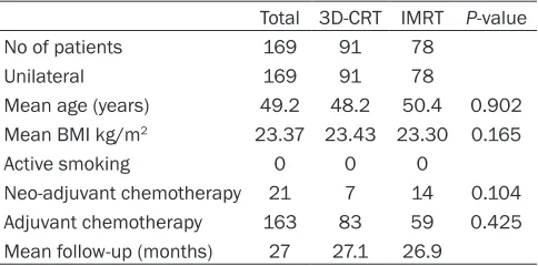
Table 1. Patient characteristics and demographics of patients with 3D-CRT or IMRT treatments
Total 3D-CRT IMRT P-value
No of patients 169 91 78
Unilateral 169 91 78
Mean age (years) 49.2 48.2 50.4 0.902 Mean BMI kg/m2 23.37 23.43 23.30 0.165
Active smoking 0 0 0
Neo-adjuvant chemotherapy 21 7 14 0.104 Adjuvant chemotherapy 163 83 59 0.425 Mean follow-up (months) 27 27.1 26.9
Table 2. Summary of DVH-based analysis for planning target volume of patients with 3D-CRT or IMRT treat-ments
Parameters 3D-CRT IMRT P value
Dnear-max (Gy) 54.58 ± 0.92 54.67 ± 0.86 0.515 Dnear-min (Gy) 47.52 ± 0.61 47.48 ± 0.56 0.738 Dmean (Gy) 51.63 ± 0.58 51.59 ± 0.42 0.614
V95% 98% ± 2% 98% ± 1% 0.998
V110% 2% ± 2% 2% ± 1% 1.000
Homogeneity index (HI) 0.13 ± 0.01 0.13 ± 0.02 1.000 Conformity index (CI) 1.41 ± 0.05 1.42 ± 0.03 0.125
Data are shown as mean ± SD.
Dosimetric analysis
For treatment plan evaluation and dosi-metric analysis, the following PTV statis-tics were obtained from dose-volume histograms (DVHs): 1) Dnear-max, Dne-
ar-min and Dmean: Dnear-max is defined to be the dose to the 2% of the PTV (D2%), Dnear-min is the dose to the 98% of the PTV (D98%), and Dmean is the mean dose of the PTV; 2) V95% and V110%: percent volume receiving great
-er than 95% to 110% of prescribed dose;
3) Dose homogeneity index (HI) and con-formity index (CI): HI and CI were
calcu-lated according to definition proposed
[image:3.612.92.332.273.381.2]the two groups. A P-value less than 0.05 was
considered statistically significant.
Results
Patient characteristics
A total of 169 breast cancer patients who com-pleted PMRT at our institution were included for analysis. The IMRT group had 78 patients, and the 3D-CRT group had 91 patients. Patient characteristics in the two groups were shown in
Table 1. All our patients are unilateral, and none of them are active smokers. There is no
significant difference in the mean age and
me-an body mass index (BMI) of patients between IMRT and 3D-CRT groups (P-values are 0.902 and 0.165, respective, by T-test). Neo-adjuvant chemotherapy had been administered in 14/78
(17.9%) of the flaps in IMRT group, and in 7/91 (7.7%) of the flaps in 3D-CRT group. Adjuvant
chemotherapy had been administered in 59/78
(75.6%) of the flaps in IMRT group, and in 83/91 (91.2%) of the flaps in 3D-CRT group. By Chi-square test, there is no significant different dif -ference in number of patients administered neo-adjuvant chemotherapy or adjuvant-che-motherapy between IMRT and 3D-CRT groups. Plan evaluation
According to the planning target volume of treatment (Table 2), there was no statistically
significant difference for all the parameters
between the IMRT and 3D-CRT groups. We compared the percentage volumes of ipsilater-al lung receiving 5 Gy (V5), 10 Gy (V10), 20 Gy (V20), and 30 Gy (V30) in IMRT and 3D-CRT groups. As shown in Table 3, the V5 of
ipsilat-eral lung was lower in the 3D-CRT group (52% ± 7%) than in the IMRT group (65% ± 9%), and their difference was statistically significant (P = 0.001). Difference of V10 was not significant between the two groups (41% ± 7% vs. 44% ±
4%, P = 0.052). However, the V20 was higher in
the 3D-CRT group (32% ± 6%) than in the IMRT group (29% ± 2%), and there was a statistical significance (P = 0.001). Similarly, the V30 was higher in the 3D-CRT group (22% ± 5%) than in the IMRT group (21% ± 2%), and there was a statistical significance as well (P = 0.001).
Organ at risk (OAR) was also evaluated between IMRT and 3D-CRT plans. As shown in Table 4,
the V5 of contralateral lung was significantly lower (P = 0.022) in the 3D-CRT group (14% ± 7%) than in the IMRT group (12% ± 4%).
However, difference of Dmean (Gy) at
contralat-eral lung were not significant between the two
groups. The V5 and V10 of heart (left-sided
lesions) were significantly lower (both P <
0.001) in the 3D-CRT group than in the IMRT group. Interestingly, at higher dosages, differ-ence of V20 and V30 of heart OAR were
insig-nificant between 3D-CRT and IMRT groups. For
other organs such as spinal cord, ipsilateral humeral head, and esophagus, dosage differ-ence between IMRT and 3D-CRT groups were
insignificant.
Side effects
When patients finished the radiation therapy,
radiation pneumonitis (RP) was diagnosed
with-in 6 months after treatment. 21/91 (23.1%)
patients in 3D CRT group were diagnosed with
radiation pneumonitis (RP) with Grade ≥ 2. 5/78 (6.4%) patients in IMRT group were diag
-nosed with RP (Grade ≥ 2). Incidence of RP has no significant difference between the two groups (P = 0.223). Furthermore, there were 13/91 (14.3%) patients who developed moist
desquamation in the 3D-CRT group, while there
were 3/78 (3.8%) patients who developed
moist desquamation in the IMRT group. In- cidence of moist desquamation cases in the
3D-CRT group was significantly higher than in the IMRT group (P = 0.021). The median time of
moist desquamation was 6 weeks after treat-ment, and mostly occurred within 1-2 weeks. The sites of moist desquamation frequently occurred in anterior axillary fold and in chest wall. Both groups had no severe radiation pneu-monitis. No other severe acute toxicities were observed.
Discussion
[image:4.612.89.288.107.174.2]With the development of economy and chang-ing of life style, cancers have been a severe
Table 3. Summary of DVH-based analysis of ipsilateral lung of patients with 3D-CRT or IMRT treatments
3D-CRT IMRT P value
V5 52% ± 7% 65% ± 9% 0.001
V10 41% ± 7% 44% ± 4% 0.052
V20 32% ± 6% 29% ± 2% < 0.001 V30 22% ± 5% 21% ± 2% < 0.001
burden of society in China [14]. The improve-ment of overall survival remains the ultimate goal for anti-cancer treatment, an adjuvant and primary radiation aims to improve overall sur-vival by treating tumor and area at risk with local regional therapy. In BC treatment, PMRT could reduce the risk of local-regional failure (LRF), with its potential physical and psycho-logic morbidity, as well as a reduction in the risks of distant relapse and death [15]. In addi-tion, PMRT for local advanced and lymph node positive breast cancer has been studied in large randomized trials [6, 16], these research
indicated that PMRT definitely improve local
regional control and overall survival [17]. The conventional PMRT treatment generally includes two opposed tangential photon beams
for chest wall, and separate anterior fields for
regional nodes with mixed photon-electron beams. This treatment has several disadvan-tages. First, the tissue between the chest wall and supraclavicular region +/- IMN may be under or overdosed. Because of the junction or overlap between the tangents and anterior
fields, conventional PMRT potentially increases
normal tissues toxicities or reduces tumor con-trol probability. Although there were several studies reported to address the junction issue between chest wall and supraclavicular region
[18, 19], it’s difficult to eliminate the overlap
between chest wall and IMN with geometric matching method. Second, the use of mixed beams for regional nodes may be associated with inhomogeneous dose distribution. In addi-tion, the maximum depth of
supra/infraclavicu-cer patients aged 35 years or younger with four or more positive nodes [22]. A retrospective analysis performed by Orecchia et al demon-strated that post-mastectomy radiotherapy reduces recurrence and mortality of breast cancer [23]. Although chest wall and regional nodes delineation techniques have been dis-cussed with available contouring guidelines [9-11], computed tomography (CT)-based plan-ning to treat chest wall and nodal regions as a whole PTV has not yet been adopted into rou-tine practice. It was reported that radiation of
SC fields using prescriptions of radiation dose
to empiric depths often leads to suboptimal coverage of targeted volumes, unnecessary degrees of dose in homogeneity, or both [21]. Optimized CT-based treatment plan generates appropriate target coverage and dose homoge-neity. However, the biggest concern of IMRT treatment is the dose-volume of ipsilateral lung. We conducted a population-based study comparing the dose-volume of ipsilateral lung and acute lung injury in patients undergoing PMRT using a linac IMRT technique versus con-ventional technique. We found that the V5 of ipsilateral lung was lower in the 3D-CRT group than in the IMRT group. However, the V20 and
V30 were significantly higher in the 3D-CRT
group than in the IMRT group (P < 0.001). These
findings demonstrated that IMRT reduce the
high dose of the ipsilateral lung, only increasing the V5.
[image:5.612.92.408.95.255.2]Radiation pneumonitis (RP) is the most com-mon side effect following PMRT. In contrast to 3D-CRT plan, distinct dosimetric parameters
Table 4. Summary of DVH-based analysis for organ at risk (OAR) evalua-tion of patients with 3D-CRT or IMRT treatments
Organ Parameters 3D-CRT IMRT P value
Contralateral lung V5 12% ± 4% 14% ± 7% 0.022 Dmean (Gy) 2.19 ± 1.03 2.25 ± 1.43 0.752 Heart (left-sided lesions) V5 45% ± 16% 56% ± 14% < 0.001
V10 26% ± 9% 31% ± 8% < 0.001
V20 15% ± 7% 14% ± 6% 0.325
V30 6% ± 5% 7% ± 3% 0.125
Dmean (Gy) 9.32 ± 1.14 8.71 ± 1.51 0.003 Spinal cord Dmax (Gy) 36.10 ± 4.93 37.25 ± 6.43 0.191 Ipsilateral humeral head Dmean (Gy) 23.98 ± 6.26 25.15 ± 5.70 0.209 Esophagus Dnear-max (Gy) 40.29 ± 9.16 42.17 ± 7.54 0.151 Dmean (Gy) 10.14 ± 2.19 10.27 ± 1.52 0.660
Data are shown as mean ± SD.
can-should be considered in IMRT planning. In our
study, the incidence of ≥ 2 Grade RP is 15.9%
in patients receiving PMRT. The incidence of RP varies widely among reports because of differ-ences in radiation techniques, evaluation of symptoms, and method of reporting. For patients treated with 3D-CRT, the volume of lung receiving 20 Gy has consistently been found to predict the risk of symptomatic RP. The frequency of symptomatic RP was
report-edly 1-7% after local and regional nodes were
treated to a total dose of 45-50 Gy with conven-tional techniques [24-26]. In the current study,
number of RP cases had no statistical signifi -cance between 3D-CRT and IMRT group. For other side reactions and OAR evaluations, we observed that in V5 and V10, IMRT induced
sig-nificantly higher percentage volumes of contra -lateral lung and heart compared to 3D-CRT, indicating IMRT technique may cause more heart lesions at low dosage range.
Our study demonstrated that IMRT reduce the high dose of the ipsilateral lung. There is no sta-tistical difference in the incidence of radiation pneumonitis after treatment. This study dem-onstrates that both 3-DCRT and IMRT tech-niques are feasible for PMRT.
Disclosure of conflict of interest
None.
Address correspondence to: Jinli Ma, Department of Radiation Oncology, Fudan University Shang- hai Cancer Center; Department of Oncology, Shanghai Medical College, Fudan University, 270 Dong’an Road, Shanghai, China. Tel: 86-21-64175590; E-mail: jlma997@163.com
References
[1] Yu KD, Di GH, Wu J, Lu JS, Shen KW, Shen ZZ and Shao ZM. Development and trends of sur-gical modalities for breast cancer in China: a review of 16-year data. Ann Surg Oncol 2007; 14: 2502-2509.
[2] Murawa P, Murawa D, Adamczyk B and Polom K. Breast cancer: Actual methods of treatment and future trends. Rep Pract Oncol Radiother 2014; 19: 165-172.
[3] Novikov SN, Kanaev SV, Semiglazov VF, Jukova LA and Krzhivitckiy PI. Comparison of two treat-ment strategies for irradiation of regional lymph nodes in patients with breast cancer: Lymph flow guided portals versus standard ra
-diation fields. Rep Pract Oncol Radiother 2015; 20: 27-31.
[4] Offersen BV, Brodersen HJ, Nielsen MM, Over-gaard J and OverOver-gaard M. Should Postmastec-tomy Radiotherapy to the Chest Wall and Re-gional Lymph Nodes Be Standard for Patients with 1-3 Positive Lymph Nodes? Breast Care (Basel) 2011; 6: 347-351.
[5] Qiao XY, Song YZ, Geng CZ, Gao W, Li CX and Zhou ZG. The value of radiotherapy in patients with T1 and T2 breast cancer with one to three positive nodes after modified radical mastec -tomy. Chin J Cancer 2010; 29: 436-440. [6] Overgaard M, Hansen PS, Overgaard J, Rose C,
Andersson M, Bach F, Kjaer M, Gadeberg CC, Mouridsen HT, Jensen MB and Zedeler K. Post-operative radiotherapy in high-risk premeno-pausal women with breast cancer who receive adjuvant chemotherapy. Danish Breast Cancer Cooperative Group 82b Trial. N Engl J Med 1997; 337: 949-955.
[7] Recht A, Gray R, Davidson NE, Fowble BL, Solin LJ, Cummings FJ, Falkson G, Falkson HC, Taylor SGt and Tormey DC. Locoregional failure 10 years after mastectomy and adjuvant chemo-therapy with or without tamoxifen without irra-diation: experience of the Eastern Cooperative Oncology Group. J Clin Oncol 1999; 17: 1689-1700.
[8] Ragaz J, Olivotto IA, Spinelli JJ, Phillips N, Jack-son SM, WilJack-son KS, Knowling MA, Coppin CM, Weir L, Gelmon K, Le N, Durand R, Coldman AJ and Manji M. Locoregional radiation therapy in patients with high-risk breast cancer receiving adjuvant chemotherapy: 20-year results of the British Columbia randomized trial. J Natl Can-cer Inst 2005; 97: 116-126.
[9] Verhoeven K, Weltens C, Remouchamps V, Mahjoubi K, Veldeman L, Lengele B, Hortoba-gyi E and Kirkove C. Vessel based delineation guidelines for the elective lymph node regions in breast cancer radiation therapy - PROCAB guidelines. Radiother Oncol 2015; 114: 11-6. [10] Atean I, Pointreau Y, Ouldamer L, Monghal C,
Bougnoux A, Bera G and Barillot I. A simplified CT-based definition of the supraclavicular and infraclavicular nodal volumes in breast cancer. Cancer Radiother 2013; 17: 39-43.
[11] Ma J, Li J, Xie J, Chen J, Zhu C, Cai G, Zhang Z, Guo X and Chen J. Post mastectomy linac IMRT irradiation of chest wall and regional nodes: dosimetry data and acute toxicities. Radiat On-col 2013; 8: 81.
[13] Fontanilla HP, Woodward WA, Lindberg ME, Kanke JE, Arora G, Durbin RR, Yu TK, Zhang L, Sharp HJ, Strom EA, Salehpour M, White J, Bu-chholz TA and Dong L. Current clinical cover-age of Radiation Therapy Oncology Group-de-fined target volumes for postmastectomy radiation therapy. Pract Radiat Oncol 2012; 2: 201-209.
[14] Ferlay J, Soerjomataram I, Dikshit R, Eser S, Mathers C, Rebelo M, Parkin DM, Forman D and Bray F. Cancer incidence and mortality worldwide: Sources, methods and major pat-terns in GLOBOCAN 2012. Int J Cancer 2015; 136: E359-386.
[15] Recht A, Edge SB, Solin LJ, Robinson DS, Esta-brook A, Fine RE, Fleming GF, Formenti S, Hu-dis C, Kirshner JJ, Krause DA, Kuske RR, Langer AS, Sledge GW Jr, Whelan TJ, Pfister DG; American Society of Clinical Oncology. Postmastectomy radiotherapy: clinical practice guidelines of the American Society of Clinical Oncology. J Clin Oncol 2001; 19: 1539-1569. [16] Overgaard M, Jensen MB, Overgaard J, Hansen
PS, Rose C, Andersson M, Kamby C, Kjaer M, Gadeberg CC, Rasmussen BB, Blichert-Toft M and Mouridsen HT. Postoperative radiotherapy in high-risk postmenopausal breast-cancer pa-tients given adjuvant tamoxifen: Danish Breast Cancer Cooperative Group DBCG 82c ran-domised trial. Lancet 1999; 353: 1641-1648. [17] Yu JB, Wilson LD, Dasgupta T, Castrucci WA
and Weidhaas JB. Postmastectomy radiation therapy for lymph node-negative, locally ad-vanced breast cancer after modified radical mastectomy: analysis of the NCI Surveillance, Epidemiology, and End Results database. Can-cer 2008; 113: 38-47.
[18] Klein EE, Taylor M, Michaletz-Lorenz M, Zoeller D and Umfleet W. A mono isocentric technique for breast and regional nodal therapy using dual asymmetric jaws. Int J Radiat Oncol Biol Phys 1994; 28: 753-760.
[19] Hernandez V, Arenas M, Pons F and Sempau J. Clinical applications of geometrical field matching in radiotherapy based on a new ana-lytical solution. Med Dosim 2011; 36: 160-165.
[20] Bentel GC, Marks LB, Hardenbergh PH and Prosnitz LR. Variability of the depth of supra-clavicular and axillary lymph nodes in patients with breast cancer: is a posterior axillary boost field necessary? Int J Radiat Oncol Biol Phys 2000; 47: 755-758.
[21] Liengsawangwong R, Yu TK, Sun TL, Erasmus JJ, Perkins GH, Tereffe W, Oh JL, Woodward WA, Strom EA, Salephour M and Buchholz TA. Treatment optimization using computed to-mography-delineated targets should be used for supraclavicular irradiation for breast can-cer. Int J Radiat Oncol Biol Phys 2007; 69: 711-715.
[22] Wu S, Li Q, Zhou J, Sun J, Li F, Lin Q and He Z. Post-mastectomy radiotherapy can improve survival in breast cancer patients aged 35 years or younger with four or more positive nodes but not in one to three positive nodes. Ther Clin Risk Manag 2014; 10: 867-874. [23] Orecchia R. Breast cancer: post-mastectomy
radiotherapy reduces recurrence and mortali-ty. Nat Rev Clin Oncol 2014; 11: 382-384. [24] Matzinger O, Heimsoth I, Poortmans P, Collette
L, Struikmans H, Van Den Bogaert W, Fourquet A, Bartelink H, Ataman F, Gulyban A, Pierart M, Van Tienhoven G, Oncology ER; Breast Cancer G. Toxicity at three years with and without irra-diation of the internal mammary and medial supraclavicular lymph node chain in stage I to III breast cancer (EORTC trial 22922/10925). Acta Oncol 2010; 49: 24-34.
[25] Wennberg B, Gagliardi G, Sundbom L, Svane G and Lind P. Early response of lung in breast cancer irradiation: radiologic density changes measured by CT and symptomatic radiation pneumonitis. Int J Radiat Oncol Biol Phys 2002; 52: 1196-1206.