Original Article
The sensitivity and specificity of single photon emission
computed tomography (SPECT) in the diagnosis of
coronary artery disease (CAD): a meta-analysis
Xichun Sun
1*, Aibo Liu
2*, Zhonghua Jiang
11Department of Medical Image, Yantai Hospital of Traditional Chinese Medicine, Yantai 264000, Shandong,
China; 2Department of Radiology, Yantaishan Hospital, Yantai 264001, Shandong, China. *Equal contributors. Received March 27, 2016; Accepted December 16, 2016; Epub April 15, 2017; Published April 30, 2017
Abstract: Background: Coronary artery disease (CAD) is a serious threat to health with effective diagnostic methods significantly reducing its risk. While single photon emission computed tomography (SPECT), is an effective non-invasive diagnostic test with broad prospects for development, its sensitivity (SEN) and specificity (SPE) remain to be proven. Methods: All publications were searched through PubMed, Embase and Cochrane Library. Studies com-paring SEN and false positive rate (FPR) of SPECT in the diagnosis of CAD were included. Data regarding relative outcomes in each study were extracted, based on SEN and FPR with 95% confidence intervals (CI). A meta-analysis was performed with several subgroup analyses. Further, the summary receiver operating characteristic (SROC) curve was plotted and its area under the curve (AUC) was calculated. Results: According to the results of meta-analysis, thallium-201 (Tl-201) is regarded as the best radiotracer with the largest partial AUC 0.86, SEN and FPR 0.82 (0.79, 0.85) and 0.25 (0.21, 0.29). Technetium-99m (Tc-99m) performs closely to Tl-201, as SEN of Tc-99m was 0.84 (0.82, 0.86), FPR 0.30 (0.27, 0.33), and partial AUC 0.85. However, there was no observed competitive advantage in combining Tl-201 together with Tc-99m. Although dual labeled compounds have highest SEN (0.85 (0.77, 0.91)), it FPR is lowest with FPR of 0.32 (0.25 to 0.39), partial AUC only 0.80. Conclusion: Tl-201 FPR was demonstrated to be the optimal radiotracer of SPECT in the diagnoses of CAD, and low-level exercise combined with pharmacologic agents was the preferable choice of SPECT stress inducer. To sum up, based on its SEN and FPR, SPECT can be widely used in CAD diagnosis.
Keywords: Coronary artery disease, single photon emission computed tomography, coronary angiography, techne-tium-99m, thallium-201, meta-analysis
Introduction
Coronary artery disease (CAD), also known as
ischemic heart disease, is caused by insuffi
-cient myocardial blood supply, leading to stable
angina, unstable angina, myocardial infarction,
and sudden coronary death [1]. Since coronary
circulation consists of the blood-supply vessels
of the heart muscle myocardium, as so-called
“end circulation”, there are no compensatory or
secondary branch vessels, which causes
seri-ous problems when any blockage occurs.
Hy-pertension, smoking, hyperlipidemia, diabetes
and physical inactivity are considered to be
high risk factors [2], while familial inheritance is
also one of the main causes. Atherosclerosis of
coronary artery is the direct catalyst leading to
CAD [3]. According to previous studies [4, 5],
6222
Int J Clin Exp Med 2017;10(4):6221-6236
present, based on the advice of National Heart,
Lung, and Blood Institute, electrocardiogram
(EKG), echocardiography, chest X-ray, single
photon emission computed tomography (SP-
ECT) and coronary angiography (CA) are the
most common cardio logical diagnostic tests
and procedures. All of the test methods can be
classified into two groups: invasive (CA), and
non-invasive, (EKG, X-ray, SPECT, etc). [9].
Since 1960, when the first CA was somewhat
accidently carried out by Mason Sones [10], it
has developed gradually and been accepted as
the golden standard in consideration of its
accuracy. Both the main and branch of
coro-nary artery are visible under X-ray for the
detec-tion of occlusion, stenosis, restenosis, or thro-
mbosis with the help of special coronary
cath-eter puncturing into the artery and injection of
radio contrast. Even though it is convenient to
diagnosis, not everyone can tolerate radio
con-trast, and due to its invasive, this does not
seem to be the optimal choice for patients with
suspected CAD.
SPECT, one of the non-invasive methods, is
increasing in popularity due to its general
appli-cability and relative harmlessness. Computed
tomography images can be obtained by varying
the intensity of γ rays from different organs or
parts of the body. According to the distinction
between images under stress and at rest,
doc-tors can diagnose myocardial ischemia,
evalu-ate the lesion range and assess the degree of
CAD [11].
In addition, there is much research on the
sen-sibility (SEN) and false positive rate (FPR) of
this meta-analysis would involve updated
cred-ible studies, and discuss the potential influen
-tial factors including tracer atom, stress and
ethnicity.
Methods
Literature identification
By searching on PubMed, Embase and Coch-
rane Library, relevant literature identification
was completed without language restriction.
Meanwhile, keywords such as “coronary artery
disease”, “coronary angiography”, “single
pho-ton emission computed tomography” and their
synonyms were combined to use as searching
terms. Besides this, reference lists of relavant
literatures were searched and examined in
case of any omission. All the potential
litera-tures were assessed by two reviewers sepa-
rately.
Inclusion criteria
Studies were included if they satisfied the fol
-lowing criteria: (i) all subjects included were
suspected as CAD, with no limitation of patients’
ethnicity; (ii) SPECT must be used as one of the
diagnostic tools, irrespective of type of
radioac-tive tracer or stress inducer; (iii) the gold
stan-dard must be CA, irrespective of interpretation;
(iv) a comparison must be performed in the
study between SPECT and CA.
Data extraction
[image:2.612.90.370.73.246.2]Independently, two reviewers screened the
titles and abstracts of retrieved literatures, and
in some cases, the full text was examined for
Figure 1. Flow chart.
more details. Any disagreements regarding in-
clusion were solved under discussion. The fol
-lowing information was extracted: author, year
of publication, country, diagnosed disease, the
diagnostic technique employed, isotope, stress
inducer and interpretation of SPECT, inclusion
of CA as gold standard, and finally outcomes
which represented data as true positive, false
positive, true negative and false negative, and
these were obtained to fulfill the 2 × 2 contin
-gency table.
Statistical analysis
SEN and SPE of each test method were
calcu-lated, based on data showing in both true and
false positives and negatives (2 × 2) contingen
-cy tables. Meanwhile, the heterogeneity among
selected studies was tested through Coch-
ran’s Q, to find if the data could be pooled
together. Usually, when
P
< 0.01 there was
sig-nificant heterogeneity, and it was necessary to
perform subgroup analyses. Moreover, if
pri-mary subgroup did not work very well, the
sec-ondary subgroup was used.
Furthermore, meta-analysis model of pooling
all studies and of subgroup categorized by
potential influencing factors were fitted for SEN
and SPE with software R® version 3.2.1 (Ma-
thSoft, Cambridge, Massachusetts). The SEN
and the SPE of SPECT compared with gold stan
-dard were expressed in the number less than 1
with 95% confidence intervals (CI). According to
these data, a summary receiver operating
char-acteristic (SROC) curve was performed, and the
area under the SROC curve (AUC) was
comput-ed through the relative integral formula to
reflect the SEN and SPE among different sub
-groups. The AUC values were in the range of 0
to 1, and if a diagnostic method was clearly
superior, its AUC would be 1; and vice versa.
Besides, to make our SROC curves more
visi-ble, instead of SPE, the FPR were used as
abscissa. The conversion adopted was:
FPR =
1-SPE
.
Results
Included studies
[image:3.612.91.523.84.323.2]This meta-analysis involved 184 studies col
-lected from 145 articles with 19,182 subjects
[12, 13, 17-159], since these all satisfied the
criteria mentioned above. At first, 1,017 litera
-tures contained the searching terms were
iden-tified and retrieved, after eliminating 13 dupli
-cates. By skimming the title and abstract of
identified articles, 846 were selected and the
full texts were reviewed. Among them, 701
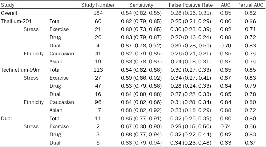
Table 1.
Main results of meta-analysis
Study Study Number Sensitivity False Positive Rate AUC Partial AUC
Overall 184 0.84 (0.82, 0.85) 0.28 (0.26, 0.31) 0.85 0.82
Thallium-201 Total 60 0.82 (0.79, 0.85) 0.25 (0.21, 0.29) 0.86 0.86
Stress Exercise 21 0.80 (0.73, 0.85) 0.30 (0.23, 0.39) 0.82 0.74
Drug 26 0.83 (0.79, 0.87) 0.20 (0.16, 0.24) 0.88 0.72
Dual 4 0.87 (0.78, 0.92) 0.39 (0.28, 0.51) 0.76 0.83
Ethnicity Caucasian 41 0.82 (0.79, 0.85) 0.26 (0.21, 0.31) 0.85 0.76
Asian 19 0.83 (0.78, 0.87) 0.24 (0.18, 0.31) 0.87 0.76
Technetium-99m Total 113 0.84 (0.82, 0.86) 0.30 (0.27, 0.33) 0.85 0.85
Stress Exercise 27 0.89 (0.86, 0.92) 0.34 (0.27, 0.41) 0.87 0.83
Drug 47 0.83 (0.79, 0.86) 0.28 (0.24, 0.33) 0.84 0.79
Dual 16 0.84 (0.80, 0.88) 0.27 (0.22, 0.33) 0.85 0.78
Ethnicity Caucasian 96 0.84 (0.82, 0.86) 0.31 (0.28, 0.34) 0.84 0.80
Asian 17 0.88 (0.82, 0.92) 0.23 (0.18, 0.29) 0.88 0.72
Dual Total 11 0.85 (0.77, 0.91) 0.32 (0.25, 0.39) 0.80 0.80
Stress Exercise 2 0.67 (0.30, 0.90) 0.29 (0.15, 0.50) 0.74 0.66
Drug 3 0.88 (0.77, 0.94) 0.32 (0.22, 0.44) 0.82 0.83
Dual 6 0.88 (0.79, 0.94) 0.34 (0.23, 0.48) 0.83 0.87
6224
Int J Clin Exp Med 2017;10(4):6221-6236
studies were excluded, since they did not
men-tion the gold standard, or did not make explicit
the technology of SPECT, or were aimed at
cost-effectiveness (
Figure 1
).
Characteristics of included studies
Among these 184 studies, 36 were targeted on
Asian patients, while other studies were
target-ed on Caucasian ethnicities. Three types of
[image:4.612.91.374.74.273.2]ra-rms of stress inducer and eth
nicity. Therefore,
in this meta-analysis, to reduce error between
different SPECT equipment and patients’ race,
two-class subgroup analyses were used, which
meant all the studies were divided into three
first-class subgroups, first with Tc-99m, Tl-201
and dual, and after that each primary subgroup
was classified as pharmacologic stress induc
-er, exercise and both, or as Asian and
Caucasian, as shown in
Figure 2
.
Figure 2. Stratified network diagram for each tracer atom: each tracer atom is stratified either by method of stress or ethnicity.
Figure 3. The summary receiver operator characteristic (SROC) curves shows the diagnostic power of three imaging agents to coronary artery disease.
diotracer were involved, wi-
th technetium-99m (Tc-99m)
the most common. Besides
this, thallium-201 (Th-201)
and Tc-99m plus Th-201
(Du-al) were also used in some
studies. Pharmacologic age-
nts, exercise and both (Dual)
were used to produce cardiac
load. Adenosine, dipyridamole
and dobutamine were empl-
oyed as pharmacologic stress
inducer. In addition, all the
studies took CA as gold
stan-dard reference. Details of
selected studies were shown
in
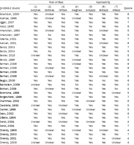
Table S1. The quality of
included studies using QUAD-
AS-2 was shown in
Table S2
.
Total SPECT
SEN and SPE of SPECT
we-re compawe-red with CA in all
included studies. Pooling all
data together, the overall SEN
turned out to be 0.84 (0.82,
0.85), while the FPR was 0.28
(0.26, 0.31), and its partial
AUC was 0.82, seen in
Table
1
.
[image:4.612.91.375.294.571.2]Thallium-201
There were 60 studies related to Tl-201 includ
-ed with SEN 0.82 (0.79, 0.85), lower than the
overall SEN. But the SPE of Tl-201 was higher
than the pooled data average, with FPR 0.25
(0.21, 0.29). Additionally, based on the SROC
curve for total Tl-201 shown in
Figure 3
, its
par-tial AUC was calculated as 0.86.
With regards to the SEN of secondary subgroup
(stress inducer), exercise was 0.80 (0.73, 0.85),
drug was 0.83 (0.79, 0.87), and combination of
both components was 0.87 (0.78, 0.92). FPR
for exercise was 0.30 with 95% CI 0.23 to 0.39,
for drug 0.20 (0.16, 0.24), and for dual 0.39
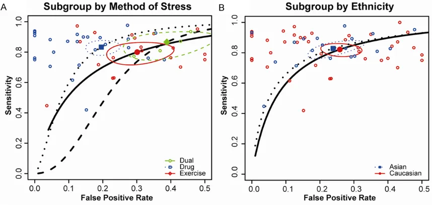
(0.28, 0.51). According to SROC curve demon
-strated in
Figure 4A
, the partial AUC for each
inducer was 0.74, 0.71 and 0.83.
As for ethnicity, SEN was 0.82 with 95% CI
ranging from 0.79 to 0.85, and 0.83 with 95%
CI from 0.78 to 0.87 respectively for Caucasian
and Asian respectively. FPR was 0.26 with 95%
CI 0.21 to 0.31 for Caucasian, and 0.24 with
95% CI 0.18 to 0.31 for Asian. The partial AUC
of the two ethnicities were both 0.76 as shown
in
Figure 4B
.
Technetium-99m
Tc-99m was the most common isotope atom
used as radiotracer, so 113 studies with 10,746
patients were involved. The SEN and FPR of
total data were 0.84 with narrow 95% CI from
0.82 to 0.86, 0.30 with 95% CI from 0.27 to
0.33. Partial AUC shown in
Figure 3
was 0.85.
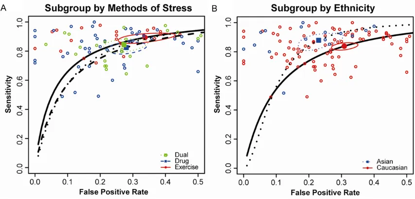
Categorized by stress inducer, exercise had the
best SEN 0.89 with 95% CI from 0.86 to 0.92,
but the worst SPE, since it FPR was 0.34 with
95% CI from 0.27 to 0.41. Additional, SEN and
SPE of dual were both better than drug alone,
since SEN of drug was 0.83 with 95% CI 0.79 to
0.86, of dual was 0.84 with 95% CI 0.80 to
0.88; FPR of drug was 0.28 with 95% CI 0.24 to
0.33, and of dual was 0.27 with 95% CI 0.22 to
0.33. However, considering these two aspects
and after correction, the order was exercise,
drug and dual, with partial AUC 0.83, 0.79 and
0.78 respectively, as shown in
Figure 5A
.
In view of the influence of ethnicity, the second
-ary subgroup was performed as Caucasian and
Asian, and SEN was 0.84 with 95% CI from
0.82 to 0.86, 0.88 with 95% CI from 0.82 to
0.92, and FPR was 0.31 with 95% CI between
0.28 and 0.34, 0.23 with 95% CI between 0.18
and 0.29. Referring to SROC curve
Figure 5B
,
the partial AUC was 0.80 for Caucasian and
0.72 for Asian.
Combination
[image:5.612.92.521.73.277.2]The subgroup analysis of combination, which
used both Tc-99m and Tl-201, included only 11
6226
Int J Clin Exp Med 2017;10(4):6221-6236
studies, and all subjects were Caucasian, with
only second-class stress inducer being
con-ducted. The merged data of dual showed that
SEN was 0.85 and its 95% CI with limitation of
0.77 to 0.91, and FPR was 0.32, and 95% CI
was 0.25 to 0.39. As illustrated in
Figure 3
, the
partial AUC was 0.80.
In the secondary subgroup, both drug and dual
stress inducers showed good performance in
SEN, as their SEN was 0.88 with 95% CI 0.77 to
0.94, and 0.88 with 95% CI 0.79 to 0.94. FPR
of each was 0.32 with 95% CI 0.22 to 0.44, and
0.34 with 95% CI 0.23 to 0.48. Although the
SEN of exercise was 0.67 with wide 95% CI
from 0.30 to 0.90, much lower than drug and
dual, its FPR was 0.29 with 95% CI 0.15 to
0.50. To sum up, drug performed better than
exercise, but worse than combination, as their
partial AUC was 0.83, 0.66, and 0.87 in SROC
curve.
Discussion
This meta-analysis gathered data from 184
studies of 145 articles with 19,182 total sub
-jects, who were suspected to suffer from CAD,
and all studies involved assessed the SEN and
SPE of SPECT, compared with golden reference
CA. Three types of radiotracers, three kinds of
stress inducer, and two different ethnicities
were tested and to minimize the probable influ
-ence generated by different SPECT equipment
and races, they were categorized into two-class
subgroups. The primary subgroup was Tc-99m,
Tl-201 and dual, and the secondary subgroup
was pharmacologic agent, exercise and both,
or Asian and Caucasian. Due to
non-invasive-ness of ultrasound, this analysis was designed
to investigate if the SEN and SPE of SPECT were
high enough to be used widely in CAD diagnosis
with isotope and stress inducer being the
opti-mal choices. Additionally, this meta-analysis
tried to asserta in whether the performance of
SPECT had a significant difference among vari
-ous ethnicities.
According to partial AUC of SROC curve for
Tl-201, Tc-99m, and combination shown in
Fi-gure 2
, Tl-201 seems to be the optimal radio
-tracer with partial AUC of 0.86, larger than over
-all AUC 0.82. Tc-99m was slightly behind Tl-201,
of which the partial AUC was 0.85. Although
combination had best performance in SEN, this
constituted the lowest SPE and partial AUC
0.80 still indicated its lower performance.
Tl-201 showed no significant difference in vari
-ous ethnic groups, however, the partial AUC of
Caucasian was 0.80 higher than 0.72 of Asian
with reference to Tc-99m.
[image:6.612.93.520.71.276.2]Radiotracers were crucial to SPECT perfor
-mance, since it determined the SEN and SPE of
SPECT directly, and currently Tl-201 and
Tc-99m, including Tc-99m sestamibiand Tc-99m
tetrofosmin, were quite common in the studies.
Tl-201, a radioactive isotope with 81 protons
and 201 neutrons, was widely used as a
radio-tracer in the field of medicine. The half-life of
Tl-201 is 74 hours, gamma decay with 80 keV
emission energy. As for Tc-99m, a meta stable
nuclear isomer of Tc-99, its half-life was around
6 hours, and it decays and emits 140 keV
gamma rays. Both isotopes can be extracted by
normally functional myocardial cells selectively.
The first-pass extraction of Tl-201 can reach
nearly 85%, while the first-pass extraction of
Tc-99m was less. Besides this, due to the lack
of redistribution, two injections at stress and
rest separately were required when Tc-99m
was served as radiotracer [160]. To avoid the
interference on second test of standard
one-day single-isotope SPECT and shorten diagnos
-tic time [56], some studies used dual-isotope,
which meant Tl-201 for rest and Tc-99m for
stress. However, this protocol did not work suc
-cessfully. Probably due to immature
dual-iso-tope SPECT technique and equipment, it had
the highest SEN with lowest SPE and wide
con-fidential interval leading to its smallest partial
AUC.
As for stress inducing method, exercise,
phar-macologic agent and combination were
fre-quently used to raise heartbeat and blood
pres-sure to an adequate level. In consideration of
side-effect and cost-efficiency, exercise was a
convenient choice. But in some cases, as pa-
tients had certain concomitants, like high-risk
unstable angina, uncontrolled hypertension. In
conditions such as these where exercise is
inadvisable, pharmacologic agents must be
used. Common medicines used as stress in-
ducer include adenosine, dipyridamole and
dobutamine. Different drugs have different
mechanisms, so medicine choice depends on
patients’ condition. In general, a combination
of low-level exercise with drug infusion can
decrease the drug side-effects and improve
image quality [160]. Therefore, using the same
radiotracer, dual stress inducers combined had
better performance.
The diversities among isotope extraction,
ab-sorption of background tissues and organs,
and clearance of organism of different
ethnici-ties can all influence the performance of SPECT.
This meta-analysis compared SPECT with gold
standard CA and indicated the SEN and SPE of
three radiotracers and stress inducers applied
to SPECT, and of two ethnicities in diagnosing
CAD. However there were still some limitations:
(i) to guarantee the quantity of selected
stud-ies, all the studies which met the criteria were
included, so some data might have seen
restric-tion from any accidental error, such as the influ
-ence of cell extraction generated by myocardial
disease, and γ ray decay caused by excess fat
or breast tissue, both of these factors can lead
to misdiagnosis and increase false positive
rate; (ii) the sample size of selected studies
were varied. The smallest studies had just 16
subjects [150], while the largest one involved
785 patients [28]. So the small sample studies
was easily diluted, which meant the large-size
studies were dominant; (iii) although two-class
subgroup analyses were performed,
heteroge-neity among each group was still extant, which
might be resultant from the technique of SPECT,
such as ECG-gated SPECT and attenuation cor
-rection SPECT, or method of SPECT image inter
-pretation, as visual, semi-quantitative and
quantitative.
Conclusions
Based on the present studies, Tl-201 seemed
the optimal SPECT radiotracer aimed atmyocar
-dial perfusion scintigraphy with highest SPE,
and combined low-level exercise with
pharma-cologic agent was the preferable choice of
stress inducer. However, the application of
stress inducer of SPECT in practical clinical
diagnosis needs to adequately consider the
patient’s condition. Additionally, extant studies
were not enough to state the explicit
connec-tion between ethnicity and SPECT performance.
Although SPECT was performed well in both
SEN and SPE and can be widely used in CAD
diagnosis, whether it can take the place of CA
requires further research.
Disclosure of conflict of interest
None.
Address correspondence to: Zhonghua Jiang, De- partment of Medical Image, Yantai Hospital of Traditional Chinese Medicine, No. 39 Xingfu Road, Yantai 264000, Shandong, China. E-mail: huangyan-liuvip@163.com
References
6228
Int J Clin Exp Med 2017;10(4):6221-6236
[2] Mehta PK, Wei J and Wenger NK. Ischemic heart disease in women: a focus on risk fac-tors. Trends Cardiovasc Med 2015; 25: 140-151.
[3] Hwang SJ, Ballantyne CM, Sharrett AR, Smith LC, Davis CE, Gotto AM Jr and Boerwinkle E. Circulating adhesion molecules VCAM-1, ICAM-1, and E-selectin in carotid atherosclerosis and incident coronary heart disease cases: the atherosclerosis risk in communities (ARIC) study. Circulation 1997; 96: 4219-4225. [4] Kaski JC. Pathophysiology and management of
patients with chest pain and normal coronary arteriograms (cardiac syndrome X). Circulation 2004; 109: 568-572.
[5] Koopman L P CN, Slorach C, et al.//CIRCULA-TION. 530 WALNUT ST, PHILADELPHIA, PA 19106-3621 USA: LIPPINCOTT WILLIAMS & WILKINS, 2010, 122(21). Epidemiology and Prevention of CV Disease: Physiology, Pharma -cology and Lifestyle. CIRCULATION 2010; 122: 530.
[6] GBD 2013 Mortality and Causes of Death Col-laborators. Global, regional, and national age-sex specific all-cause and cause-specific mor -tality for 240 causes of death, 1990-2013: a systematic analysis for the Global Burden of Disease Study 2013. Lancet 2015; 385: 117-171.
[7] Moran AE, Forouzanfar MH, Roth GA, Mensah GA, Ezzati M, Murray CJ and Naghavi M. Tem -poral trends in ischemic heart disease mortal-ity in 21 world regions, 1980 to 2010: the Global Burden of Disease 2010 study. Circula-tion 2014; 129: 1483-1492.
[8] Bugiardini R, Manfrini O, Pizzi C, Fontana F and Morgagni G. Endothelial function predicts fu-ture development of coronary artery disease: a study of women with chest pain and normal coronary angiograms. Circulation 2004; 109: 2518-2523.
[9] Hwang IC, Kim YJ, Kim KH, Shin DH, Lee SP, Kim HK and Sohn DW. Diagnostic yield of coro -nary angiography in patients with acute chest pain: role of noninvasive test. Am J Emerg Med 2014; 32: 1-6.
[10] Nissen S. Coronary angiography and intravas-cular ultrasound. Am J Cardiol 2001; 87: 15A-20A.
[11] Zhou T, Yang LF, Zhai JL, Li J, Wang QM, Zhang RJ, Wang S, Peng ZH, Li M and Sun G. SPECT myocardial perfusion versus fractional flow re -serve for evaluation of functional ischemia: a meta analysis. Eur J Radiol 2014; 83: 951-956.
[12] Ogilby JD, Kegel JG, Heo J and Iskandrian AE. Correlation between hemodynamic changes and tomographic sestamibi imaging during
di-pyridamole-induced coronary hyperemia. J Am Coll Cardiol 1998; 31: 75-82.
[13] Miller DD, Younis LT, Chaitman BR and Strat -mann H. Diagnostic accuracy of dipyridamole technetium 99m-labeled sestamibi myocardial tomography for detection of coronary artery disease. J Nucl Cardiol 1997; 4: 18-24. [14] Single photon emission computed tomography
for the diagnosis of coronary artery disease: an evidence-based analysis. Ont Health Tech -nol Assess Ser 2010; 10: 1-64.
[15] Parker MW, Iskandar A, Limone B, Perugini A, Kim H, Jones C, Calamari B, Coleman CI and Heller GV. Diagnostic accuracy of cardiac posi -tron emission tomography versus single pho-ton emission computed tomography for coro-nary artery disease: a bivariate meta-analysis. Circ Cardiovasc Imaging 2012; 5: 700-707. [16] Takx RA, Blomberg BA, El Aidi H, Habets J, de
Jong PA, Nagel E, Hoffmann U and Leiner T. Di -agnostic accuracy of stress myocardial perfu-sion imaging compared to invasive coronary angiography with fractional flow reserve meta-analysis. Circ Cardiovasc Imaging 2015; 8. [17] Abramson BL, Ruddy TD, deKemp RA, Lara
-mee LA, Marquis JF and Beanlands RS. Stress perfusion/metabolism imaging: a pilot study for a potential new approach to the diagnosis of coronary disease in women. J Nucl Cardiol 2000; 7: 205-212.
[18] Acampa W, Cuocolo A, Sullo P, Varrone A, Nico -lai E, Pace L, Petretta M and Salvatore M. Di-rect comparison of technetium 99m-sestamibi and technetium 99m-tetrofosmin cardiac sin-gle photon emission computed tomography in patients with coronary artery disease. J Nucl Cardiol 1998; 5: 265-274.
[19] Aggeli C, Christoforatou E, Giannopoulos G, Roussakis G, Kokkinakis C, Barbetseas J, Vla -chopoulos C and Stefanadis C. The diagnostic value of adenosine stress-contrast echocar-diography for diagnosis of coronary artery dis-ease in hypertensive patients: comparison to Tl-201 single-photon emission computed to -mography. Am J Hypertens 2007; 20: 533-538.
[20] Aksut SV, Pancholy S, Cassel D, Cave V, Heo J and Iskandrian AS. Results of adenosine single photon emission computed tomography thalli-um-201 imaging in hemodynamic nonre-sponders. Am Heart J 1995; 130: 67-70. [21] Amanullah AM, Berman DS, Hachamovitch R,
Kiat H, Kang X and Friedman JD. Identification of severe or extensive coronary artery disease in women by adenosine technetium-99m ses-tamibi SPECT. Am J Cardiol 1997; 80: 132-137. [22] Amanullah AM, Bevegard S, Lindvall K and
echocardiography and myocardial perfusion by technetium-99m sestamibi tomography during adenosine-induced coronary vasodilation and comparison with coronary angiography. Am J Cardiol 1993; 72: 983-989.
[23] Astarita C, Palinkas A, Nicolai E, Maresca FS, Varga A and Picano E. Dipyridamole-atropine stress echocardiography versus exercise SPECT scintigraphy for detection of coronary artery disease in hypertensives with positive exercise test. J Hypertens 2001; 19: 495-502. [24] Avakian SD, Grinberg M, Meneguetti JC,
Ramires JA and Mansur AP. SPECT dipyridam -ole scintigraphy for detecting coronary artery disease in patients with isolated severe aortic stenosis. Int J Cardiol 2001; 81: 21-27. [25] Banzo I, Pena FJ, Allende RH, Quirce R and Car
-ril JM. Prospective clinical comparison of non-corrected and attenuation- and scatter-correct-ed myocardial perfusion SPECT in patients with suspicion of coronary artery disease. Nucl Med Commun 2003; 24: 995-1002.
[26] Becker M, Hundemer A, Zwicker C, Altiok E, Krohn T, Mottaghy FM, Lente C, Kelm M, Marx N and Hoffmann R. Detection of coronary ar -tery disease in postmenopausal women: the significance of integrated stress imaging tests in a 4-year prognostic study. Clin Res Cardiol 2015; 104: 258-271.
[27] Benoit T, Vivegnis D, Lahiri A, Itti R, Braat S and Rigo P. Tomographic myocardial imaging with technetium-99m tetrofosmin. Comparison with tetrofosmin and thallium planar imaging and with angiography. Eur Heart J 1996; 17: 635-642.
[28] Berman DS, Kang X, Nishina H, Slomka PJ, Shaw LJ, Hayes SW, Cohen I, Friedman JD, Ger -lach J and Germano G. Diagnostic accuracy of gated Tc-99m sestamibi stress myocardial per -fusion SPECT with combined supine and prone acquisitions to detect coronary artery disease in obese and nonobese patients. J Nucl Cardiol 2006; 13: 191-201.
[29] Beygui F, Le Feuvre C, Maunoury C, Helft G, Antonietti T, Metzger JP and Vacheron A. De -tection of coronary restenosis by exercise elec-trocardiography thallium-201 perfusion imag-ing and coronary angiography in asymptomatic patients after percutaneous transluminal coro-nary angioplasty. Am J Cardiol 2000; 86: 35-40.
[30] Blankstein R, Shturman LD, Rogers IS, Rocha-Filho JA, Okada DR, Sarwar A, Soni AV, Bezerra H, Ghoshhajra BB, Petranovic M, Loureiro R, Feuchtner G, Gewirtz H, Hoffmann U, Mamuya WS, Brady TJ and Cury RC. Adenosine-induced stress myocardial perfusion imaging using du-al-source cardiac computed tomography. J Am Coll Cardiol 2009; 54: 1072-1084.
[31] Bokhari S, Shahzad A and Bergmann SR. Su-periority of exercise myocardial perfusion im-aging compared with the exercise ECG in the diagnosis of coronary artery disease. Coron Artery Dis 2008; 19: 399-404.
[32] Boomsma MM, Niemeyer MG, van der Wall EE, van Eck-Smit BL, Zwinderman AH, Boomsma JH and Pauwels EK. Tc-99m tetrofosmin myo -cardial SPECT perfusion imaging: comparison of rest-stress and stress-rest protocols. Int J Card Imaging 1998; 14: 105-111.
[33] Borges-Neto S, Mahmarian JJ, Jain A, Roberts R and Verani MS. Quantitative thallium-201 single photon emission computed tomography after oral dipyridamole for assessing the pres-ence, anatomic location and severity of coro-nary artery disease. J Am Coll Cardiol 1988; 11: 962-969.
[34] Chammas E, Yatim A, Hage C, Sokhn K, Tarcha W and Ghanem G. Evaluation of Tc-99m tetro -fosmin scan for coronary artery disease diag-nosis. Asian Cardiovasc Thorac Ann 2002; 10: 244-247.
[35] Chen L, Wang X, Bao J, Geng C, Xia Y and Wang J. Direct comparison of cardiovascular mag-netic resonance and single-photon emission computed tomography for detection of coro-nary artery disease: a meta-analysis. PLoS One 2014; 9: e88402.
[36] Ciavolella M, Tomai F, Vicchio D, Ruscitti G, Gi -annitti C, Scali D, Schad N and Reale A. Single-day combined evaluation of regional myocar-dial perfusion and function at rest and peak exercise with 99mTc-MIBI in patients with coro -nary artery disease. Int J Card Imaging 1993; 9: 299-311.
[37] Cramer MJ, Verzijlbergen JF, van der Wall EE, Vermeersch PH, Niemeyer MG, Zwinderman AH, Ascoop CA and Pauwels EK. Comparison of adenosine and high-dose dipyridamole both combined with low-level exercise stress for 99Tcm-MIBI SPET myocardial perfusion imag -ing. Nucl Med Commun 1996; 17: 97-104. [38] Daou D, Delahaye N, Vilain D, Lebtahi R, Far
-aggi M and Le Guludec D. Identification of ex -tensive coronary artery disease: incremental value of exercise Tl-201 SPECT to clinical and stress test variables. J Nucl Cardiol 2002; 9: 161-168.
6230
Int J Clin Exp Med 2017;10(4):6221-6236
[40] Dondi M, Fagioli G, Salgarello M, Zoboli S, Nan-ni C and Cidda C. Myocardial SPECT: what do we gain from attenuation correction (and when)? Q J Nucl Med Mol Imaging 2004; 48: 181-187.
[41] Elhendy A, Schinkel AF, Bax JJ, van Domburg RT, Valkema R, Biagini E, Feringa HH and Pol -dermans D. Accuracy of stress Tc-99m tetro -fosmin myocardial perfusion tomography for the diagnosis and localization of coronary ar-tery disease in women. J Nucl Cardiol 2006; 13: 629-634.
[42] Elhendy A, Schinkel AF, van Domberg RT, Bax JJ, Valkema R and Poldermans D. Non-invasive diagnosis of in stent stenosis by stress 99m technetium tetrofosmin myocardial perfusion imaging. Int J Cardiovasc Imaging 2006; 22: 657-662.
[43] Elhendy A, van Domburg RT, Bax JJ, Nierop PR, Geleijnse ML, Ibrahim MM and Roelandt JR. Noninvasive diagnosis of coronary artery ste-nosis in women with limited exercise capacity: comparison of dobutamine stress echocar-diography and 99mTc sestamibi single-photon emission CT. Chest 1998; 114: 1097-1104. [44] Elhendy A, van Domburg RT, Bax JJ, Polder
-mans D, Sozzi FB and Roelandt JR. Accuracy of dobutamine technetium 99m sestamibi SPECT imaging for the diagnosis of single-vessel coro-nary artery disease: comparison with echocar-diography. Am Heart J 2000; 139: 224-230. [45] Elhendy A, van Domburg RT, Sozzi FB, Polder
-mans D, Bax JJ and Roelandt JR. Impact of hy-pertension on the accuracy of exercise stress myocardial perfusion imaging for the diagnosis of coronary artery disease. Heart 2001; 85: 655-661.
[46] Emmett L, Iwanochko RM, Freeman MR, Baro-let A, Lee DS and Husain M. Reversible region -al w-all motion abnorm-alities on exercise tech-netium-99m-gated cardiac single photon emi- ssion computed tomography predict high-grade angiographic stenoses. J Am Coll Cardiol 2002; 39: 991-998.
[47] Esteves FP, Galt JR, Folks RD, Verdes L and Garcia EV. Diagnostic performance of low-dose rest/stress Tc-99m tetrofosmin myocardial perfusion SPECT using the 530c CZT camera: quantitative vs visual analysis. J Nucl Cardiol 2014; 21: 158-165.
[48] Fagret D, Marie PY, Brunotte F, Giganti M, Le Guludec D, Bertrand A, Wolf JE, Piffanelli A, Chossat F, Bekhechi D, et al. Myocardial perfu-sion imaging with technetium-99m-Tc NOET: comparison with thallium-201 and coronary angiography. J Nucl Med 1995; 36: 936-943. [49] Ficaro EP, Fessler JA, Shreve PD, Kritzman JN,
Rose PA and Corbett JR. Simultaneous trans-mission/emission myocardial perfusion
to-mography. Diagnostic accuracy of attenuation-corrected 99mTc-sestamibi single-photon emission computed tomography. Circulation 1996; 93: 463-473.
[50] Forster S, Rieber J, Ubleis C, Weiss M, Bartens -tein P, Cumming P, Klauss V and Hacker M. Tc-99m sestamibi single photon emission com-puted tomography for guiding percutaneous coronary intervention in patients with multives-sel disease: a comparison with quantitative coronary angiography and fractional flow re -serve. Int J Cardiovasc Imaging 2010; 26: 203-213.
[51] Fragasso G, Lu C, Dabrowski P, Pagnotta P, Sheiban I and Chierchia SL. Comparison of stress/rest myocardial perfusion tomography, dipyridamole and dobutamine stress echocar-diography for the detection of coronary dis-ease in hypertensive patients with chest pain and positive exercise test. J Am Coll Cardiol 1999; 34: 441-447.
[52] Gallowitsch HJ, Sykora J, Mikosch P, Kresnik E, Unterweger O, Molnar M, Grimm G and Lind P. Attenuation-corrected thallium-201 single-pho-ton emission tomography using a gadolini-um-153 moving line source: clinical value and the impact of attenuation correction on the extent and severity of perfusion abnormalities. Eur J Nucl Med 1998; 25: 220-228.
[53] Gentile R, Vitarelli A, Schillaci O, Lagana B, Gi -anni C, Rossi-Fanelli F and Fedele F. Diagnostic accuracy and prognostic implications of stress testing for coronary artery disease in the el-derly. Ital Heart J 2001; 2: 539-545.
[54] Gonzalez P, Massardo T, Jofre MJ, Yovanovich J, Prat H, Munoz A, Arriagada M, Anzoategui W and Carmona AR. 201Tl myocardial SPECT de -tects significant coronary artery disease be -tween 50% and 75% angiogram stenosis. Rev Esp Med Nucl 2005; 24: 305-311.
[55] Grossman GB, Garcia EV, Bateman TM, Heller GV, Johnson LL, Folks RD, Cullom SJ, Galt JR, Case JA, Santana CA and Halkar RK. Quantita -tive Tc-99m sestamibi attenuation-corrected SPECT: development and multicenter trial vali -dation of myocardial perfusion stress gender-independent normal database in an obese population. J Nucl Cardiol 2004; 11: 263-272. [56] Groutars RG, Verzijlbergen JF, Tiel-van Buul
MM, Zwinderman AH, Ascoop CA, van Hemel NM and van der Wall EE. The accuracy of 1-day dual-isotope myocardial SPECT in a population with high prevalence of coronary artery dis-ease. Int J Cardiovasc Imaging 2003; 19: 229-238.
arm exercise. Am Heart J 1994; 127: 1516-1520.
[58] Hacker M, Jakobs T, Matthiesen F, Vollmar C, Nikolaou K, Becker C, Knez A, Pfluger T, Reiser M, Hahn K and Tiling R. Comparison of spiral multidetector CT angiography and myocardial perfusion imaging in the noninvasive detection of functionally relevant coronary artery lesions: first clinical experiences. J Nucl Med 2005; 46: 1294-1300.
[59] Hambye AS, Vervaet AM and Dobbeleir AA. Head-to-head comparison of uncorrected and scatter corrected, summed and end diastolic myocardial perfusion SPECT in coronary artery disease. Nucl Med Commun 2004; 25: 347-353.
[60] Hannoush H, Shaar K, Alam S, Nasrallah A, Sawaya J and Dakik HA. Analysis of referral patterns, predictive accuracy, and impact on patient management of myocardial perfusion imaging in a new nuclear cardiology laboratory. J Nucl Cardiol 2003; 10: 148-153.
[61] He Q, Yao Z, Yu X, Qu W, Sun F, Ji F, Xu F and Qian Y. Evaluation of (99m)Tc-MIBI myocardial perfusion imaging with intravenous infusion of adenosine triphosphate in diagnosis of coro-nary artery disease. Chin Med J (Engl) 2002; 115: 1603-1607.
[62] He ZX, Iskandrian AS, Gupta NC and Verani MS. Assessing coronary artery disease with di-pyridamole technetium-99m-tetrofosmin SPE- CT: a multicenter trial. J Nucl Med 1997; 38: 44-48.
[63] He ZX, Shi RF, Wu YJ, Tian YQ, Liu XJ, Wang SW, Shen R, Qin XW, Gao RL, Narula J and Jain D. Direct imaging of exercise-induced myocardial ischemia with fluorine-18-labeled deoxyglu -cose and Tc-99m-sestamibi in coronary artery disease. Circulation 2003; 108: 1208-1213. [64] Hecht HS, DeBord L, Shaw R, Chin H, Dunlap R,
Ryan C and Myler RK. Supine bicycle stress echocardiography versus tomographic thalli-um-201 exercise imaging for the detection of coronary artery disease. J Am Soc Echocar-diogr 1993; 6: 177-185.
[65] Heiba SI, Hayat NJ, Salman HS, Higazy E, Sayed ME, Saleh Z, Khalaf AI, Naeem M and Bourosly S. Technetium-99m-MIBI myocardial SPECT: supine versus right lateral imaging and comparison with coronary arteriography. J Nucl Med 1997; 38: 1510-1514.
[66] Hida S, Chikamori T, Tanaka H, Igarashi Y, Hat -ano T, Usui Y, Miyagi M and Yamashina A. Diag -nostic value of left ventricular function after adenosine triphosphate loading and at rest in the detection of multi-vessel coronary artery disease using myocardial perfusion imaging. J Nucl Cardiol 2009; 16: 20-27.
[67] Ho FM, Huang PJ, Liau CS, Lee FK, Chieng PU, Su CT and Lee YT. Dobutamine stress echocar -diography compared with dipyridamole thalli-um-201 single-photon emission computed to-mography in detecting coronary artery disease. Eur Heart J 1995; 16: 570-575.
[68] Ho YL, Wu CC, Huang PJ, Tseng WK, Lin LC, Chieng PU, Chen MF and Lee YT. Dobutamine stress echocardiography compared with exer-cise thallium-201 single-photon emission com-puted tomography in detecting coronary artery disease-effect of exercise level on accuracy. Cardiology 1997; 88: 379-385.
[69] Huang PJ, Ho YL, Wu CC, Chao CL, Chen MF, Chieng PU and Lee YT. Simultaneous dobuta -mine stress echocardiography and thalli-um-201 perfusion imaging for the detection of coronary artery disease. Cardiology 1997; 88: 556-562.
[70] Huang PJ, Yen RF, Chieng PU, Chen ML and Su CT. Do beta-blockers affect the diagnostic sen -sitivity of dobutamine stress thallium-201 sin-gle photon emission computed tomographic imaging? J Nucl Cardiol 1998; 5: 34-39. [71] Hung GU, Lee KW, Chen CP, Yang KT and Lin
WY. Worsening of left ventricular ejection frac -tion induced by dipyridamole on Tl-201 gated myocardial perfusion imaging predicts signifi -cant coronary artery disease. J Nucl Cardiol 2006; 13: 225-232.
[72] Iftikhar I, Koutelou M, Mahmarian JJ and Vera -ni MS. Simultaneous perfusion tomography and radionuclide angiography during dobuta-mine stress. J Nucl Med 1996; 37: 1306-1310. [73] Isaaz K, Afif Z, Prevot N, Cerisier A, Lamaud M,
Richard L, Faure E, Granjon D, Robin C, Hassan MS, Da Costa A and Dubois F. The value of stress single-photon emission computed to-mography imaging performed routinely at 6 months in asymptomatic patients for predict-ing angiographic restenosis after successful direct percutaneous intervention for acute ST elevation myocardial infarction. Coron Artery Dis 2008; 19: 89-97.
[74] Iskandrian AS, Heo J, Nguyen T, Beer SG, Cave V, Ogilby JD, Untereker W and Segal BL. As -sessment of coronary artery disease using single-photon emission computed tomography with thallium-201 during adenosine-induced coronary hyperemia. Am J Cardiol 1991; 67: 1190-1194.
6232
Int J Clin Exp Med 2017;10(4):6221-6236
[76] Johansen A, Hoilund-Carlsen PF, Christensen HW, Vach W, Jorgensen HB, Veje A and Hagh -felt T. Diagnostic accuracy of myocardial perfu -sion imaging in a study population without post-test referral bias. J Nucl Cardiol 2005; 12: 530-537.
[77] Kajinami K, Seki H, Takekoshi N and Mabuchi H. Noninvasive prediction of coronary athero -sclerosis by quantification of coronary artery calcification using electron beam computed tomography: comparison with electrocardio-graphic and thallium exercise stress test re-sults. J Am Coll Cardiol 1995; 26: 1209-1221. [78] Katayama T, Ogata N and Tsuruya Y. Diagnostic
accuracy of supine and prone thallium-201 stress myocardial perfusion single-photon emission computed tomography to detect cor-onary artery disease in inferior wall of left ven-tricle. Ann Nucl Med 2008; 22: 317-321. [79] Khattar RS, Senior R, Joseph D and Lahiri A.
Comparison of arbutamine stress 99mTc-la -beled sestamibi single-photon emission com-puted tomographic imaging and echocardiog-raphy for detection of the extent and severity of coronary artery disease and inducible isch-emia. J Nucl Cardiol 1997; 4: 211-216. [80] Khattar RS, Senior R and Lahiri A. Assessment
of myocardial perfusion and contractile func-tion by inotropic stress Tc-99m sestamibi SPECT imaging and echocardiography for opti -mal detection of multivessel coronary artery disease. Heart 1998; 79: 274-280.
[81] Kisacik HL, Ozdemir K, Altinyay E, Oguzhan A, Kural T, Kir M, Kutuk E and Goksel S. Compari -son of exercise stress testing with simultane-ous dobutamine stress echocardiography and technetium-99m isonitrile single-photon emis-sion computerized tomography for diagnosis of coronary artery disease. Eur Heart J 1996; 17: 113-119.
[82] Korosoglou G, Dubart AE, DaSilva KG Jr, La-badze N, Hardt S, Hansen A, Bekeredjian R, Zugck C, Zehelein J, Katus HA and Kuecherer H. Real-time myocardial perfusion imaging for pharmacologic stress testing: added value to single photon emission computed tomography. Am Heart J 2006; 151: 131-138.
[83] Kupari M, Virtanen KS, Turto H, Viitasalo M, Manttari M, Lindroos M, Koskela E, Leinonen H, Pohjola-Sintonen S and Heikkila J. Exclusion of coronary artery disease by exercise thalli-um-201 tomography in patients with aortic valve stenosis. Am J Cardiol 1992; 70: 635-640.
[84] Lancellotti P, Benoit T, Rigo P and Pierard LA. Dobutamine stress echocardiography versus quantitative technetium-99m sestamibi SPECT for detecting residual stenosis and multivessel
disease after myocardial infarction. Heart 2001; 86: 510-515.
[85] Lima RS, Watson DD, Goode AR, Siadaty MS, Ragosta M, Beller GA and Samady H. Incre -mental value of combined perfusion and func-tion over perfusion alone by gated SPECT myo -cardial perfusion imaging for detection of severe three-vessel coronary artery disease. J Am Coll Cardiol 2003; 42: 64-70.
[86] Lin SL, Chiou KR, Huang WC, Peng NJ, Tsay DG and Liu CP. Detection of coronary artery dis-ease using real-time myocardial contrast echo-cardiography: a comparison with dual-isotope resting thallium-201/stress technectium-99m sestamibi single-photon emission computed tomography. Heart Vessels 2006; 21: 226-235.
[87] Lipiec P, Wejner-Mik P, Krzeminska-Pakula M, Kusmierek J, Plachcinska A, Szuminski R, Pe-ruga JZ and Kasprzak JD. Accelerated stress real-time myocardial contrast echocardiogra-phy for the detection of coronary artery dis-ease: comparison with 99mTc single photon emission computed tomography. J Am Soc Echocardiogr 2008; 21: 941-947.
[88] Liu YB, Huang PJ, Su CT, Chieng PU, Wu CC and Ho YL. Comparison of S-T segment/heart rate slope with exercise thallium imaging and con-ventional S-T segment criteria in detecting cor -onary artery disease: effect of exercise level on accuracy. Cardiology 1998; 89: 229-234. [89] Mahmarian JJ, Pratt CM, Cocanougher MK and
Verani MS. Altered myocardial perfusion in pa -tients with angina pectoris or silent ischemia during exercise as assessed by quantitative thallium-201 single-photon emission comput-ed tomography. Circulation 1990; 82: 1305-1315.
[90] Mak KH, Ang ES, Goh AS, Na KX, Sundram FX and Tan AT. Myocardial perfusion imaging with technetium-99m sestamibi SPECT in the eval -uation of coronary artery disease. Australas Radiol 1995; 39: 112-117.
[91] Marwick T, D’Hondt AM, Baudhuin T, Willemart B, Wijns W, Detry JM and Melin J. Optimal use of dobutamine stress for the detection and evaluation of coronary artery disease: combi-nation with echocardiography or scintigraphy, or both? J Am Coll Cardiol 1993; 22: 159-167. [92] Marwick TH, Lafont A, Go RT, Underwood DA,
Saha GB and MacIntyre WJ. Identification of recurrent ischemia after coronary artery by-pass surgery: a comparison of positron emis-sion tomography and single photon emisemis-sion computed tomography. Int J Cardiol 1992; 35: 33-41.
-tion of SPECT attenua-tion correc-tion using x-ray computed tomography-derived attenuation maps: multicenter clinical trial with angio-graphic correlation. J Nucl Cardiol 2005; 12: 676-686.
[94] Matsumoto N, Sato Y, Suzuki Y, Yoda S, Kuni-masa T, Kato M, Tadehara F, Lewin HC, Hyun MC and Saito S. Usefulness of rapid low-dose/ high-dose 1-day 99mTc-sestamibi ECG-gated myocardial perfusion single-photon emission computed tomography. Circ J 2006; 70: 1585-1589.
[95] McClellan JR, Dugan TM and Heller GV. Pat -terns of use and clinical utility of exercise thal-lium-201 single photon emission-computed tomography in a community hospital. Cardiol-ogy 1996; 87: 134-140.
[96] Melikian N, De Bondt P, Tonino P, De Winter O, Wyffels E, Bartunek J, Heyndrickx GR, Fearon WF, Pijls NH, Wijns W and De Bruyne B. Frac -tional flow reserve and myocardial perfusion imaging in patients with angiographic multi-vessel coronary artery disease. JACC Cardio-vasc Interv 2010; 3: 307-314.
[97] Michaelides AP, Psomadaki ZD, Dilaveris PE, Richter DJ, Andrikopoulos GK, Aggeli KD, Stefa-nadis CI and Toutouzas PK. Improved detec -tion of coronary artery disease by exercise electrocardiography with the use of right pre-cordial leads. N Engl J Med 1999; 340: 340-345.
[98] Mohiuddin SM, Ravage CK, Esterbrooks DJ, Lu-cas BD Jr and Hilleman DE. The comparative safety and diagnostic accuracy of adenosine myocardial perfusion imaging in women ver-sus men. Pharmacotherapy 1996; 16: 646-651.
[99] Montz R, Perez-Castejon MJ, Jurado JA, Martin-Comin J, Esplugues E, Salgado L, Ventosa A, Cantinho G, Sa EP, Fonseca AT and Vieira MR. Technetium-99m tetrofosmin rest/stress myo -cardial SPET with a same-day 2-hour protocol: comparison with coronary angiography. A Spanish-Portuguese multicentre clinical trial. Eur J Nucl Med 1996; 23: 639-647.
[100] Morishima T, Chikamori T, Hatano T, Tanaka N, Takazawa K and Yamashina A. Correlation be -tween myocardial uptake of technetium-99m-sestamibi and pressure-derived myocardial fractional flow reserve. J Cardiol 2004; 43: 155-163.
[101] Mouden M, Ottervanger JP, Knollema S, Tim -mer JR, Reiffers S, Oostdijk AH, de Boer MJ and Jager PL. Myocardial perfusion imaging with a cadmium zinc telluride-based gamma camera versus invasive fractional flow reserve. Eur J Nucl Med Mol Imaging 2014; 41: 956-962.
[102] Nallamothu N, Ghods M, Heo J and Iskandrian AS. Comparison of thallium-201 single-photon emission computed tomography and electro-cardiographic response during exercise in pa-tients with normal rest electrocardiographic results. J Am Coll Cardiol 1995; 25: 830-836. [103] Nguyen T, Heo J, Ogilby JD and Iskandrian AS.
Single photon emission computed tomography with thallium-201 during adenosine-induced coronary hyperemia: correlation with coronary arteriography, exercise thallium imaging and two-dimensional echocardiography. J Am Coll Cardiol 1990; 16: 1375-1383.
[104] Nishimura S, Mahmarian JJ, Boyce TM and Ve -rani MS. Quantitative thallium-201 single-pho-ton emission computed tomography during maximal pharmacologic coronary vasodilation with adenosine for assessing coronary artery disease. J Am Coll Cardiol 1991; 18: 736-745. [105] Nishiyama Y, Miyagawa M, Kawaguchi N, Na-kamura M, Kido T, Kurata A, Kido T, Ogimoto A, Higaki J and Mochizuki T. Combined supine and prone myocardial perfusion single-photon emission computed tomography with a cadmi-um zinc telluride camera for detection of coro-nary artery disease. Circ J 2014; 78: 1169-1175.
[106] Ogilby JD, Iskandrian AS, Untereker WJ, Heo J, Nguyen TN and Mercuro J. Effect of intrave -nous adenosine infusion on myocardial perfu-sion and function. Hemodynamic/angiograph -ic and scintigraph-ic study. Circulation 1992; 86: 887-895.
[107] Palmas W, Friedman JD, Diamond GA, Silber H, Kiat H and Berman DS. Incremental value of simultaneous assessment of myocardial func-tion and perfusion with technetium-99m ses-tamibi for prediction of extent of coronary ar-tery disease. J Am Coll Cardiol 1995; 25: 1024-1031.
[108] Patil HR, Bateman TM, McGhie AI, Burgett EV, Courter SA, Case JA and Heller GV. Diagnostic accuracy of high-resolution attenuation-cor-rected Anger-camera SPECT in the detection of coronary artery disease. J Nucl Cardiol 2014; 21: 127-134.
[109] Patsilinakos SP, Spanodimos S, Rontoyanni F, Kranidis A, Antonelis IP, Sotirellos K, Antonatos D, Tsaglis E, Nikolaou N and Tsigas D. Adenos -ine stress myocardial perfusion tomographic imaging in patients with significant aortic ste -nosis. J Nucl Cardiol 2004; 11: 20-25. [110] Peltier M, Vancraeynest D, Pasquet A, Ay T, Ro
tomog-6234
Int J Clin Exp Med 2017;10(4):6221-6236
raphy and quantitative coronary angiography. J Am Coll Cardiol 2004; 43: 257-264.
[111] Previtali M, Cannizzaro G, Lanzarini L, Calsami-glia G, Poli A and Fetiveau R. Comparison of dobutamine stress echocardiography and ex-ercise stress Thallium-201 SPECT for detection of myocardial ischemia after acute myocardial infarction treated with thrombolysis. Int J Card Imaging 1999; 15: 195-204.
[112] Psirropoulos D, Efthimiadis A, Boudonas G, Pa-padopoulos I, PaPa-padopoulos G, Ekklisiarchos D, Parthenis M, Constantinidis T and Lefkos N. Detection of myocardial ischemia in the elderly versus the young by stress thallium-201 scin-tigraphy and its relation to important coronary artery disease. Heart Vessels 2002; 16: 131-136.
[113] Rieber J, Jung P, Erhard I, Koenig A, Hacker M, Schiele TM, Segmiller T, Stempfle HU, Theisen K, Siebert U and Klauss V. Comparison of pres -sure mea-surement, dobutamine contrast stress echocardiography and SPECT for the evaluation of intermediate coronary stenoses. The COMPRESS trial. Int J Cardiovasc Intervent 2004; 6: 142-147.
[114] Roach PJ, Hansen PS, Scott AM, Cooper RA, Hoschl R, Wiseman JC, Bernar A and Edwards AC. Comparison of optimised planar scintigra-phy with SPECT thallium, exercise ECG and an -giography in the detection of coronary artery disease. Aust N Z J Med 1996; 26: 806-812. [115] Rollan MJ, San Roman JA, Vilacosta I, Ortega
JR and Bratos JL. Dobutamine stress echocar-diography in the diagnosis of coronary artery disease in women with chest pain: comparison with different noninvasive tests. Clin Cardiol 2002; 25: 559-564.
[116] Rosenkranz S, Voth E, Larosee K, Baer FM, Kettering K, Smolarz K, Moka D, Schicha H, Erdmann E and Deutsch HJ. Identification of hemodynamically significant restenosis after percutaneous transluminal coronary angio-plasty in acute myocardial infarction by trans-esophageal dobutamine stress echocardiogra-phy and comparison with myocardial single photon emission computed tomography. J In-terv Cardiol 2001; 14: 271-282.
[117] Rubello D, Zanco P, Candelpergher G, Borsato N, Chierichetti F, Saitta B, Minello S and Ferlin G. Usefulness of 99mTc-MIBI stress myocardi -al SPECT bull’s-eye quantification in coronary artery disease. Q J Nucl Med 1995; 39: 111-115.
[118] Sahiner I, Akdemir UO, Kocaman SA, Sahi-narslan A, Timurkaynak T and Unlu M. Quanti -tative evaluation improves specificity of myo -cardial perfusion SPECT in the assessment of functionally significant intermediate coronary artery stenoses: a comparative study with
frac-tional flow reserve measurements. Ann Nucl Med 2013; 27: 132-139.
[119] Sakuma H, Suzawa N, Ichikawa Y, Makino K, Hirano T, Kitagawa K and Takeda K. Diagnostic accuracy of stress first-pass contrast-en -hanced myocardial perfusion MRI compared with stress myocardial perfusion scintigraphy. AJR Am J Roentgenol 2005; 185: 95-102. [120] San Roman JA, Vilacosta I, Castillo JA, Rollan
MJ, Hernandez M, Peral V, Garcimartin I, de la Torre MM and Fernandez-Aviles F. Selection of the optimal stress test for the diagnosis of coronary artery disease. Heart 1998; 80: 370-376.
[121] Santana Boado C, Candell Riera J, Aguade Bruix S, Castell Conesa J, Bermejo Fraile B, Canela Coll T, Valenzuela Juan H, Missorici M and Soler Soler J. [Quantification of myocardial ischemia in regions dependent on occluded coronary arteries in patients without previous infarction]. Rev Esp Cardiol 1998; 51: 388-395.
[122] Santoro GM, Sciagra R, Buonamici P, Consoli N, Mazzoni V, Zerauschek F, Bisi G and Fazzini PF. Head-to-head comparison of exercise stress testing, pharmacologic stress echocar-diography, and perfusion tomography as first-line examination for chest pain in patients without history of coronary artery disease. J Nucl Cardiol 1998; 5: 19-27.
[123] Schaap J, Kauling RM, Boekholdt SM, Post MC, Van der Heyden JA, de Kroon TL, van Es HW, Rensing BJ and Verzijlbergen JF. Usefulness of coronary calcium scoring to myocardial perfu-sion SPECT in the diagnosis of coronary artery disease in a predominantly high risk popula-tion. Int J Cardiovasc Imaging 2013; 29: 677-684.
[124] Schepis T, Gaemperli O, Koepfli P, Namdar M, Valenta I, Scheffel H, Leschka S, Husmann L, Eberli FR, Luscher TF, Alkadhi H and Kaufmann PA. Added value of coronary artery calcium score as an adjunct to gated SPECT for the evaluation of coronary artery disease in an in-termediate-risk population. J Nucl Med 2007; 48: 1424-1430.
[125] Schillaci O, Moroni C, Scopinaro F, Tavolaro R, Danieli R, Bossini A, Cassone R and Colella AC. Technetium-99m sestamibi myocardial tomog -raphy based on dipyridamole echocardiogra-phy testing in hypertensive patients with chest pain. Eur J Nucl Med 1997; 24: 774-778. [126] Senior R, Lepper W, Pasquet A, Chung G, Hoff
-sion computed tomography. Am Heart J 2004; 147: 1100-1105.
[127] Senior R, Moreo A, Gaibazzi N, Agati L, Tie -mann K, Shivalkar B, von Bardeleben S, Galiu-to L, Lardoux H, Trocino G, Carrio I, Le Guludec D, Sambuceti G, Becher H, Colonna P, Ten Cate F, Bramucci E, Cohen A, Bezante G, Aggeli C and Kasprzak JD. Comparison of sulfur hexa-fluoride microbubble (SonoVue)-enhanced myocardial contrast echocardiography with gated single-photon emission computed to-mography for detection of significant coronary artery disease: a large European multicenter study. J Am Coll Cardiol 2013; 62: 1353-1361. [128] Senior R, Sridhara BS, Anagnostou E, Handler
C, Raftery EB and Lahiri A. Synergistic value of simultaneous stress dobutamine sestamibi single-photon-emission computerized tomog-raphy and echocardiogtomog-raphy in the detection of coronary artery disease. Am Heart J 1994; 128: 713-718.
[129] Shelley S, Sathyamurthy I, Madhavan, Subra-manyan K, Najeeb OM and Ramachandran P. Adenosine myocardial SPECT--its efficacy and safety and correlation with coronary angio-gram. J Assoc Physicians India 2003; 51: 557-560.
[130] Shin JH, Pokharna HK, Williams KA, Mehta R and Ward RP. SPECT myocardial perfusion im -aging with prone-only acquisitions: correlation with coronary angiography. J Nucl Cardiol 2009; 16: 590-596.
[131] Shirai N, Yamagishi H, Yoshiyama M, Teragaki M, Akioka K, Takeuchi K, Yoshikawa J and Ochi H. Incremental value of assessment of region -al w-all motion for detection of multivessel coro-nary artery disease in exercise (201)Tl gated myocardial perfusion imaging. J Nucl Med 2002; 43: 443-450.
[132] Slavich GA, Guerra UP, Morocutti G, Fioretti PM, Fresco C, Orlandi C, Orsolon PG, Forster T and Feruglio GA. Feasibility of simultaneous Tc99m sestamibi and 2D-echo cardiac imag -ing dur-ing dobutamine pharmacologic stress. Preliminary results in a female population. Int J Card Imaging 1996; 12: 113-118.
[133] Slomka PJ, Fish MB, Lorenzo S, Nishina H, Ger -lach J, Berman DS and Germano G. Simplified normal limits and automated quantitative as-sessment for attenuation-corrected myocardi-al perfusion SPECT. J Nucl Cardiol 2006; 13: 642-651.
[134] Smanio PE, Carvalho AC, Tebexreni AS, Thom A, Rodrigues F, Meneghelo R, Mastrocolla L, Alves A, Piegas LS and Paola AA. Coronary ar-tery disease in asymptomatic type-2 diabetic women. A comparative study between exercise test, cardiopulmonary exercise test, and dipyri-damole myocardial perfusion scintigraphy in
the identification of ischemia. Arq Bras Cardiol 2007; 89: 263-269, 290-267.
[135] Smart SC, Bhatia A, Hellman R, Stoiber T, Kras -now A, Collier BD and Sagar KB. Dobutamine-atropine stress echocardiography and dipyri-damole sestamibi scintigraphy for the dete- ction of coronary artery disease: limitations and concordance. J Am Coll Cardiol 2000; 36: 1265-1273.
[136] Soman P, Khattar R, Senior R and Lahiri A. Ino-tropic stress with arbutamine is superior to va-sodilator stress with dipyridamole for the de-tection of reversible ischemia with Tc-99m sestamibi single-photon emission computed tomography. J Nucl Cardiol 1997; 4: 364-371. [137] Srimahachota S, Limpijankit T, Boonyaratavej
S, Tepmongkol S, Udayachalerm W, Suithichai -yakul T and Ngarmukos P. Detection of reste -nosis after percutaneous transluminal coro-nary angioplasty using the exercise treadmill test and technetium 99m-sestamibi scintigra-phy. J Med Assoc Thai 2001; 84: 307-313. [138] Stewart RE, Schwaiger M, Molina E, Popma J,
Gacioch GM, Kalus M, Squicciarini S, al-Aouar ZR, Schork A and Kuhl DE. Comparison of ru-bidium-82 positron emission tomography and thallium-201 SPECT imaging for detection of coronary artery disease. Am J Cardiol 1991; 67: 1303-1310.
[139] Suzuki Y, Slomka PJ, Wolak A, Ohba M, Suzuki S, De Yang L, Germano G and Berman DS. Mo-tion-frozen myocardial perfusion SPECT im -proves detection of coronary artery disease in obese patients. J Nucl Med 2008; 49: 1075-1079.
[140] Tadehara F, Yamamoto H, Tsujiyama S, Hinoi T, Matsuo S, Matsumoto N, Sato Y and Kohno N. Feasibility of a rapid protocol of 1-day single-isotope rest/adenosine stress Tc-99m sesta -mibi ECG-gated myocardial perfusion imaging. J Nucl Cardiol 2008; 15: 35-41.
[141] Taillefer R, DePuey EG, Udelson JE, Beller GA, Benjamin C and Gagnon A. Comparison be-tween the end-diastolic images and the summed images of gated 99mTc-sestamibi SPECT perfusion study in detection of coronary artery disease in women. J Nucl Cardiol 1999; 6: 169-176.
[142] Taillefer R, DePuey EG, Udelson JE, Beller GA, Latour Y and Reeves F. Comparative diagnostic accuracy of Tl-201 and Tc-99m sestamibi SPECT imaging (perfusion and ECG-gated SPECT) in detecting coronary artery disease in women. J Am Coll Cardiol 1997; 29: 69-77. [143] Takeishi Y, Takahashi N, Fujiwara S, Atsumi H,
6236
Int J Clin Exp Med 2017;10(4):6221-6236
[144] Takeuchi M, Araki M, Nakashima Y and Kuroi -wa A. Comparison of dobutamine stress echo-cardiography and stress thallium-201 single-photon emission computed tomography for detecting coronary artery disease. J Am Soc Echocardiogr 1993; 6: 593-602.
[145] Takeuchi M, Miura Y, Toyokawa T, Araki M, Na -kashima Y and Kuroiwa A. The comparative di -agnostic value of dobutamine stress echocar-diography and thallium stress tomography for detecting restenosis after coronary angioplas-ty. J Am Soc Echocardiogr 1995; 8: 696-702. [146] Takeuchi M, Sonoda S, Miura Y and Kuroiwa A.
Comparative diagnostic value of dobutamine stress echocardiography and stress thalli-um-201 single-photon-emission computed to-mography for detecting coronary artery dis-ease in women. Coron Artery Dis 1996; 7: 831-835.
[147] Thompson RC, Heller GV, Johnson LL, Case JA, Cullom SJ, Garcia EV, Jones PG, Moutray KL and Bateman TM. Value of attenuation correc -tion on ECG-gated SPECT myocardial perfusion imaging related to body mass index. J Nucl Car-diol 2005; 12: 195-202.
[148] Tsai MF, Kao PF and Tzen KY. Improved diag -nostic performance of thallium-201 myocardi-al perfusion scintigraphy in coronary artery disease: from planar to single photon emission computed tomography imaging. Chang Gung Med J 2002; 25: 522-530.
[149] Wang FP, Amanullah AM, Kiat H, Friedman JD and Berman DS. Diagnostic efficacy of stress technetium 99m-labeled sestamibi myocardial perfusion single-photon emission computed tomography in detection of coronary artery dis-ease among patients over age 80. J Nucl Car -diol 1995; 2: 380-388.
[150] Warner MF, Pippin JJ, DiSciascio G, Paulsen WH, Arrowood JA, Tatum JL, Goudreau E and Vetrovec GW. Assessment of thallium scintigra -phy and echocardiogra-phy during dobutamine infusion for the detection of coronary artery disease. Cathet Cardiovasc Diagn 1993; 29: 122-127.
[151] Watanabe K, Sekiya M, Ikeda S, Miyagawa M, Kinoshita M and Kumano S. Comparison of ad-enosine triphosphate and dipyridamole in di-agnosis by thallium-201 myocardial scintigra-phy. J Nucl Med 1997; 38: 577-581.
[152] Wolak A, Slomka PJ, Fish MB, Lorenzo S, Acam -pa W, Berman DS and Germano G. Quantita -tive myocardial-perfusion SPECT: comparison of three state-of-the-art software packages. J Nucl Cardiol 2008; 15: 27-34.
[153] Wolak A, Slomka PJ, Fish MB, Lorenzo S, Ber -man DS and Ger-mano G. Quantitative diagnos-tic performance of myocardial perfusion SPECT with attenuation correction in women. J Nucl Med 2008; 49: 915-922.
[154] Wu MC, Chin KC, Lin KH and Chiu NT. Diagnos -tic efficacy of a low-dose 32-projection SPECT 99mTc-sestamibi myocardial perfusion imag -ing protocol in routine practice. Nucl Med Com-mun 2009; 30: 140-147.
[155] Yao Z, Liu XJ, Shi R, Dai R, Zhang S, Liu Y, Li S, Tian Y and Zhang X. A comparison of 99mTc-MIBI myocardial SPET with electron beam com -puted tomography in the assessment of coro-nary artery disease. Eur J Nucl Med 1997; 24: 1115-1120.
[156] Yao Z, Liu XJ, Shi RF, Dai R, Zhang S, Liu YZ, Tian YQ and Zhang XL. A comparison of 99Tcm-MIBI myocardial SPET and electron beam com -puted tomography in the assessment of coro-nary artery disease in two different age groups. Nucl Med Commun 2000; 21: 43-48.
[157] Yao ZM, Li W, Qu WY, Zhou C, He Q and Ji FS. Comparison of (99)mTc-methoxyisobutylisoni -trile myocardial single-photon emission com-puted tomography and electron beam comput-ed tomography for detecting coronary artery disease in patients with no myocardial infarc-tion. Chin Med J (Engl) 2004; 117: 700-705. [158] Yeih DF, Huang PJ and Ho YL. Enhanced diag
-nosis of coronary artery disease in women by dobutamine thallium-201 ST-segment/heart rate slope and thallium-201 myocardial SPECT. J Formos Med Assoc 2007; 106: 832-839. [159] Yoon AJ, Melduni RM, Duncan SA, Ostfeld RJ
and Travin MI. The effect of beta-blockers on the diagnostic accuracy of vasodilator pharma-cologic SPECT myocardial perfusion imaging. J Nucl Cardiol 2009; 16: 358-367.
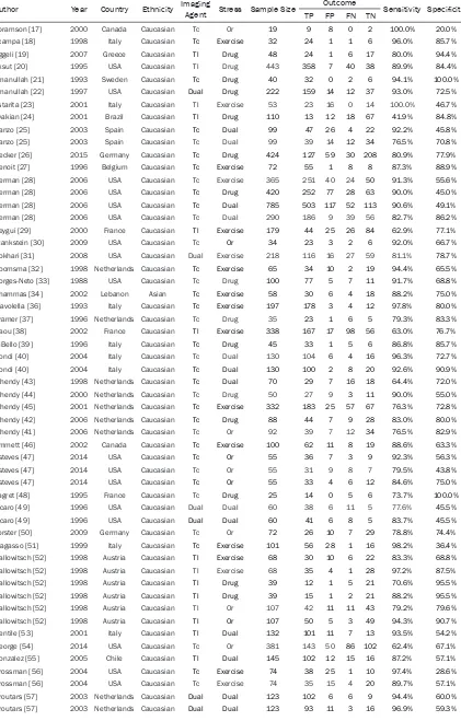
Table S1.
Main characteristics of included studies
Author Year Country Ethnicity Imaging Agent Stress Sample Size Outcome Sensitivity Specificity TP FP FN TN
Abramson [17] 2000 Canada Caucasian Tc Or 19 9 8 0 2 100.0% 20.0%
Acampa [18] 1998 Italy Caucasian Tc Exercise 32 24 1 1 6 96.0% 85.7%
Aggeli [19] 2007 Greece Caucasian Tl Drug 48 24 1 6 17 80.0% 94.4%
Aksut [20] 1995 USA Caucasian Tl Drug 443 358 7 40 38 89.9% 84.4% Amanullah [21] 1993 Sweden Caucasian Tc Drug 40 32 0 2 6 94.1% 100.0%
Amanullah [22] 1997 USA Caucasian Dual Drug 222 159 14 12 37 93.0% 72.5%
Astarita [23] 2001 Italy Caucasian Tl Exercise 53 23 16 0 14 100.0% 46.7%
Avakian [24] 2001 Brazil Caucasian Tl Drug 110 13 12 18 67 41.9% 84.8% Banzo [25] 2003 Spain Caucasian Tc Dual 99 47 26 4 22 92.2% 45.8% Banzo [25] 2003 Spain Caucasian Tc Dual 99 39 14 12 34 76.5% 70.8% Becker [26] 2015 Germany Caucasian Tc Drug 424 127 59 30 208 80.9% 77.9%
Benoit [27] 1996 Belgium Caucasian Tc Exercise 72 55 1 8 8 87.3% 88.9%
Berman [28] 2006 USA Caucasian Tc Exercise 365 251 40 24 50 91.3% 55.6%
Berman [28] 2006 USA Caucasian Tc Drug 420 252 77 28 63 90.0% 45.0%
Berman [28] 2006 USA Caucasian Tc Dual 785 503 117 52 113 90.6% 49.1%
Berman [28] 2006 USA Caucasian Tc Dual 290 186 9 39 56 82.7% 86.2%
Beygui [29] 2000 France Caucasian Tl Exercise 179 44 25 26 84 62.9% 77.1%
Blankstein [30] 2009 USA Caucasian Tc Or 34 23 3 2 6 92.0% 66.7%
Bokhari [31] 2008 USA Caucasian Dual Exercise 218 116 16 27 59 81.1% 78.7% Boomsma [32] 1998 Netherlands Caucasian Tc Exercise 65 34 10 2 19 94.4% 65.5%
Borges-Neto [33] 1988 USA Caucasian Tc Drug 100 77 5 7 11 91.7% 68.8% Chammas [34] 2002 Lebanon Asian Tc Exercise 58 30 6 4 18 88.2% 75.0%
Ciavolella [36] 1993 Italy Caucasian Tc Exercise 197 178 3 4 12 97.8% 80.0% Cramer [37] 1996 Netherlands Caucasian Tc Drug 35 23 1 6 5 79.3% 83.3%
Daou [38] 2002 France Caucasian Tl Exercise 338 167 17 98 56 63.0% 76.7%
DiBello [39] 1996 Italy Caucasian Tc Drug 45 33 1 5 6 86.8% 85.7% Dondi [40] 2004 Italy Caucasian Tc Dual 130 104 6 4 16 96.3% 72.7%
Dondi [40] 2004 Italy Caucasian Tc Dual 130 100 2 8 20 92.6% 90.9%
Elhendy [43] 1998 Netherlands Caucasian Tc Dual 70 29 7 16 18 64.4% 72.0%
Elhendy [44] 2000 Netherlands Caucasian Tc Drug 50 27 9 3 11 90.0% 55.0%
Elhendy [45] 2001 Netherlands Caucasian Tc Exercise 332 183 25 57 67 76.3% 72.8% Elhendy [42] 2006 Netherlands Caucasian Tc Drug 88 44 7 9 28 83.0% 80.0% Elhendy [41] 2006 Netherlands Caucasian Tc Or 92 39 7 12 34 76.5% 82.9% Emmett [46] 2002 Canada Caucasian Tc Exercise 100 62 11 8 19 88.6% 63.3%
Esteves [47] 2014 USA Caucasian Tc Or 55 36 7 3 9 92.3% 56.3%
Esteves [47] 2014 USA Caucasian Tc Or 55 31 9 8 7 79.5% 43.8% Esteves [47] 2014 USA Caucasian Tc Or 55 33 4 6 12 84.6% 75.0%
Fagret [48] 1995 France Caucasian Tc Drug 25 14 0 5 6 73.7% 100.0%
Ficaro [49] 1996 USA Caucasian Dual Dual 60 38 6 11 5 77.6% 45.5%
Ficaro [49] 1996 USA Caucasian Dual Dual 60 41 6 8 5 83.7% 45.5%
Forster [50] 2009 Germany Caucasian Tc Or 72 26 10 7 29 78.8% 74.4%
Fragasso [51] 1999 Italy Caucasian Tc Exercise 101 56 28 1 16 98.2% 36.4%
Gallowitsch [52] 1998 Austria Caucasian Tl Exercise 68 30 10 6 22 83.3% 68.8% Gallowitsch [52] 1998 Austria Caucasian Tl Exercise 68 35 4 1 28 97.2% 87.5% Gallowitsch [52] 1998 Austria Caucasian Tl Drug 39 12 1 5 21 70.6% 95.5%
Gallowitsch [52] 1998 Austria Caucasian Tl Drug 39 15 1 2 21 88.2% 95.5%
Gallowitsch [52] 1998 Austria Caucasian Tl Or 107 42 11 11 43 79.2% 79.6%
Gallowitsch [52] 1998 Austria Caucasian Tl Or 107 50 5 3 49 94.3% 90.7%
Gentile [53] 2001 Italy Caucasian Tl Dual 132 101 11 7 13 93.5% 54.2%
George [54] 2014 USA Caucasian Tc Or 381 143 50 86 102 62.4% 67.1%
Gonzalez [55] 2005 Chile Caucasian Tl Dual 145 102 12 15 16 87.2% 57.1%
Grossman [56] 2004 USA Caucasian Tc Exercise 74 38 25 1 10 97.4% 28.6% Grossman [56] 2004 USA Caucasian Tc Exercise 74 35 15 4 20 89.7% 57.1%
Groutars [57] 2003 Netherlands Caucasian Dual Dual 123 102 6 6 9 94.4% 60.0%