Original Article
Application of laparoscopy in urology: a study on the
learning curve in experience at a large center
including 4707 cases
Weiquan Guo1,2*,Liang Gao1*,Min Zhu3, Tianyong Fan1, Lu Yang1, Qiang Wei1
1Department of Urology, West China Hospital, Sichuan University, Chengdu, China; 2Department of Urology, Forestry Center Hospital of Sichuan Province, Chengdu, China; 3Department of Surgery, Chengdu University of TCM, Chengdu, China. *Equal contributors.
Received August 26, 2015; Accepted December 5, 2015; Epub February 15, 2016; Published February 29, 2016
Abstract: Purpose: To analyze the effects of a learning curve in laparoscopic approaches in urological surgery and the prevalence of complications in a large medical center. Materials and methods: Clinical data of all patients who underwent laparoscopic surgery at the Department of Urology, West China Hospital, from March 2003 to December 2012 were retrospectively reviewed. All postoperative complications were recorded and classified according to the Clavien-Dindo system, and the parameters of average operating time (OT), estimated blood loss (EBL), length of hospital stay after operation (LOSO), and rate of complication (RC) were analyzed statistically. Results: Of a total of 4707 patients selected, 4293 (91.2%) were treated by a retroperitoneal approach and 414 were treated by a transperitoneal approach. With the gradual maturity of technologies, OT, EBL, LOSO, and RC significantly decreased (P < 0.001). RC reached a plateau after the 3rd year. Furthermore, all postoperative complications according to the
Clavien-Dindo system revealed that complications occurred in 778 (16.5%) cases, 78 during surgery and 700 after surgery. Conclusions: Trends in OT, EBL, LOSO, and intra- and postoperative RCs significantly decreased through -out the study period. The 3rd year was regarded as a turning point in the learning curve for laparoscopic surgery. However, the applied range expanded along with an increase in difficult and complicated pRCedures. Complications may be efficiently reduced by summarizing RC and analysis of treatments and characteristics of the learning curve.
Keywords: Clavien-dindo, laparoscopy, learning curve, urology
Introduction
The application of Laparoscopic techniques will decrease the extent of trauma, the rate of com-plication (RC), and the length of hospital stay after operation (LOSO) [1]. Although the original intention of adopting laparoscopic techniques in urological surgery was to minimize invasive-ness, an increasing number of perioperative complications occur because of the greater prevalence of complicated surgeries encoun-tered along with the maturity of technologies [2, 3]. Parsons et al. [4] reported a RC of 4.4%-19.0%, but this rate could have been easily
influenced by multiple factors, such as the pro
-ficiency of the surgeon, level of difficulty, and
number of surgical sites.
In 1992, a hierarchy of postoperative complica-tions was proposed by Clavien et al. [5], which
classify postoperative complications into 5 grades (I-V) according to severity from slightest pain to death, in which grades III and IV were further divided into two subgroups [5, 6]. Further, grades I and II were merged as minor complications, whereas grades III and IV are considered as major complications [7].
Here we summarize a learning curve in laparo-scopic techniques in urological surgery since
first adopting these techniques in March 2003
to December 2012 to share our experience and
provide possible guidance for the classification
of postoperative complications according to the Clavien-Dindo system.
Materials and methods
sur-Surgical technique
All patients received general anesthesia by tra-cheal cannulation and were assigned to one of two groups according to the surgical approach, i.e., transperitoneal group and retroperitoneal group. In the transperitoneal group, patients were placed in a horizontal position for surger-ies targeting the bladder, prostate, spermatic cords, etc., or a semi-lateral position for surger-ies targeting the kidneys, ureters, adrenals, and retroperitoneal cavity. Initially, a vertical 3-5-cm incision below the umbilicus was made through the peritoneum into the abdominal cavity. A 10-mm Trocar sheath was then imbed-ded, after which pneumoperitoneum was established with carbon dioxide at a pressure of 12-15 mmHg. An additional 2 or 3 sheathes were placed within sight of the laparoscope. For the retroperitoneal group, the waist was elevated to expose the affected side after pro-cedures in the lateral position were completed. The main port was placed on the midaxillary line 2-3 cm cephalad to the iliac crest. A 2-3-cm incision was made through the skin and the three muscle layers of the abdominal wall were separated using a vessel clamp to expose the exterior Gerota’s fascia. When necessary, the
surgeon could extend the space by using a fin -ger. Then, 5- and 10-mm Trocar sheathes were placed into the extended retro-peritoneal space at the subcostal points of the posterior and anterior axillary lines, respectively. Finally, the
main sheath was implanted and then fixed with
sutures to prevent air leakage. The pneumo-peritoneum was maintained at a pressure of <12-15 mmHg. The incisions were extended according to the size of the excised specimens.
years). Rates of converting to open surgery in three phases were compared using the chi-squared test. Linear regression analysis was performed to identify trends in operating time (OT), estimated blood loss (EBL), and LOSO. Changes in trends in intra- and postoperative complications were assessed using the chi-squared test. A probability (P) value <0.05 was
considered statistically significant.
Moreover, data were recorded separately for surgeries involving multiple organs unilaterally or bilaterally and for repeated or staging proce-dures for the same patient. For multiple compli-cations in 1 patient, the most severe complica-tion was included for analysis.
Results
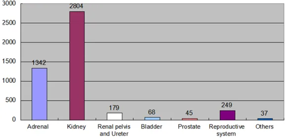
A total of 4724 surgeries were performed for 4707 patients, 4293 (91.2%) were treated by a retroperitoneal approach and 414 (8.8%) by a transperitoneal approach. Of these, 1342 (28.4%) focus on adrenal, 2804 (59.3%) for kid-ney, 179 (3.8%) for renal pelvis and ureter, 68 (1.4%) for bladder, 45 (1.0%) for prostate, 249 (5.3%) for reproductive system disease and others 37 (0.8%) (Figure 1). Seventeen cases underwent 2 surgeries because lesions in two sites were treated simultaneously. No repeated or staging procedures were performed. All basic patient data are summarized in Tables 1 and 2.
Complications were encountered in 778 (16.5%) cases and laparoscopy was converted to open surgery in 90 (1.9%). Regression
[image:2.612.89.380.73.213.2]analy-sis showed significant differences and decreas -ing trends in OT, EBL, and LOSO (t = -4.580, P <
Figure 1. Composition of surgeries in each location.
Statistical analysis
SPSS 17.0 software (IBM SPSS, Inc., Chicago, IL, USA) was used for all statistical analyses. All results are pre-sented as means (± standard deviations; SD) and rates. To analyze complications sys-tematically, we arbitrarily divided the 10-year period of 2003-2012 into three
phas-es: phase I (the first 3 years),
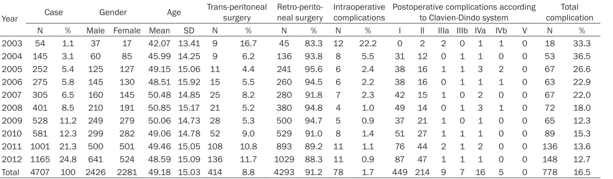
Table 1. The basic information, operation approaches and complications of patients
Year Case Gender Age
Trans-peritoneal
surgery neal surgeryRetro-perito- Intraoperative complications Postoperative complications according to Clavien-Dindo system complicationTotal
N % Male Female Mean SD N % N % N % I II IIIa IIIb IVa IVb V N %
2003 54 1.1 37 17 42.07 13.41 9 16.7 45 83.3 12 22.2 0 2 2 0 1 1 0 18 33.3
2004 145 3.1 60 85 45.99 14.25 9 6.2 136 93.8 8 5.5 31 12 0 1 1 0 0 53 36.5
2005 252 5.4 125 127 49.15 15.06 11 4.4 241 95.6 6 2.4 38 16 1 1 3 2 0 67 26.6
2006 275 5.8 145 130 48.51 15.92 15 5.5 260 94.5 6 2.2 38 16 0 1 1 1 0 63 22.9
2007 305 6.5 160 145 50.48 14.85 25 8.2 280 91.8 7 2.3 42 15 1 0 2 0 0 67 22.0
2008 401 8.5 210 191 50.85 15.17 21 5.2 380 94.8 4 1.0 49 14 0 1 3 1 0 72 18.0
2009 528 11.2 249 279 50.06 14.73 28 5.3 500 94.7 5 0.9 37 21 1 0 1 0 0 65 12.3
2010 581 12.3 299 282 49.06 14.78 52 9.0 529 91.0 8 1.4 51 27 1 1 1 0 0 89 15.3
2011 1001 21.3 500 501 49.46 15.05 108 10.8 893 89.2 11 1.1 76 44 2 1 2 0 0 136 13.6
2012 1165 24.8 641 524 48.59 15.09 136 11.7 1029 88.3 11 0.9 87 47 1 1 1 0 0 148 12.7 Total 4707 100 2426 2281 49.18 15.03 414 8.8 4293 91.2 78 1.7 449 214 9 7 16 5 0 778 16.5
0.001; t = -4.438, P < 0.001; and t = -10.410, P < 0.001, respectively). The chi-squared test
revealed significant differences in the inci -dence of intra- and postoperative
complica-tions (χ² = 46.611, P < 0.001 and χ² = 56.843,
P < 0.001, respectively) with a significant pla -teau exhibited after the 3rd year, indicating a
turning point. Further, a second plateau was observed in the incidence of postoperative complications after phase II (the 2nd 3-year
[image:4.612.89.405.96.360.2]peri-od) (Figure 2). Percentages of reconstruction and lower urinary tract surgeries were increased because of continuously extended ranges (Table 2).
Prevalence and treatment of intraoperative complications
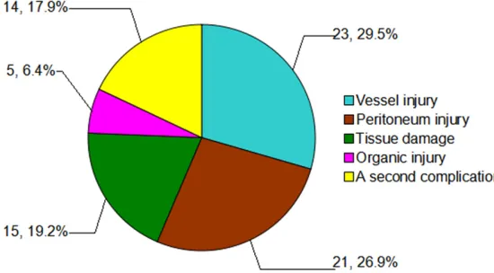
At least one complication occurred in 78 cases, accounting for 1.7% of surgeries, which includ-ed the following (Figure 3).
Vessel injury in 23 cases (7 involving the in- ferior vena cava, 6 of which were repaired by open surgery; 7 involving the renal vess- els, which were all repaired by open surgery; 2 involving the central adrenal veins, 1 of which was repaired by converting the proce-dure to open surgery; 2 involving the iliac
ves-Severe synechia and tissue damage occurr- ed in 15 patients, for which a hand-assisted approach was adopted in 4 cases, while in 11 patients the procedure was converted to open surgery.
Organic injuries occurred in 5 cases (renal parenchyma injury in 1 patient with a cyst and left adrenal mass and liver injury in 1 patient with right adrenal pheochromocytoma), all of which were repaired by conversion to open sur-gery. Moreover, gastric and rectal injury occurred in 1 patient with a large retroperito-neal mass and in 1 during radical prostatecto-my, respectively, both of which were repaired without using laparoscopy.
A second complication occurred in 14 patients, including three instances of hypercapnia, 2 of translocation of calculus, 3 of pleural damage,
and 1 of fluctuation of blood pressure.
In our series, in 16 (3.5%) cases the procedure was converted to open surgery during stage I, in 18 (1.8%) during stage II, and in 56 (1.7%) during stage III. Further, pairwise comparisons
revealed a significant difference between stag
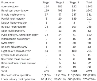
-es I and III (χ² = 7.178, P = 0.028). Table 2. Number of laparoscopic cases, reconstruction operation ratio,
lower urinary tract operation ratio by stage
Procedures Stage I Stage II Stage III Total
Adrenalectomy 134 286 922 1342
Renal cyst decortication 199 499 944 1642
Simple nephrectomy 37 57 587 681
Partial nephrectomy 3 20 189 212
Duplex kidney excision 1 3 3 7
Radical nephrectomy 23 26 197 246
Nephroureterectomy 4 13 36 53
Pyelolithotomy/Ureterolithotomy 25 24 61 110
laparoscopic pyeloplasty 2 2 16 20
Cystectomy 1 9 58 68
Radical prostatectomy 1 0 42 43
Ligation of spermatic vein 14 21 180 215
Lymph node dissection 3 3 5 11
Spermatic mass excision 3 5 8 16
Retroperitoneal mass excision 3 5 14 22
Others 2 15 19 36
Total 455 988 3281 4724
Reconstruction operation 6 (1.3%) 12 (1.2%) 115 (3.5%) 133 (2.8%) Lower urinary tract operation 20 (4.4%) 50 (5.1%) 305 (9.3%) 375 (7.9%)
sels, 1 of which was repaired by open sur-gery; and 5 cases of neoplastic bleeding, of which 4 were repaired converting the proce-dure to open surgery). Emergency intraopera-tive transfusion was necessary in 16 cases (mean, 3.6 U; range, 2-10 U).
Prevalence and treatment of postoperative complications
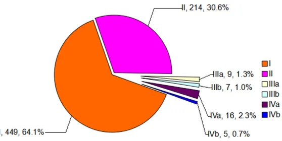
Postoperative complications occurred in a total
of 700 patients, which were classified accord
-ing to a modification of the Clavien-Dindo sys -tem. Of these, 663 (94.7%) were regarded as minor complications [449 (64.1%) grade I and 214 (30.6%) grade II] and 37 as major compli-cations [9 (1.3%), 7 (1.0%), 16 (2.3%), and 5 (0.7%) were categorized as grade IIIa, IIIb, IVa, and IVb, respectively] (Figure 4) as described below.
Grade I: Three hundred and ninety cases
classi-fied as grade I were treated with drugs, which
included antiemetics, antipyretics, analgesics, diuretics, and electrolytes. Incision infection,
titis was observed in 7 and 4 cases, respective-ly. Bowel obstruction was treated by gastric intubation or use of an anal tube in 19 cases. Moreover, transfusions were necessary in 15 cases. Deep venous thrombosis of the lower limbs and muscular venous thrombosis was diagnosed in 1 patient each, respectively. Other complications exceeding grade I, which were treated by drug administration, were observed in 27 cases.
[image:5.612.95.521.74.258.2]Grade IIIa: Urinary leakage occurred in 3 patients after ureterolithotomy due to discon-nection of a double-J catheter. All instances of urinary leakage were treated by cystoscopic-assisted ureteral stent implantation. Hydro- pneumothorax developed in 1 patient after cys-tectomy of the left kidney. Ectopia of a double-J
Figure 2. Trends of average operation time (OT), estimated blood loss (EBL), length of hospital stay after operation (LOSO), intra- and postoperative complication rate gradually.
Figure 3. Constitution of intraoperative complications.
non-infectious diarrhea, deliri-um, bowel obstruction, subcu-taneous emphysema, pulmo-nary atelectasis, and atrial
fibrillation occurred in 15, 10,
10, 9, 6, 5, and 4 cases, respectively.
[image:5.612.90.365.312.464.2]kera-catheter was observed in 1 patient following
radical cystectomy and ileal outflow tract
obstruction, which was adjusted by radiological guidance. Fracture of a drainage tube occurred in 1 case, which was removed by reoperation. Continuous hematuria derived from the renal segmental artery occurred in 1 patient, which was treated by interventional embolization. Anastomotic leakage between the bladder and urethra was observed in 1 patient following radical prostatectomy, which was recovered by conservative management.
Grade IIIb: Hemorrhaging developed in 3 cases after adrenalectomy, 1 after radical nephrecto-my and 1 after radical nephroureterectonephrecto-my, all of which were cured by an open approach. Reoperation was performed in 1 patient because of leakage after cystectomy of the left kidney, whereas herniorrhaphy of the ileal con-duit was performed in 1 patient following by radical cystectomy.
Grade IVa: Cardiovascular dysfunction devel-oped in 5 patients, pulmonary dysfunction in 4, adrenal crisis in 2, and kidney failure requiring dialysis in 1 after adrenalectomy. Pulmonary dysfunction after radical cystectomy, kidney failure requiring dialysis after radical nephrec-tomy, and heart failure after simple nephrecto-my occurred in 1, 2, and 1 patient, respectively.
Grade IVb: Multiple organ dysfunction syn-drome was diagnosed in 2 patients after radi-cal nephrectomy, 2 after adrenalectomy, and 1 after simple nephrectomy, who were all treated in intensive care units. There was no instance of procedure-related mortality.
results of our study, 16.5% for overall complica-tions and 1.96% for cases in which the proce-dure was converted to open surgery, were in accordance with the results reported by Inoue et al. (14.6% and 1.9%, respectively) [16].
It is anticipated that the RC will significantly
decrease with the acquisition of surgical experi-ence [16-18]. In our study, the 3rd year was
regarded as a turning point, as similarly report-ed by Akin et al. [18]. In addition, a study con-ducted in 4 centers reported a RC of 13.3%
after the first 100 laparoscopic surgeries,
which rapidly decreased to 3.6% thereafter [3]. On the other hand, the application of laparo-scopic surgery was also changed besides the increase in the number of cases. In our study, percentages of 1.3%, 1.2%, 3.5% in surgeries referring to reconstruction and 4.4%, 5.1%, 9.3% in surgeries focusing on the lower urinary tract in three stages were respectively report-ed. It showed that, with the accumulating of
surgical proficiency, tendencies of challenging more difficult reconstructive and lower tract
surgeries were gradually favored. More impor-tantly, increasing OT, EBL, LOSO, and RC could not be presented. Meanwhile, the rate of
con-version to open surgery decreased significantly.
Moreover, the intra- and postoperative RCs pla-teaued after the second 3-year period, which is the reason we divided the 10-year study period into three phases. However, these tendencies
would be largely influenced by updated equip -ment and improved cooperation between sur-geons and nurses apart from surgical skills. Approximately 15 years ago, Guillonneau et al. [19] and Ghavamian et al. [20] reported a
sig-Figure 4. Postoperative complications according to the Clavien-Dindo clas-sification system.
Discussion
[image:6.612.92.371.70.210.2]nificant decrease in OT in laparoscopic prosta -tectomy. However, this trend could be similar to the results of our study, which showed that a
longer OT may be associated with insufficient skills during stage I, and a significant decrease
in OT was shown in stage II because of accumu-lating experience. Furthermore, the slight increase in OT in stage III compared with stage
II could be caused by encountering more diffi -cult diseases. Moreover, similar decreasing trends were observed in EBL and LOSO [21]; however, there was no increase in EBL or LOSO
in stages II and III even though more difficult
procedures were performed.
Although laparoscopy is considered very safe, complications remain common, regardless of experience. The concepts of minimally invasive surgery and open surgery should not be con-fused, as the main complications can be fatal [8]. Although the overall intraoperative compli-cation rate was only 10% (78 cases) in this study, vascular injury was the most common and serious complication, often requiring con-version to open surgery [22]. In our study, vas-cular injury, mostly involving the great vessels, occurred in 23 cases, and in 19 of which the procedure was converted to open surgery. In our opinion, laparoscopic-assisted suturing could be attempted to stop small venous bleed-ing, during which an appropriate increase in pneumoperitoneum pressure could be helpful. However, this procedure is suitable only for experienced surgeons. On the other hand, con-version to open surgery should be considered
as the first choice for arterial bleeding or mas -sive hemorrhaging after rapid clamping. In this series, no patient died due to vascular injury. However, organic injuries occurred in 5 patients during surgery, while synechia and severe tis-sue damage occurred in 15 cases, in 4 of which the procedure was completed using hand-assisted procedures, while in the other 11 the procedure was converted to open surgery. In our opinion, a semi-open laparoscopic approach
with hand-assisted procedures is an efficient
method initially and could also accelerate the learning process. Through the acquisition of
experience, open surgery could be also the first
choice for patients with unclear anatomical structures caused by severe adhesions, which could shorten OT and decrease EBL and RCs. Unlike other countries, most urological proce-dures in China employ an extraperitoneal
approach [23], through which less influence on
abdominal viscera could be considered com-pared with those through transperitoneal approach. A previous retrospective study of 883 cases in our center to compare extraperi-toneal and transperiextraperi-toneal approaches con-cluded that differences in complication rates
between these two approaches were insignifi -cant, and longer OT and LOSO were observed in the transperitoneal group [24]. In our study, 4293 (91.2%) surgeries were performed through an extraperitoneal approach and peri-toneal injury was observed in only 21 cases performed through the retroperitoneal app- roach, of which only 1 was due to a puncture caused by hand-manipulation of a sheath [8, 25]. Furthermore, in cases where the peritone-um is opened carelessly, the surgery can be continued without conversion to open surgery. For large lesions of the adrenals, kidneys, or retroperitoneal cavity, or cases of intraopera-tive peritoneal injury, we formerly adopted a translumbar/peritoneal joint approach with bal-anced pressure after expanding the incision to the peritoneum.
The Clavien-Dindo system is increasingly used to classify complications following urological procedures. An increasing number of centers have begun to classify complications based on this system to standardize data and promote the quality of evaluation [26-31]. All complica-tions in our study were evaluated using the Clavien-Dindo system, in which 663 (94.7%) cases were considered minor complications, which was a higher percentage than 76.6% as reported by Akin et al. [18]. However, multiple factors may have contributed to this result. First, the administration of data may have been
easily influenced because the data was collect -ed from different centers and surgeons. This is particularly true for minor complications, which may be easily overlooked. Second, this evalua-tion system is not quantitative, which could
induce classification bias. Furthermore, differ -ences in therapeutic choices between develop-ing and developed countries may affect conclu-sions [31]. Besides, different constituent ratios in morbid composition may contribute to
clas-sification complications. For example, 3665
Conclusion
Parameters of OT, EBL, LOSO, and prevalence of intra- and postoperative complications are
expected to significantly decrease with the
maturity of skills. A plateau in complications was observed after the 3rd year, which could be
regarded as a turning point in the learning curve. Moreover, the incidence of puncture-related complications could be effectively decreased through the application of hand-guided techniques in retroperitoneal surgeries. For large retroperitoneal lesions or intraopera-tive peritoneal injury, a translumbar/peritoneal joint approach with balanced pressure after expanding trauma at peritoneum should be
considered. The Clavien-Dindo system is effi
-cient for classification of postoperative compli -cations and helpful to summarize and guide the choice of laparoscopic procedures.
Acknowledgements
This paper was supported by the Pillar Program from Science and Technology Department of Sichuan Province (Grant No. 2012SZ0009), Prostate Cancer Foundation Young Investigator Award 2013 and the Project of Natural Science Foundation of China (NSFC) (Grant No. 81300627).
Discloser of conflict of interest
None.
Address correspondence to: Drs. Lu Yang and Qiang Wei, Department of Urology, West China Hospital, Sichuan University, No. 37 Guoxue Xiang, Chengdu 610041, China. E-mail: [email protected] (LY); [email protected] (QW)
References
[1] Hedican SP. Laparoscopy in urology. Surg Clin North Am 2000; 80: 1465-1485.
[2] Guillonneau B, Vallencien G. Laparoscopic radical prostatectomy. The Montsouris tech-nique. J Urol 2000; 163: 1643-1649.
[3] Fahlenkamp D, Rassweiler J, Fornara P, Frede T, Loening SA. Complications of laparoscopic procedures in urology: experience with 2,407 procedures at 4 German centers. J Urol 1999; 162: 765-771.
[4] Parsons JK, Varkarakis I, Rha KH, Jarrett TW, Pinto PA, Kavoussi LR. Complications of ab-dominal urologic laparoscopy: longitudinal five-year analysis. Urology 2004; 63: 27-32.
[5] Clavien PA, Sanabria JR, Strasberg SM. Proposed classification of complications of surgery with examples of utility in cholecystec-tomy. Surgery 1992; 111: 518-526.
[6] Clavien PA, Barkun J, de Oliviera ML, Vauthey JN, Dindo D, Schulick RD, de Santibañes E, Pekolj J, Slankamenac K, Bassi C, Graf R, Vonlanthen R, Padbury R, Cameron JL, Makuuchi M. The Clavien-Dindo classification of surgical complications: Five-year experi-ence. Ann Surg 2009; 250: 187-196.
[7] Hruza MO, Pini G, Gozen SA, Schulze M, Teber D, Rassweiler JJ. Complications in 2200 con-secutive laparoscopic radical prostatectomies: standardised evaluation and analysis of learn-ing curves. Eur Urol 2010; 58: 733-741. [8] Soulie M, Salomon L, Seguin P, Mervant C,
Mouly P, Hoznek A, Antiphon P, Plante P, Abbou CC. Multi-institutional study of complications in 1085 laparoscopic urologic procedures. Urology 2001; 58: 899-903.
[9] Shao PF, Yin CJ, Meng XX, Ju XB, Lü Q, Li J. Modified transperitoneal laparoscopic radical prostatectomy: technique and clinical out-comes. Zhonghua Wai Ke Za Zhi 2011; 1: 542-545.
[10] Asimakopoulos AD, Annino F, D’Orazio A, Pereira CF, Mugnier C, Hoepffner JL, Piechaud T, Gaston R. Complete periprostatic anatomy preservation during robot-assisted laparoscop-ic radlaparoscop-ical prostatectomy (RALP): the new pubo-vesical complex-sparing technique. Eur Urol 2010; 58: 407-417.
[11] Moreira SG, Ordorica RC, Sakti D. History of laparoscopy: an odyssey of innovations. In: Gill IS, editor. Textbook of laparoscopic urology. New York: Informa Healthcare Inc; 2006. pp. 3-10.
[12] Na YQ. Laparoscopic operation. Textbook of Wu Jieping mi niao wai ke xue. Edited by Wu JP. (Chi) Ji’nan: Shandong Science and Technology Press; 2004. pp. 2132-2134.
[13] Rassweiler JJ, Stolzenburg J, Sulser T, Deger S, Zumbé J, Hofmockel G, John H, Janetschek G, Fehr JL, Hatzinger M, Probst M, Rothenberger KH, Poulakis V, Truss M, Popken G, Westphal J, Alles U, Fornara P. Laparoscopic radical prosta-tectomy-the experience of the German Laparoscopic Working Group. Eur Urol 2006; 49: 113-119.
[14] Guillonneau B, Vallancien G. Laparoscopic radical prostatectomy: the Montsouris experi-ence. J Urol 2000; 163: 418-422.
[15] Wong MT, Ng KH, Lim JF, Ooi BS, Tang CL, Eu KW. 418 cases of laparoscopic colorectal re-sections: a single-institution experience and literature review. Singapore Med J 2010; 51: 650-654.
Complications of urologic laparoscopic sur-gery: a single institute experience of 1017 pro-cedures. J Endourol 2010; 24: 253-260. [17] Siow A, Nikam YA, Ng C, Su MC. Urological
com-plications of laparoscopic hysterectomy: a four-year review at KK Women’s and Children’s Hospital, Singapore. Singapore Med J 2007; 48: 217-221.
[18] Akin Y, Ates M, Celik O, Ucar M, Yucel S, Erdogru T. Complications of urologic laparoscopic sur-gery: A center surgeon’s experience involving 601 procedures including the learning curve. Kaohsiung J Med Sci 2013; 29: 275-279. [19] Guillonneau B, Rozet F, Barret E, Cathelineau
X, Vallancien G. Laparoscopic radical prosta-tectomy: assessment after 240 procedures. Urol Clin North Am 2001; 28: 189-202. [20] Ghavamian R, Schenk G, Hoenig DM, Williot P,
Melman A. Overcoming the steep learning curve of laparoscopic radical prostatectomy: single-surgeon experience. J Endourol 2004; 18: 567-571.
[21] Simforoosh N, Soltani MH, Basiri A, Tabibi A, Gooran S, Sharifi SH, Shakibi MH. Evolution of Laparoscopic Live Donor Nephrectomy: A Single-Center Experience with 1510 Cases over 14 Years. J Endourol 2014; 28: 34-39. [22] Hsu TH, Su LM, Ratner LE, Kavoussi LR.
Renovascular complications of laparoscopic donor nephrectomy. Urology 2002; 60: 811-815.
[23] Wang GX, Feng L, Cao RF. The prevention and treatment of complications of retroperitoneo-scopic technique in urology. (Chi) Journal of Clinical Urology 2006; 21: 811-815.
[24] Han DJ, Fan TY, Wei Q. The prevention and treatment of urological laparoscopy complica-tions. Journal of Clinical Urology 2009; 24: 664-668.
[25] Abbou CC, Cicco A, Gasman D, Hoznek A, Antiphon P, Chopin DK, Salomon L. Retroperitoneal laparoscopic versus open radi-cal nephrectomy. J Urol 1999; 161: 1776-1780.
[26] Thomas AA, Aron M, Hernandez AV, Lane BR, Gill IS. Laparoscopic partial nephrectomy in oc-togenarians. Urology 2009; 74: 1042-1046. [27] Hsu TH, Gill IS, Fazeli-Matin S, Soble JJ, Sung
GT, Schweizer D, Novick AC. Radical nephrec-tomy and nephroureterecnephrec-tomy in the octoge-narian and nonageoctoge-narian: Comparison of lapa-roscopic and open approaches. Urology 1999; 53: 1121-1125.
[28] Gong EM, Orvieto MA, Lyon MB, Gong EM, Orvieto MA, Lyon MB, Lucioni A, Gerber GS, Shalhav AL. Analysis of impact of body mass index on outcomes of laparoscopic renal sur-gery. Urology 2007; 69: 38-43.
[29] Harper JD, Breda A, Leppert JT, Veale JL, Gritsch HA, Schulam PG. Experience with 750 consecutive patients laparoscopic donor ne-phrectomies-is it time to use a standardized classification of complications? J Urol 2010; 183: 1941-1946.
[30] Kapoor A, Nassir A, Chew B, Gillis A, Luke P, Whelan P. Comparison of laparoscopic radical renal surgery in morbidly obese and non-obese patients. J Endourol 2004; 18: 657-660. [31] Khan A, Palit V, Myatt A, Cartledge JJ, Browning