Original Article
Positive interferon-gamma release assay
results are correlated with paradoxical
reaction in tuberculous meningitis
Tingting Lu1*, Xinqing Lin2*, Yaqing Shu1, Qing Tian1, Yuge Wang1, Zhengqi Lu1, Yongqiang Dai1
1Department of Neurology, The Third Affiliated Hospital of Sun Yat-sen University, Guangzhou, China; 2State
Key Laboratory of Respiratory Disease, National Clinical Research Center for Respiratory Disease, Guangzhou Institute of Respiratory Disease, First Affiliated Hospital, Guangzhou Medical University, Guangzhou, China. *Equal
contributors.
Received December 3, 2016; Accepted August 12, 2017; Epub September 15, 2017; Published September 30, 2017
Abstract: Background: Evidence suggests that T cell-based interferon-γ release assays (IGRAs) may be useful for the diagnosis of tuberculosis. The present study aims to investigate the clinical significance of blood T-SPOT.TB assay, a commercially available IGRAs, in tuberculous meningitis (TBM) patients. Methods: Records of consecutive TBM and non-TBM meningitis patients admitted from July 2011 to March 2016 at a tertiary hospital were retrospec -tively reviewed. Clinical and magnetic resonance (MR) findings, as well as T-SPOT.TB results were comprehensively assessed. Results: A total of 61 TBM patients and 85 non-TBM meningitis patients were enrolled. The sensitivity and specificity of T-SPOT.TB for TBM patients were 62.3% and 72.9%, respectively. Positive T-SPOT.TB results were related to diagnostic category (P=0.015), TB outside of the central nervous system (CNS) (P=0.002), active TB outside of CNS (P=0.009), hydrocephalus (P=0.039) and basal exudates (P=0.031) on MR, and paradoxical reac -tion to anti-tuberculosis drugs (P=0.032). The logistic regression analysis showed that TB outside of CNS was the only independent predictor for positive T-SPOT.TB (P=0.013). Conclusion: These results collectively indicate that blood T-SPOT.TB should be supplementary for the diagnosis of TBM, rather than serving as a single test to diagnose or exclude the disease. However, positive T-SPOT.TB results may reflect typical clinical and MR features of TBM. Moreover, positive results are associated with paradoxical reaction to treatment, which is related to immune status to tuberculosis infection. Thus, T-SPOT.TB results provide potentially useful information for the consideration of immune-modulating therapy.
Keywords: Interferon-gamma release assay, tuberculous meningitis, paradoxical reaction, T-SPOT.TB
Introduction
Tuberculosis is considered as one of the lead-ing causes of death due to infectious diseases worldwide [1]. Although tuberculous meningitis (TBM) represents only approximately 1% of all tuberculosis cases, it kills or disables about half of the people affected [2]. Treatment delay has been long recognized as the strongest risk factor for poor prognosis. However, early diag-nosis of TBM is notoriously difficult because clinical features in the early stage are non-spe-cific and laboratory tests are insensitive [3]. Definitive diagnosis of TBM mainly relies on the confirmation of M. tuberculosis inside the cen-tral nervous system (CNS). Unfortunately,
sen-sitivities for CSF Ziehl-Neelsen staining and nucleic acid amplification techniques in TBM are both less than 60% [2]. In clinical practice, the majority of clinically diagnosed TBM cases were classified as probable or possible, particu-larly for HIV-negative patients.
Table 1. Baseline clinical characteristics in suspected meningitis
Characteristics TBM (n=61) Non-TBM (n=85) P Value
Age, mean years ± SE 39.34±1.886 42.02±1.931 0.337#
Male sex, n (%) 38 (62.3) 57 (67.1) 0.552
With tuberculosis outside of CNS, n (%) 29 (47.5) 13 (15.3) <0.001
Underlying condition or illness, n (%)
No underlying illness 27 (44.3) 34 (40.0) 0.607
Diabetes mellitus 9 (14.8) 12 (14.1) 0.960
Metabolic syndrome and related diseases* 11 (18.0) 24 (28.2) 0.859
Autoimmune diseases 7 (11.5) 6 (7.1) 0.109
Chronic hepatitis B virus infection 9 (14.8) 13 (15.3) 0.332
Pregnancy and related conditions** 3 (4.9) 1 (1.2) 0.128##
Dysfunction of major organs 2 (3.3) 3 (3.5) 1.000##
Prior trauma and/or surgery 3 (4.9) 1 (1.2) 0.113##
Receiving immunosuppressive treatment 5 (8.2) 7 (8.2) 0.959
Immunosuppressed condition*** 9 (14.8) 13 (15.3) 0.911
*Including hypertension, abnormal glucose tolerance, overweight and obesity, lipid metabolism disorder, hepatic adipose infil
-tration, and arteriosclerotic artery disease, et al. **Including application of assisted reproductive technology and pathological
abortion. ***Defined as patients with underlying diseases such as malignancy, liver cirrhosis, chronic renal failure, pregnancy or
patients receiving immunosuppressive treatment. #Student’s t test. ##Fisher’s exact test.
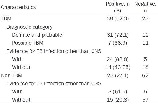
Table 2. Results of blood T-SPOT.TB in different subgroups of patients with meningitis
Characteristics Positive, n (%) Negative, n
TBM 38 (62.3) 23
Diagnostic category
Definite and probable 31 (72.1) 12
Possible TBM 7 (38.9) 11
Evidence for TB infection other than CNS
With 24 (82.8) 5
Without 14 (43.75) 18
Non-TBM 23 (27.1) 62
Evidence for TB infection other than CNS
With 8 (61.5) 5
Without 15 (20.8) 57
IGRAs was highest in active pulmonary TB, with specificity depending on the burden of latent TB infection. Researches also suggest that IGRAs are particularly useful for the diagnosis of extrapulmonary TB, but the sensitivity is rela-tively lower, with a range of 79.8-89% [4]. There was limited information for IGRA-based TBM diagnosis, especially in high-burden countries. As a commercially available IGRA, T-SPOT.TB assay detects IFN-γ induced by ESAT-6 and CFP-10. Thus, the present study attempted to investigate the clinical significance of T-SPOT. TB assay in TBM.
Materials and methods
Patients
[image:2.612.91.348.373.542.2]Table 3. Diagnostic performance of the blood T-SPOT.TB in tuberculous meningitis
Sensitivity* %
(95% CI) Specificity
** %
(95% CI) Positive predictive value % (95% CI) Negative predictive value % (95% CI) Positive likelihood ratio (95% CI) Negative likelihood ratio (95% CI)
All cases 62.3 (48.9-74.1) 72.9 (62.0-81.7) 62.3 (48.9-74.1) 72.9 (62.0-81.7) 2.30 (1.54-3.43) 0.517 (0.371-0.719)
Diagnostic category
Definite and probable TBM 72.1 (56.1-84.2) 72.9 (62.0-81.7) 57.4 (43.3-70.5) 83.8 (73.0-91.0) 2.66 (1.79-3.96) 0.382 (0.235-0.624)
Possible TBM 38.9 (18.3-63.8) 72.9 (62.0-81.7) 23.3 (10.6-42.7) 84.9 (74.2-91.9) 1.44 (0.731-2.82) 0.838 (0.575-1.22)
Evidence for TB infection other than CNS
Without TB outside of CNS 43.7 (26.8-62.1) 79.2 (67.7-87.5) 48.3 (29.9-67.1) 76.0 (64.5-84.8) 2.10 (1.15-3.82) 0.710 (0.520-0.971) With TB outside of CNS 82.8 (63.5-93.5) 38.5 (15.1-67.7) 75.0 (56.2-87.9) 50.0 (20.1-79.8) 1.34 (0.848-2.13) 0.448 (0.159-1.27)
culosis treatment: the worsening of a preexist-ing insults, the appearance of new insults on MR, and/or transient worsening of CSF param-eters [9]. Paradoxical reactions on MR included the appearance of new tuberculomas, hydro-cephalus, infarction, vasculitis, optochiasmatic and spinal arachnoiditis and the expansion of an existing insult. Paradoxical reactions during the first three months of treatment were docu-mented. Disability status was recorded at time of enrollment and 6 months post-admission. The follow-up outcome was evaluated with the modified Barthel index (MBI), and patients were classified into two categories including (1) poor outcome, i.e., MBI score lower than 50 or death; and (2) good outcome, i.e., MBI higher than 50.
Statistical analysis
Statistical analysis was performed using SPSS 19.0 under the Windows environment (SPSS Inc., Chicago, IL, USA). The diagnostic value of T-SPOT.TB assay was expressed in terms of sensitivity, specificity, positive predictive value, negative predictive value, positive likelihood ratio, and negative likelihood ratio. Ninety-five percent confidence intervals (CIs) were calcu-lated using the Wilson score method. The rela-tionship between T-SPOT.TB results and various clinical and MRI findings was assessed. Qua- litative data were analyzed with the X2 tests or
Fisher’s exact test, and quantitative data were analyzed using two-tailed Mann-Whitney U- tests or student’s t test. In addition, logistic regression analysis was applied to identify the independent predictive factors of positive T- SPOT.TB.
Results
Patient characteristics
A total of 61 patients with TBM and 85 patients with other meningitis were reviewed in the pres-ent study. All patipres-ents were HIV-negative. In the patient group with TBM, there were 2 patients with “definite” TBM, 41 patients with “proba-ble” TBM, and 18 patients with “possi“proba-ble” TBM. In the non-TBM meningitis patient group, there were 38 patients with cryptococcal meningitis, 34 patients with viral meningitis, and 13 pa- tients with purulent meningitis. Their clinical characteristics at time of admission are sum-marized by patient group in Table 1.
procedures of the study. All patients provided written informed consent.
Clinical, laboratory and MRI assessments
The age, gender, medical history, and clinical presentation of each patient were retrieved from the patient’s medical records. Disturbance of consciousness was defined by a Glasgow Coma Scale rating below 8. Cranial nerve palsy, movement impairment, and the presence of seizures were also recorded. Severity of TBM was graded into three stages, including stage 1: fully conscious and without specific symp-toms; stage 2: lethargy or cranial nerve pal- sies; and stage 3: stupor, severe illness, gross paralysis or paresis [6]. HIV antibody tests, chest radiographs, and other necessary exami-nations for extra-CNS tuberculosis were carried out. Opening pressure, cells, protein, glucose, and chloride were examined in CSF, which was also subjected to pathogenic culture.
Brain MRI was performed using a 1.5-Tesla MR scanner (GE Health care, Milwaukee, USA). T1, T2, fluid attenuated inversion recovery (FLAIR), diffusion weighted imaging (DWI), T1 contrast and magnetic resonance angiography (MRA) images were obtained in the first week of hospi-talization. Evidence of infarcts, hydrocephalus, basal exudates, tuberculomas and vasculitis was recorded.
T-SPOT.TB
The T-SPOT.TB assay was performed upon en- rollment and interpreted according to the man-ufacturer’s insert guidelines [7, 8]. Briefly, pe- ripheral blood mononuclear cells (PBMC) were separated from peripheral venous blood, and 2.5×10⁵ PBMC were plated per well in wells
precoated with anti-human IFN-γ antibody. The PBMC were cultured at 37°C for 18 h. The num-ber of spot-forming cells (SFCs) in each well was counted automatically. Laboratory staffs were blind to the clinical characteristics of the patients.
Treatments and prognosis
antituber-Figure 1. Scatter plots displaying counts of spot-forming cells (SFCs) in peripheral blood mononuclear cells using the T-SPOT.TB according to the following criteria: A. Type of meningitis; B. Diagnostic category of tuberculous meningitis (TBM); C. With or without tuberculosis (TB) outside of the central nervous system (CNS); and D. Presence or absence of paradoxical reaction. Differences between patient groups were assessed using Mann-Whitney U-tests.
Diagnostic value of the blood T-SPOT.TB for TBM
Thirty-eight TBM patients (out of 61, 62.3%) were T-SPOT.TB positive, while 23 non-TBM patients (out of 85, 27.1%) were positive (P=0.000) (Table 2). As shown in Table 3, the sensitivity and specificity of T-SPOT for diagnos-ing TBM were 62.3% (95% CI; 48.9-74.1%) and 72.9% (95% CI; 62.0-81.7%), respectively. The sensitivity of the blood T-SPOT.TB for diagnos-ing TBM differed between patient groups; patients with possible TBM and definite/proba-ble TBM were identified with sensitivities of 38.9% and 72.1%, respectively. The sensitivity of T-SPOT.TB in patients with TB infection out-side CNS was higher than in patients without TB outside CNS (82.8% and 43.7%, respective-ly), though the specificity was lower (38.5% and 79.2%, respectively).
As displayed in Figure 1A-C, patients with TBM showed higher numbers of SFCs for ESAT-6 and CFP-10 than non-TBM meningitis patients (both P=0.000). The numbers of SFCs for ESAT-6 and CFP-10 were compared in TBM patients with different diagnostic categories and presence of tuberculosis outside of CNS. Patients with
defi-nite and probable TBM had significantly higher SFCs numbers for ESAT-6 (P=0.013) and CFP-10 (P=0.0CFP-10) than patients with possible TBM. In addition, TBM patients with TB outside of CNS exhibited significantly higher SFCs num-bers for ESAT-6 and CFP-10 (P=0.000 and P=0.001, respectively).
Factors associated with positive T-SPOT.TB results in TBM
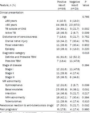
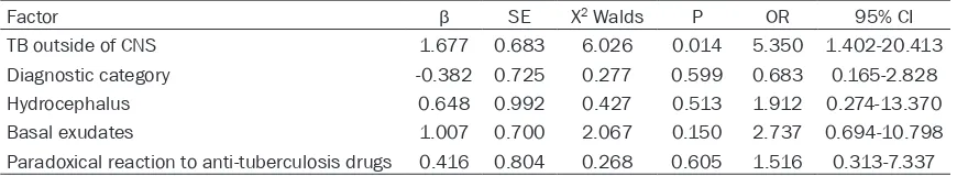
Correlation tests between T-SPOT.TB results and clinical and MR manifestations in TBM patients are displayed in Table 4. Positive T-SPOT.TB results were associated with TB out-side of CNS (P=0.002), active TB outout-side of CNS (P=0.009), diagnostic category (P=0.015), and hydrocephalus (P=0.039) and basal exu-dates (P=0.031) on MR. As shown in logistic regression analysis, TB outside of CNS was the only independent predictor for positive T-SPOT. TB results (P=0.013) (Table 5).
Correlations between positive T-SPOT.TB and prognosis
[image:5.612.89.514.74.330.2]anti-Table 4. Factors associated with positive result of T-SPOT.TB in tuber-culous meningitis
Feature, n (%) Positive result
(n=38)
Negative result (n=23)
P Value
Clinical presentation
Age 0.765
≥60 years 4 (10.5) 3 (13.0)
<60 years 34 (89.5) 20 (87.0)
TB outside of CNS 24 (63.1) 5 (21.7) 0.002
Active TB 15 (39.5) 2 (8.7) 0.009
Disturbance of consciousness 7 (18.4) 5 (21.7) 0.752 Cranial nerve injury 13 (34.2) 7 (30.4) 0.761 Focal weakness 11 (28.9) 7 (30.4) 0.902
Epilepsy 10 (26.3) 3 (13.0) 0.220
Diagnostic category 0.015
Definite and Probable TBM 31 (81.6) 12 (52.2)
Possible TBM 7 (18.4) 11 (47.8)
Stage of disease 0.394
Stage I 12 (31.6) 11 (47.8)
Stage II 11 (28.9) 4 (17.4)
Stage III 15 (39.5) 8 (34.8)
MR abnormality
Hydrocephalus 12 (31.6) 2 (8.7) 0.039
Basal exudates 25 (65.8) 9 (39.1) 0.031
Infarction 14 (36.8) 5 (21.7) 0.217
MRA abnormality 16 (42.1) 7 (30.4) 0.384
Tuberculomas 11 (28.9) 4 (17.4) 0.310 Paradoxical reaction to anti-tuberculosis drugs* 17 (50.0) 5 (21.7) 0.032
Poor prognosis* 6 (17.6) 4 (17.4) 0.980
*Total N=57, including 34 T-SPOT.TB positive cases and 23 T-SPOT.TB negative cases.
tuberculosis drugs occurred in 22 patients. Positive T-SPOT.TB results were associated with paradoxical reaction (P=0.032) (Table 4). SFCs numbers for ESAT-6 and CFP-10 were also higher in patients who developed paradoxical reaction (P=0.003 and P=0.002, respectively) (Figure 1D). The prognosis was good in 47 patients, while poor in 10 patients. However, T-SPOT.TB results were not significantly associ-ated with the prognosis (Table 4).
Discussion
Still until now, the diagnosis of TBM in the clini-cal practice is made by cliniclini-cal manifestation. The majority of patients are diagnosed with probable or possible TBM, especially in HIV negative patients, and a fraction of patients are diagnosed with definite TBM (e.g., 5.9% in our
local center) [10]. Using blood T-SPOT.TB to diagno- se TBM is very convenient, since T-SPOT.TB tests can be completed within 24 hours, and is not affected by previous BCG vaccina-tion. The diagnostic value of T-SPOT.TB has been de- monstrated to be greater than TST, even in HIV pa- tients [8, 11, 12]. The pre- sent study comprehensi- vely assessed the perfor-mance of blood T-SPOT.TB assay for TBM under real clinical condition, in which-the majority of which-the TBM cases could not be etio-logically diagnosed. Alth- ough results showed that the sensitivity and speci- ficity of T-SPOT.TB were both inadequate, positive result of T-SPOT.TB could provide additional informa-tion in the diagnosis and management of TBM.
[image:6.612.94.388.98.467.2]Table 5. Multivariate logistic regression analysis of factors related to positive T-SPOT.TB results in tuberculous meningitis
Factor β SE X2 Walds P OR 95% CI
TB outside of CNS 1.677 0.683 6.026 0.014 5.350 1.402-20.413
Diagnostic category -0.382 0.725 0.277 0.599 0.683 0.165-2.828
Hydrocephalus 0.648 0.992 0.427 0.513 1.912 0.274-13.370
Basal exudates 1.007 0.700 2.067 0.150 2.737 0.694-10.798
Paradoxical reaction to anti-tuberculosis drugs 0.416 0.804 0.268 0.605 1.516 0.313-7.337
the blood brain barrier further influences the transfer of antigens, sensitivity in TBM could be even lower. The sensitivity of blood T-SPOT.TB in TBM was estimated at approximately 62.5-79% in previous studies [4, 15, 18]. According to our results, the overall sensitivity was 62.3% (95% CI, 48.9-74.1%). Our data further indicat-ed the diagnostic performance of T-SPOT.TB differed according to the diagnostic category, i.e., 72.1% for probable/definite TBM, and 38.9% for possible TBM. As indicated, the sen-sitivity and specificity for TBM were both inad-equate for application in a high-burden situa-tion. The results of IGRAs depend on antigenic load, host responsiveness to these antigens, and host-pathogen interactions, the SFCs in PBMC are higher in high antigen-load situations such as definite or probable TBM, as well as in TBM cases with TB outside of CNS.
As revealed by univariate analyses, positive re- sults of T-SPOT.TB were associated with pre-sentation of tuberculosis outside of CNS. This finding is consistent with the principle that T-SPOT.TB detects PBMCs reactive to TB-related antigens in the peripheral blood. In addition, as revealed by the logistic regression analysis, TB outside of CNS was the only predictive factor for positive T-SPOT.TB results, indicating that peripheral blood T-SPOT.TB could be consid-ered as alternative evidence for tuberculosis outside of CNS, which should be considered as supportive evidence for the diagnosis of TBM. The result should be analyzed in combination with the clinical manifestations and other diag-nostic methods, such as CSF, cranial computed tomography/MRI, and et al. However, positive T-SPOT.TB results were also associated with hydrocephalous and basal exudates on MR, which are typical manifestations of TBM. Ad- ditionally, T-SPOT.TB results were associated with diagnostic classification, which is made
upon the basis of clinical presentation. Indeed, to the authors’ knowledge, no similar informa-tion has not been reported for TBM. These results indicated that results of T-SPOT.TB could also serve as an indicator of disease state. TBM patients with positive T-SPOT.TB would be expected to have more typical pre- sentations.
Though positive T-SPOT.TB was not related to prognosis directly, our results indicated a cor-relation with paradoxical treatment reaction to anti-tuberculosis drugs. As already known, the manifestation of tuberculosis infection largely depends on the intensity of the immune re- sponse to Mycobacterium tuberculosis [19].
Mycobacterial cell wall antigens are present in
the affected brain tissues. After effective anti-tuberculosis treatment, massive amounts of
mycobacterial antigens are released and trig
-ger an exag-gerated inflammatory reaction,
whi-ch results in CSF and MR worsening and clini- cal deterioration. This phenomenon is the
so-called paradoxical reaction, which usually does
not require changes in anti-tuberculosis
thera-py and would be alleviated by continuing treat -ment. It is crucial to distinguish paradoxical reaction from diagnostic error, treatment
fail-ure, drug hypersensitivity, concomitant infec -tions and so on. Misinterpreting this benign treatment reaction could result in diagnostic confusion and treatment discontinuation, whi-
ch may lead to an unfavorable prognosis. When
paradoxical reaction in the form of acute exac-erbations becames life threatening and
dis-abling, treatment with immuno-modulatory
Conclusions
In summary, the present study demonstrated that T-SPOT.TB should be supplementary for the diagnosis of TBM, rather than being applied as a single test to diagnose or exclude the dis-ease. The results of T-SPOT.TB reflect the clini-cal features of the disease, as well as immune status to tuberculosis infection in TBM patients, thus providing useful information regarding the utility of immune-modulating therapy.
Acknowledgements
This study was supported by Science and Technology Planning Project of Guangdong Province, China (2012B031800041) and Me- dical Scientific Research Foundation of Guang- dong Province, China (A2016224).
Disclosure of conflict of interest
None.
Address correspondence to: Yongqiang Dai, De- partment of Neurology, The Third Affiliated Hos-pital of Sun Yat-sen University, Guangzhou 510-630, China. Tel: 85252336; Fax: +86-20-38284162; E-mail: daiyongqianggz@163.com
References
[1] Murray CJ, Ortblad KF, Guinovart C, Lim SS, Wolock TM, Roberts DA, Dansereau EA, Graetz N, Barber RM, Brown JC, Wang H, Duber HC, Naghavi M, Dicker D, Dandona L, Salomon JA, Heuton KR, Foreman K, Phillips DE, Fleming TD, Flaxman AD, Phillips BK, Johnson EK, Coggeshall MS, Abd-Allah F, Abera SF, Abra-ham JP, Abubakar I, Abu-Raddad LJ, Abu-Rmei-leh NM, Achoki T, Adeyemo AO, Adou AK, Ad -suar JC, Agardh EE, Akena D, Al Kahbouri MJ, Alasfoor D, Albittar MI, Alcala-Cerra G, Alegretti MA, Alemu ZA, Alfonso-Cristancho R, Alhabib S, Ali R, Alla F, Allen PJ, Alsharif U, Alvarez E, Alvis-Guzman N, Amankwaa AA, Amare AT, Amini H, Ammar W, Anderson BO, Antonio CA, Anwari P, Arnlov J, Arsenijevic VS, Artaman A, Asghar RJ, Assadi R, Atkins LS, Badawi A, Balakrishnan K, Banerjee A, Basu S, Beardsley J, Bekele T, Bell ML, Bernabe E, Beyene TJ, Bhala N, Bhalla A, Bhutta ZA, Abdulhak AB, Binagwaho A, Blore JD, Basara BB, Bose D, Brainin M, Breitborde N, Castaneda-Orjuela CA, Catala-Lopez F, Chadha VK, Chang JC, Chiang PP, Chuang TW, Colomar M, Cooper LT, Cooper C, Courville KJ, Cowie BC, Criqui MH, Dandona R, Dayama A,
Thomson AJ, Thorne-Lyman AL, Towbin JA, Traebert J, Tran BX, Dimbuene ZT, Tsilimbaris M, Uchendu US, Ukwaja KN, Uzun SB, Vallely AJ, Vasankari TJ, Venketasubramanian N, Vio -lante FS, Vlassov VV, Vollset SE, Waller S, Wal -lin MT, Wang L, Wang X, Wang Y, Weichenthal S, Weiderpass E, Weintraub RG, Westerman R, White RA, Wilkinson JD, Williams TN, Woldeyo -hannes SM, Wong JQ, Xu G, Yang YC, Yano Y, Yentur GK, Yip P, Yonemoto N, Yoon SJ, Younis M, Yu C, Jin KY, El Sayed Zaki M, Zhao Y, Zheng Y, Zhou M, Zhu J, Zou XN, Lopez AD and Vos T. Global, regional, and national incidence and mortality for HIV, tuberculosis, and malaria during 1990-2013: a systematic analysis for the Global Burden of Disease Study 2013. Lancet 2014; 384: 1005-1070.
[2] Thwaites GE, van Toorn R and Schoeman J. Tu -berculous meningitis: more questions, still too few answers. Lancet Neurol 2013; 12: 999-1010.
[3] Baker CA, Cartwright CP, Williams DN, Nelson SM and Peterson PK. Early detection of central nervous system tuberculosis with the gen-probe nucleic acid amplification assay: utility in an inner city hospital. Clin Infect Dis 2002; 35: 339-342.
[4] Cho OH, Park KH, Kim SM, Park SJ, Moon SM, Chong YP, Sung H, Kim MN, Jeong JY, Lee SO, Choi SH, Woo JH, Kim YS and Kim SH. Diagnos -tic performance of T-SPOT.TB for extrapulmo -nary tuberculosis according to the site of infec -tion. J Infect 2011; 63: 362-369.
[5] Marais S, Thwaites G, Schoeman JF, Torok ME, Misra UK, Prasad K, Donald PR, Wilkinson RJ and Marais BJ. Tuberculous meningitis: a uni-form case definition for use in clinical research. Lancet Infect Dis 2010; 10: 803-812.
[6] Brancusi F, Farrar J and Heemskerk D. Tuber-culous meningitis in adults: a review of a de-cade of developments focusing on prognostic factors for outcome. Future Microbiol 2012; 7: 1101-1116.
[7] Lee YM, Kim SM, Park SJ, Park KH, Lee SO, Choi SH, Kim YS, Woo JH and Kim SH. Indeter -minate T-SPOT.TB test results in patients with suspected extrapulmonary tuberculosis in rou -tine clinical practice. Infect Chemother 2013; 45: 44-50.
[8] Ramos JM, Robledano C, Masia M, Belda S, Padilla S, Rodriguez JC and Gutierrez F. Contri -bution of interferon gamma release assays testing to the diagnosis of latent tuberculosis infection in HIV-infected patients: a compari -son of QuantiFERON-TB Gold In Tube, T-SPOT. TB and tuberculin skin test. BMC Infect Dis 2012; 12: 169.
[9] Garg RK, Malhotra HS and Kumar N. Paradoxi -cal reaction in HIV negative tuberculous men -ingitis. J Neurol Sci 2014; 340: 26-36.
[10] Lu TT, Lin XQ, Zhang L, Cai W, Dai YQ, Lu ZZ, Wu AM, Bao J, Yang Y, Hu XQ and Lu ZQ. Magnetic resonance angiography manifestations and prognostic significance in HIV-negative tuber -culosis meningitis. Int J Tuberc Lung Dis 2015; 19: 1448-1454.
[11] Danielsen AV, Floe A, Lillebaek T, Hoffmann HJ and Hilberg O. An interferon-gamma release assay test performs well in routine screening for tuberculosis. Dan Med J 2014; 61: A4856. [12] Redelman-Sidi G and Sepkowitz KA. IFN-gam -ma release assays in the diagnosis of latent tuberculosis infection among immunocompro-mised adults. Am J Respir Crit Care Med 2013; 188: 422-431.
[13] National Technic Steering Group Of The Epide -miological Sampling Survey For T and Duanmu H. [Report on fourth national epidemiological sampling survey of tuberculosis]. Zhonghua Jie He He Hu Xi Za Zhi 2002; 25: 3-7.
[14] Kim SH, Chu K, Choi SJ, Song KH, Kim HB, Kim NJ, Park SH, Yoon BW, Oh MD and Choe KW. Diagnosis of central nervous system tuberculo -sis by T-cell-based assays on peripheral blood and cerebrospinal fluid mononuclear cells. Clin Vaccine Immunol 2008; 15: 1356-1362. [15] Kim SH, Cho OH, Park SJ, Lee EM, Kim MN,
Lee SO, Choi SH, Kim YS, Woo JH, Lee SA and Kang JK. Rapid diagnosis of tuberculous men -ingitis by T cell-based assays on peripheral blood and cerebrospinal fluid mononuclear cells. Clin Infect Dis 2010; 50: 1349-1358. [16] Yan L, Xiao H, Han M and Zhang Q. Diagnostic
value of T-SPOT.TB interferon-gamma release assays for active tuberculosis. Exp Ther Med 2015; 10: 345-351.
[17] Fan L, Chen Z, Hao XH, Hu ZY and Xiao HP. In-terferon-gamma release assays for the diagno -sis of extrapulmonary tuberculo-sis: a system -atic review and meta-analysis. FEMS Immunol Med Microbiol 2012; 65: 456-466.
[18] Chen X, Yang Q, Zhang M, Graner M, Zhu X, Larmonier N, Liao M, Yu W, Deng Q and Zhou B. Diagnosis of active tuberculosis in China us-ing an in-house gamma interferon enzyme-linked immunospot assay. Clin Vaccine Immu -nol 2009; 16: 879-884.