1026
https://doi.org/10.1107/S2056989019008569 Acta Cryst.(2019). E75, 1026–1029research communications
Received 15 February 2019 Accepted 16 June 2019
Edited by W. T. A. Harrison, University of Aberdeen, Scotland
Keywords:Thiourea; benzamide derivative; crystal structure; Hirshfeld surface.
CCDC references:1923234; 1923233
Supporting information:this article has supporting information at journals.iucr.org/e
Crystal structure and Hirshfeld surface analysis of
N
-(2-chlorophenylcarbamothioyl)-4-fluoro-benzamide and
N
-(4-bromophenylcarbamothioyl)-4-fluorobenzamide
Sidra Akhter, Muhammad Iqbal Choudhary, Hina Siddiqui and Sammer Yousuf*
H.E.J. Research Institute Of Chemistry, International Center for Chemical and Biological Sciences, University of Karachi, Karachi 75270, Pakistan. *Correspondence e-mail: [email protected]
The title compounds, C14H10ClFN2OS (1) and C14H10BrFN2OS (2), were
synthesized by two-step reactions. The dihedral angles between the aromatic rings are 31.99 (3) and 9.17 (5)for1and2, respectively. Compound1features
an intramolecular bifurcated N—H (O,Cl) link due to the presence of the
ortho-Cl atom on the benzene ring, whereas2features an intramolecular N— H O hydrogen bond. In the crystal of1, inversion dimers linked by pairs of N—H S hydrogen bonds generateR22(8) loops. The extended structure of 2
features the same motif but an additional weak C—H S interaction links the inversion dimers into [100] double columns. Hirshfeld surface analyses indicate that the most important contributors towards the crystal packing are H H (26.6%), S H/H.S (13.8%) and Cl H/H Cl (9.5%) contacts for 1 and H H (19.7%), C H/H C (14.8%) and Br H/H Br (12.4%) contacts for
2.
1. Chemical context
Thiourea and its derivatives show a broad range of biological activities (Solmazet al., 2018; Saeedet al., 2018; Pandeyet al., 2019). The crystal structures of many thiourea derivatives and their metal complexes have been reported (Lai et al.,2018; Contreras Aguilaret al., 2018; Fakharet al., 2018; Mitorajet al., 2018; Pervezet al., 2018; Hashimet al., 2017 Ghazalet al., 2019; Zhanget al., 2019). As part of our studies in this area, we now describe the syntheses, crystal structures and Hirshfeld surface analyses of the thiourea derivatives N -(2-chlorophenyl-carbamothioyl)-4-fluorobenzamide (C14H10ClFN2OS, 1)
and N-(4-bromophenylcarbamothioyl)-4-fluorobenzamide (C14H10BrFN2OS, 2). The biological activities of these
compounds were previously reported by Khanet al.(2018).
2. Structural commentary
Compound1(Fig. 1) is composed of apara-fluoro-substituted [C—F = 1.3579 (16) A˚ ] benzoyl ring linked to aortho -chloro-substituted phenyl ring [C—Cl = 1.7387 (14) A˚ ] in while in2
(Fig. 2), a para-fluoro-substituted [C—F = 1.350 (2) A˚ ] benzoyl ring is linked to apara-bromo-substituted phenyl ring
[C—Br = 1.8991 (17) A˚ ]viaa thiourea (S1/N1/N2/C8) linkage. The benzoyl (O1/C1–C7) and phenyl rings (C9–C14) are arranged about the thiourea moiety in anantifashion having torsion angles C8—N1—C7—C6 = 170.22 (13) and C9— N2—C8—S1 = 4.5 (2) in compound 1, with corresponding
values of176.01 (16) and 3.8 (3), respectively, in compound
2. The dihedral angles between the phenyl rings are 31.99 (3) and 9.17 (5)in1and2, respectively. Compound1features an intramoleclar bifurcated N—H (O,Cl) hydrogen bond (Table 1) due to the presence of theortho-Cl atom whereas2
has an intramolecular N—H O link (Table 2). Both struc-tures feature an intramolecular C—H S bond, which closes an S(6) ring. These intramolecular hydrogen bonds may be responsible for the anti arrangement of the aromatic rings about the thiourea linker.
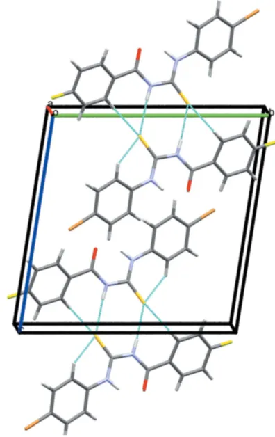
3. Supramolecular features
In the crystal of1, inversion dimers linked by pairwise N1— H1A S1 hydrogen bonds (Table 1) generate R2
2(8) loops
(Fig. 3). The crystal of2features the same motif (Table 2), but an additional weak C—H S bond links the dimers into double columns propagating in the [100] direction (Fig. 4).
research communications
Acta Cryst.(2019). E75, 1026–1029 Akhteret al. C
[image:2.610.46.298.70.191.2]14H10ClFN2OS and C14H10BrFN2OS
1027
Table 1Hydrogen-bond geometry (A˚ ,) for1.
D—H A D—H H A D A D—H A
C10—H10 S1 0.95 2.57 3.1945 (14) 124
N2—H1B Cl 0.87 (2) 2.482 (19) 2.9246 (12) 112.3 (14) N2—H1B O1 0.87 (2) 1.924 (19) 2.6600 (14) 141.6 (17) N1—H1A S1i 0.85 (2) 2.67 (2) 3.4031 (13) 145.2 (16)
Symmetry code: (i)xþ1;yþ1;zþ1.
Table 2
Hydrogen-bond geometry (A˚ ,) for
2.
D—H A D—H H A D A D—H A
N1—H1A S1i 0.88 2.69 3.5081 (15) 154
N2—H1B O1 0.88 1.88 2.610 (2) 139
C10—H10 S1 0.95 2.65 3.2319 (18) 120
C1—H1 S1ii 0.95 2.81 3.7312 (18) 165
[image:2.610.313.564.208.266.2] [image:2.610.47.296.242.350.2]Symmetry codes: (i)xþ1;yþ1;z; (ii)x;yþ1;z.
Figure 3
Partial packing diagram for 1. Light-blue lines indicate directional interactions
Figure 1
The molecular structure of1showing 50% displacement ellipsoids; the blue lines represent the intramolecular interactions.
Figure 2
The molecular structure of2showing 50% displacement ellipsoids; the blue lines represent the intramolecular interactions.
Figure 4
[image:2.610.338.536.404.718.2] [image:2.610.45.292.608.714.2]4. Database survey
A search of Cambridge Structural Database (CSD version 5.39, update of February 2018) for compounds related to1and
2 yielded hits for N -{[4-chloro-3-(trifluoromethyl)phen-yl]carbamothioyl}-3-methylbenzamide (CCDC deposition No. 1840069) and 4-chloro-N -{[4-chloro-3-(trifluoromethyl)phen-yl]carbamothioyl}benzamide (CCDC 1587395) (Zhanget al., 2019): these compounds have the same skeleton as the title compounds but with different substituents attached to the phenyl rings. In both compounds, pairwise N—H S hydrogen bonds are responsible for the formation of inversion dimers with an R2
2(8) motif, as also observed in title
compounds.
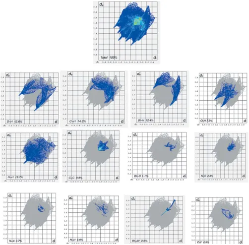
5. Hirshfeld surface analysis
In order to further analyse the close contacts and inter-molecular interactions in the crystals of 1 and 2, Hirshfeld surfaces (mapped over dnorm, curvedness and shape-index)
(Fig. 5) and two-dimensional fingerprint plots (Figs. 6 and 7)
were generated using CrystalExplorer3.1 (Mackenzie et al., 2017). The fingerprint plot for1decomposed into individual contact types indicates that the the most significant contribu-tions are from H H (van der Waals) (26.6%) contacts, followed by S H/H S (13.8%), Cl H/H Cl (9.5%) O H/H O (6.7%), F H/H F (6.6%), Cl F/F Cl (3.7%) and F C/C F (3.1%) interactions. In compound2, H H (19.7%) (van der Waals contacts) are the most signif-icant, followed by C H/H C (14.8%), S H/H S (12.6%), Br H/H Br (12.4%), C C (9.9%) and O N/ N O (7.9%) interactions.
6. Synthesis and Crystallization
Compounds1and2were synthesized by adopting a literature procedure (Binzet et al., 2018) with slight modification: we refluxed the reactants in distilled solvents for 20 min. instead of refluxing them in anhydrous solvents for 4 h. In the first step, 4-fluorobenzoyle chloride (1 mmol) and potassium thiocyanate (1 mmol) were dissolved in acetone (10 ml) at room temperature with constant stirring for 20 minutes to obtain a white precipitate of 4-fluorophenyl isothiocyanate. In the second step, 1 mmol of 2-chloro phenyl aniline (for1) or 4-bromophenyl aniline (for2) were added to the mixture and refluxed at 343 K. Hydrochloric acid (0.5N, 10 ml) was added and the solution was filtered to obtain the desired products:1
in 69% yield and 2 in 80% yield. For recrystallization, compound 1was dissolved in a mixture of dichloromethane
1028
Akhteret al. C [image:3.610.314.566.70.361.2]14H10ClFN2OS and C14H10BrFN2OS Acta Cryst.(2019). E75, 1026–1029
research communications
Figure 5
The Hirshfeld surfaces of1and2.
Figure 6
Two dimensional fingerprint plots for1.
Figure 7
[image:3.610.45.296.471.718.2]and methanol (1:1) while compound 2 was dissolved in di-chloromethane and left for slow evaporation at room temperature to obtain colourless prisms of 1 and colourless plates of2
7. Data collection and Refinement
Crystal data, data collection and structure refinement details are summarized in Table 3. The C-bound H atoms atoms were positioned with idealized geometry (C—H = 0.93–0.97 A˚ ) and refined as riding atoms. In 1, the N-bound H atoms were located in difference-Fourier maps and their positions were freely refined; in 2, the N-bound H atoms were located in difference-Fourier maps and refined as riding atoms in their as-found relative positions. The constraint Uiso(H) =
1.2Ueq(carrier) was applied in all cases.
Funding information
The authors thank the Higher Education Commission of Pakistan (HEC) for financial support through research project No. 20-2830 under the National Research Program for Universities.
References
Binzet, G., Gumus, I., Dogen, A., Flo¨rke, U., Kulcu, N. & Arslan, H. (2018).J. Mol. Struct.1161, 519–529.
Bruker (2000). APEX2, SAINT and SADABS. Bruker AXS Inc., Madison, Wisconsin, USA.
Contreras Aguilar, E., Echeverrı´a, G., Piro, O., Ulic, S., Jios, J., Tuttolomondo, M. & Pe´rez, H. (2018).Mol. Phys.116, 399–413. Fakhar, I., Hussien, N. J., Sapari, S., Bloh, A. H., Yusoff, S. F. M.,
Hasbullah, S. A., Yamin, B. M., Mutalib, S. A., Shihab, M. S. & Yousif, E. (2018).J. Mol. Struct.1159, 96–102.
Ghazal, K., Shoaib, S., Khan, M., Khan, S., Rauf, M. K., Khan, N., Badshah, A., Tahir, M. N. & Ali, I. (2019).J. Mol. Struct.1177, 12-130.
Hashim, S. N. M., Jumal, J. & Kassim, K. (2017).Adv. Sci. Lett.23, 4523–4527.
Khan, M. R., Zaib, S., Rauf, M. K., Ebihara, M., Badshah, A., Zahid, M., Nadeem, M. A. & Iqbal, J. (2018).J. Mol. Struct.1164, 354–362. Lai, L. C., Rahman, C. N. B. C. A., Tahir, M. I. M., Ravoof, T. B. S. A., Jotani, M. M. & Tiekink, E. R. T. (2018).Acta Cryst.E74, 256–260. Mackenzie, C. F., Spackman, P. R., Jayatilaka, D. & Spackman, M. A.
(2017).IUCrJ,4, 575–587.
Mitoraj, M. P., Babashkina, M. G., Isaev, A. Y., Chichigina, Y. M., Robeyns, K., Garcia, Y. & Safin, D. A. (2018).Cryst. Growth Des.
18, 5385–5397.
Pandey, S. K., Pratap, S., Tiwari, M. K., Marverti, G. & Jasinski, J. P. (2019).J. Mol. Struct.1175, 963–970.
Pervez, H., Khan, N., Iqbal, J., Zaib, S., Yaqub, M., Tahir, M. N. & Naseer, M. M. (2018).Heterocycl. Commun.24, 51–58.
Saeed, A., Mustafa, M. N., Zain-ul-Abideen, M., Shabir, G., Erben, M. F. & Flo¨rke, U. (2018).J. Sulfur Chem. pp. 1–39.
Sheldrick, G. M. (2008).Acta Cryst.A64, 112–122. Sheldrick, G. M. (2015).Acta Cryst.C71, 3–8.
Solmaz, U., Gumus, I., Binzet, G., Celik, O., Balci, G. K., Dogen, A. & Arslan, H. (2018).J. Coord. Chem.71, 200–218.
Zhang, Y., Zhang, X., Qiao, L., Ding, Z., Hang, X., Qin, B., Song, J. & Huang, J. (2019).J. Mol. Struct.1176, 335–345.
research communications
Acta Cryst.(2019). E75, 1026–1029 Akhteret al. C
[image:4.610.48.562.93.394.2]14H10ClFN2OS and C14H10BrFN2OS
1029
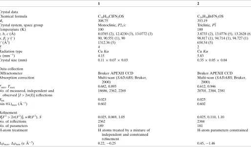
Table 3Experimental details.
1 2
Crystal data
Chemical formula C14H10ClFN2OS C14H10BrFN2OS
Mr 308.75 353.19
Crystal system, space group Monoclinic,P21/c Triclinic,P1
Temperature (K) 100 100
a,b,c(A˚ ) 8.0785 (2), 12.4230 (3), 13.0772 (3) 3.8733 (2), 13.0776 (5), 13.2628 (6)
,,(
) 90, 90.551 (1), 90 98.817 (1), 94.714 (1), 94.727 (1)
V(A˚3) 1312.36 (5) 658.54 (5)
Z 4 2
Radiation type CuK CuK
(mm1) 4.15 5.83
Crystal size (mm) 0.110.070.03 0.350.050.04
Data collection
Diffractometer Bruker APEXII CCD Bruker APEXII CCD
Absorption correction Multi-scan (SADABS; Bruker,
2000)
Multi-scan (SADABS; Bruker, 2000)
Tmin,Tmax 0.682, 0.895 0.612, 0.946
No. of measured, independent and observed [I> 2(I)] reflections
18686, 2362, 2269 20701, 2384, 2381
Rint 0.023 0.025
(sin/)max(A˚
1
) 0.602 0.602
Refinement
R[F2> 2(F2)],wR(F2),S 0.025, 0.069, 1.05 0.025, 0.110, 1.10
No. of reflections 2362 2384
No. of parameters 189 181
H-atom treatment H atoms treated by a mixture of
independent and constrained refinement
H-atom parameters constrained
max, min(e A˚
3
) 0.22,0.25 0.45,1.46
supporting information
sup-1 Acta Cryst. (2019). E75, 1026-1029
supporting information
Acta Cryst. (2019). E75, 1026-1029 [https://doi.org/10.1107/S2056989019008569]
Crystal structure and Hirshfeld surface analysis of
N
-(2-chlorophenylcarbamo-thioyl)-4-fluorobenzamide and
N
-(4-bromophenylcarbamothioyl)-4-fluoro-benzamide
Sidra Akhter, Muhammad Iqbal Choudhary, Hina Siddiqui and Sammer Yousuf
Computing details
For both structures, data collection: APEX2 (Bruker, 2000); cell refinement: SAINT (Bruker, 2000); data reduction:
SAINT (Bruker, 2000); program(s) used to solve structure: SHELXT2014 (Sheldrick, 2015a); program(s) used to refine
structure: SHELXL2016 (Sheldrick, 2015b); molecular graphics: SHELXTL (Sheldrick, 2008); software used to prepare
material for publication: SHELXTL (Sheldrick, 2008).
N-(2-Chlorophenylcarbamothioyl)-4-fluorobenzamide (1)
Crystal data
C14H10ClFN2OS
Mr = 308.75
Monoclinic, P21/c
a = 8.0785 (2) Å b = 12.4230 (3) Å c = 13.0772 (3) Å β = 90.551 (1)° V = 1312.36 (5) Å3
Z = 4
F(000) = 632 Dx = 1.563 Mg m−3
Cu Kα radiation, λ = 1.54178 Å Cell parameters from 9977 reflections θ = 4.9–68.3°
µ = 4.15 mm−1
T = 100 K Prism, colourless 0.11 × 0.07 × 0.03 mm
Data collection
Bruker APEXII CCD diffractometer ω scans
Absorption correction: multi-scan (SADABS; Bruker, 2000) Tmin = 0.682, Tmax = 0.895
18686 measured reflections
2362 independent reflections 2269 reflections with I > 2σ(I) Rint = 0.023
θmax = 68.3°, θmin = 4.9°
h = −9→9 k = −14→14 l = −15→15
Refinement
Refinement on F2
Least-squares matrix: full R[F2 > 2σ(F2)] = 0.025
wR(F2) = 0.069
S = 1.05 2362 reflections 189 parameters 0 restraints
Primary atom site location: dual
Hydrogen site location: mixed
H atoms treated by a mixture of independent and constrained refinement
w = 1/[σ2(F
o2) + (0.0347P)2 + 0.7594P]
where P = (Fo2 + 2Fc2)/3
(Δ/σ)max = 0.001
Δρmax = 0.22 e Å−3
supporting information
sup-2 Acta Cryst. (2019). E75, 1026-1029
Special details
Geometry. All esds (except the esd in the dihedral angle between two l.s. planes) are estimated using the full covariance matrix. The cell esds are taken into account individually in the estimation of esds in distances, angles and torsion angles; correlations between esds in cell parameters are only used when they are defined by crystal symmetry. An approximate (isotropic) treatment of cell esds is used for estimating esds involving l.s. planes.
Fractional atomic coordinates and isotropic or equivalent isotropic displacement parameters (Å2)
x y z Uiso*/Ueq
S1 0.73325 (5) 0.47025 (3) 0.56374 (3) 0.02331 (12) Cl 0.83162 (5) 0.03157 (3) 0.58416 (3) 0.02495 (11) F1 0.18907 (12) 0.30493 (8) 0.01782 (7) 0.0314 (2) O1 0.54889 (12) 0.15637 (8) 0.42244 (7) 0.0192 (2) N1 0.56777 (15) 0.33824 (10) 0.44484 (9) 0.0179 (3) N2 0.70992 (14) 0.25311 (9) 0.57516 (9) 0.0157 (2) C1 0.43467 (17) 0.36712 (11) 0.24179 (10) 0.0177 (3) H1 0.493438 0.425890 0.271153 0.021* C2 0.35330 (18) 0.38022 (12) 0.14870 (11) 0.0206 (3) H2 0.355095 0.447218 0.113699 0.025* C3 0.26979 (18) 0.29291 (12) 0.10866 (10) 0.0211 (3) C4 0.26446 (18) 0.19359 (12) 0.15557 (11) 0.0224 (3) H4 0.206152 0.135158 0.125243 0.027* C5 0.34669 (18) 0.18165 (11) 0.24822 (11) 0.0199 (3) H5 0.345811 0.113881 0.281860 0.024* C6 0.43099 (16) 0.26820 (11) 0.29281 (10) 0.0156 (3) C7 0.51917 (16) 0.24761 (11) 0.39152 (10) 0.0160 (3) C8 0.67071 (16) 0.34671 (11) 0.53083 (10) 0.0162 (3) C9 0.80473 (16) 0.23264 (11) 0.66453 (10) 0.0148 (3) C10 0.83157 (17) 0.30697 (11) 0.74300 (10) 0.0184 (3) H10 0.789298 0.378033 0.736600 0.022* C11 0.91962 (17) 0.27772 (12) 0.83031 (11) 0.0213 (3) H11 0.938625 0.329427 0.882642 0.026* C12 0.98017 (18) 0.17407 (12) 0.84215 (11) 0.0224 (3) H12 1.040289 0.154849 0.902214 0.027* C13 0.95246 (18) 0.09856 (12) 0.76576 (11) 0.0217 (3) H13 0.992398 0.027057 0.773432 0.026* C14 0.86603 (17) 0.12824 (11) 0.67813 (10) 0.0170 (3) H1B 0.672 (2) 0.1970 (16) 0.5432 (14) 0.028 (5)* H1A 0.528 (2) 0.3988 (16) 0.4260 (14) 0.031 (5)*
Atomic displacement parameters (Å2)
U11 U22 U33 U12 U13 U23
supporting information
sup-3 Acta Cryst. (2019). E75, 1026-1029
N2 0.0206 (6) 0.0125 (6) 0.0141 (6) 0.0000 (5) −0.0046 (4) −0.0015 (4) C1 0.0208 (7) 0.0165 (6) 0.0158 (7) 0.0002 (5) −0.0008 (5) −0.0019 (5) C2 0.0257 (7) 0.0196 (7) 0.0166 (7) 0.0034 (6) −0.0016 (6) 0.0024 (5) C3 0.0209 (7) 0.0304 (8) 0.0119 (7) 0.0041 (6) −0.0042 (5) −0.0015 (6) C4 0.0241 (7) 0.0243 (8) 0.0188 (7) −0.0040 (6) −0.0045 (6) −0.0048 (6) C5 0.0240 (7) 0.0170 (7) 0.0187 (7) −0.0012 (6) −0.0017 (5) −0.0007 (5) C6 0.0158 (6) 0.0171 (7) 0.0137 (6) 0.0017 (5) −0.0004 (5) −0.0012 (5) C7 0.0171 (6) 0.0152 (7) 0.0158 (7) 0.0000 (5) −0.0002 (5) −0.0011 (5) C8 0.0184 (6) 0.0150 (6) 0.0152 (6) 0.0020 (5) −0.0023 (5) −0.0010 (5) C9 0.0139 (6) 0.0169 (6) 0.0135 (6) −0.0009 (5) −0.0008 (5) 0.0022 (5) C10 0.0208 (7) 0.0169 (7) 0.0175 (7) −0.0002 (5) −0.0020 (5) −0.0003 (5) C11 0.0221 (7) 0.0250 (7) 0.0167 (7) −0.0030 (6) −0.0034 (5) −0.0019 (6) C12 0.0211 (7) 0.0292 (8) 0.0169 (7) 0.0001 (6) −0.0055 (5) 0.0048 (6) C13 0.0226 (7) 0.0211 (7) 0.0215 (7) 0.0039 (6) −0.0024 (6) 0.0053 (6) C14 0.0189 (6) 0.0160 (7) 0.0161 (6) 0.0000 (5) −0.0001 (5) −0.0007 (5)
Geometric parameters (Å, º)
S1—C8 1.6709 (14) C4—C5 1.384 (2) Cl—C14 1.7387 (14) C4—H4 0.9500 F1—C3 1.3579 (16) C5—C6 1.3972 (19) O1—C7 1.2264 (17) C5—H5 0.9500 N1—C7 1.3794 (18) C6—C7 1.4904 (18) N1—C8 1.3961 (17) C9—C10 1.3959 (19) N1—H1A 0.85 (2) C9—C14 1.3990 (19) N2—C8 1.3360 (18) C10—C11 1.3879 (19) N2—C9 1.4141 (17) C10—H10 0.9500 N2—H1B 0.87 (2) C11—C12 1.385 (2) C1—C2 1.3875 (19) C11—H11 0.9500 C1—C6 1.3987 (19) C12—C13 1.387 (2) C1—H1 0.9500 C12—H12 0.9500 C2—C3 1.378 (2) C13—C14 1.3860 (19) C2—H2 0.9500 C13—H13 0.9500 C3—C4 1.379 (2)
supporting information
sup-4 Acta Cryst. (2019). E75, 1026-1029
F1—C3—C4 118.09 (13) C12—C11—H11 119.6 C2—C3—C4 123.54 (13) C10—C11—H11 119.6 C3—C4—C5 117.94 (13) C11—C12—C13 119.58 (13) C3—C4—H4 121.0 C11—C12—H12 120.2 C5—C4—H4 121.0 C13—C12—H12 120.2 C4—C5—C6 120.72 (13) C14—C13—C12 119.47 (13) C4—C5—H5 119.6 C14—C13—H13 120.3 C6—C5—H5 119.6 C12—C13—H13 120.3 C5—C6—C1 119.30 (12) C13—C14—C9 121.78 (13) C5—C6—C7 117.15 (12) C13—C14—Cl 118.45 (11) C1—C6—C7 123.49 (12) C9—C14—Cl 119.76 (10)
C6—C1—C2—C3 −0.1 (2) C9—N2—C8—S1 4.5 (2) C1—C2—C3—F1 179.70 (12) C7—N1—C8—N2 −9.6 (2) C1—C2—C3—C4 −0.7 (2) C7—N1—C8—S1 168.84 (11) F1—C3—C4—C5 −179.92 (12) C8—N2—C9—C10 22.4 (2) C2—C3—C4—C5 0.5 (2) C8—N2—C9—C14 −162.20 (13) C3—C4—C5—C6 0.6 (2) C14—C9—C10—C11 1.4 (2) C4—C5—C6—C1 −1.3 (2) N2—C9—C10—C11 176.73 (13) C4—C5—C6—C7 −178.72 (13) C9—C10—C11—C12 −1.1 (2) C2—C1—C6—C5 1.1 (2) C10—C11—C12—C13 0.0 (2) C2—C1—C6—C7 178.32 (13) C11—C12—C13—C14 0.7 (2) C8—N1—C7—O1 8.6 (2) C12—C13—C14—C9 −0.3 (2) C8—N1—C7—C6 −170.22 (13) C12—C13—C14—Cl −179.45 (11) C5—C6—C7—O1 15.54 (19) C10—C9—C14—C13 −0.7 (2) C1—C6—C7—O1 −161.73 (13) N2—C9—C14—C13 −176.39 (12) C5—C6—C7—N1 −165.60 (12) C10—C9—C14—Cl 178.42 (10) C1—C6—C7—N1 17.14 (19) N2—C9—C14—Cl 2.72 (17) C9—N2—C8—N1 −177.23 (12)
Hydrogen-bond geometry (Å, º)
D—H···A D—H H···A D···A D—H···A
C10—H10···S1 0.95 2.57 3.1945 (14) 124 N2—H1B···Cl 0.87 (2) 2.482 (19) 2.9246 (12) 112.3 (14) N2—H1B···O1 0.87 (2) 1.924 (19) 2.6600 (14) 141.6 (17) N1—H1A···S1i 0.85 (2) 2.67 (2) 3.4031 (13) 145.2 (16)
Symmetry code: (i) −x+1, −y+1, −z+1.
N-(4-Bromophenylcarbamothioyl)-4-fluorobenzamide (2)
Crystal data
C14H10BrFN2OS
Mr = 353.19
Triclinic, P1 a = 3.8733 (2) Å b = 13.0776 (5) Å c = 13.2628 (6) Å α = 98.817 (1)°
β = 94.714 (1)° γ = 94.727 (1)° V = 658.54 (5) Å3
Z = 2 F(000) = 348 Dx = 1.771 Mg m−3
supporting information
sup-5 Acta Cryst. (2019). E75, 1026-1029
Cell parameters from 9945 reflections θ = 3.4–68.2°
µ = 5.83 mm−1
T = 100 K Plate, colourless 0.35 × 0.05 × 0.04 mm
Data collection
Bruker APEXII CCD diffractometer φ and ω scans
Absorption correction: multi-scan (SADABS; Bruker, 2000) Tmin = 0.612, Tmax = 0.946
20701 measured reflections
2384 independent reflections 2381 reflections with I > 2σ(I) Rint = 0.025
θmax = 68.2°, θmin = 3.4°
h = −4→4 k = −15→15 l = −15→15
Refinement
Refinement on F2
Least-squares matrix: full R[F2 > 2σ(F2)] = 0.025
wR(F2) = 0.110
S = 1.10 2384 reflections 181 parameters 0 restraints
Primary atom site location: dual
Hydrogen site location: inferred from neighbouring sites
H-atom parameters constrained w = 1/[σ2(F
o2) + (0.1P)2]
where P = (Fo2 + 2Fc2)/3
(Δ/σ)max = 0.001
Δρmax = 0.45 e Å−3
Δρmin = −1.46 e Å−3
Special details
Geometry. All esds (except the esd in the dihedral angle between two l.s. planes) are estimated using the full covariance matrix. The cell esds are taken into account individually in the estimation of esds in distances, angles and torsion angles; correlations between esds in cell parameters are only used when they are defined by crystal symmetry. An approximate (isotropic) treatment of cell esds is used for estimating esds involving l.s. planes.
Fractional atomic coordinates and isotropic or equivalent isotropic displacement parameters (Å2)
x y z Uiso*/Ueq
supporting information
sup-6 Acta Cryst. (2019). E75, 1026-1029
C7 0.4782 (5) 0.66558 (14) 0.23084 (14) 0.0105 (4) C9 0.0233 (4) 0.38653 (13) 0.30136 (13) 0.0086 (4) C8 0.2378 (4) 0.48253 (14) 0.16562 (13) 0.0085 (4) C10 −0.1837 (5) 0.30056 (14) 0.24536 (13) 0.0115 (4) H10 −0.240925 0.296827 0.173769 0.014* C11 −0.3051 (4) 0.22064 (14) 0.29513 (13) 0.0105 (4) H11 −0.444455 0.161739 0.257544 0.013* C12 −0.2224 (5) 0.22707 (13) 0.39998 (14) 0.0102 (4) C13 −0.0217 (5) 0.31221 (13) 0.45694 (13) 0.0109 (4) H13 0.031180 0.315998 0.528723 0.013* C14 0.0999 (5) 0.39136 (15) 0.40755 (14) 0.0114 (4) H14 0.237633 0.450144 0.445916 0.014*
Atomic displacement parameters (Å2)
U11 U22 U33 U12 U13 U23
Br1 0.0169 (2) 0.0096 (2) 0.0125 (2) −0.00227 (13) 0.00215 (13) 0.00588 (13) O1 0.0312 (8) 0.0122 (7) 0.0076 (7) −0.0065 (6) 0.0033 (6) 0.0011 (5) N1 0.0154 (8) 0.0108 (8) 0.0057 (7) 0.0005 (6) 0.0007 (6) 0.0022 (6) N2 0.0170 (8) 0.0073 (7) 0.0090 (7) −0.0001 (6) 0.0021 (6) 0.0019 (6) F1 0.0328 (7) 0.0078 (6) 0.0213 (7) −0.0054 (5) 0.0078 (5) 0.0051 (5) S1 0.0140 (3) 0.0082 (3) 0.0069 (3) −0.00094 (19) 0.00084 (19) 0.00050 (19) C1 0.0092 (8) 0.0080 (9) 0.0102 (8) −0.0004 (6) 0.0023 (6) 0.0005 (7) C2 0.0161 (9) 0.0103 (9) 0.0133 (9) 0.0022 (7) 0.0031 (7) 0.0031 (7) C3 0.0197 (9) 0.0068 (9) 0.0194 (10) 0.0022 (7) 0.0119 (8) 0.0033 (7) C4 0.0149 (9) 0.0097 (9) 0.0148 (9) −0.0026 (7) 0.0020 (7) −0.0021 (7) C5 0.0144 (8) 0.0095 (9) 0.0093 (8) −0.0016 (7) −0.0006 (7) −0.0012 (7) C6 0.0106 (8) 0.0103 (9) 0.0089 (9) −0.0002 (7) 0.0008 (6) 0.0008 (7) C7 0.0118 (8) 0.0104 (9) 0.0098 (9) −0.0013 (7) 0.0013 (6) 0.0037 (7) C9 0.0120 (8) 0.0070 (9) 0.0086 (8) 0.0005 (7) 0.0048 (6) 0.0047 (6) C8 0.0103 (8) 0.0086 (8) 0.0071 (8) 0.0020 (7) −0.0002 (6) 0.0026 (6) C10 0.0137 (8) 0.0120 (9) 0.0088 (8) 0.0001 (7) −0.0005 (7) 0.0028 (6) C11 0.0116 (8) 0.0092 (9) 0.0097 (8) −0.0028 (6) 0.0007 (6) 0.0004 (6) C12 0.0128 (8) 0.0080 (9) 0.0112 (8) 0.0005 (6) 0.0023 (7) 0.0056 (6) C13 0.0138 (8) 0.0132 (9) 0.0067 (8) −0.0003 (7) 0.0018 (7) 0.0048 (6) C14 0.0130 (8) 0.0092 (9) 0.0112 (9) −0.0007 (6) 0.0011 (7) 0.0002 (6)
Geometric parameters (Å, º)
supporting information
sup-7 Acta Cryst. (2019). E75, 1026-1029
S1—C8 1.6687 (18) C11—C12 1.389 (2) C1—C2 1.377 (3) C11—H11 0.9500 C1—C6 1.399 (3) C12—C13 1.385 (3) C1—H1 0.9500 C13—C14 1.379 (3) C2—C3 1.375 (3) C13—H13 0.9500 C2—H2 0.9500 C14—H14 0.9500 C3—C4 1.391 (3)
C7—N1—C8 128.20 (16) O1—C7—N1 122.08 (17) C7—N1—H1A 115.9 O1—C7—C6 121.00 (17) C8—N1—H1A 115.9 N1—C7—C6 116.92 (16) C8—N2—C9 131.19 (16) C10—C9—C14 119.30 (16) C8—N2—H1B 114.4 C10—C9—N2 124.87 (15) C9—N2—H1B 114.4 C14—C9—N2 115.78 (16) C2—C1—C6 120.62 (17) N2—C8—N1 114.66 (16) C2—C1—H1 119.7 N2—C8—S1 126.92 (15) C6—C1—H1 119.7 N1—C8—S1 118.43 (13) C3—C2—C1 118.79 (17) C11—C10—C9 119.57 (16) C3—C2—H2 120.6 C11—C10—H10 120.2 C1—C2—H2 120.6 C9—C10—H10 120.2 F1—C3—C2 119.46 (18) C12—C11—C10 119.85 (16) F1—C3—C4 117.55 (18) C12—C11—H11 120.1 C2—C3—C4 122.98 (18) C10—C11—H11 120.1 C5—C4—C3 117.70 (18) C13—C12—C11 121.42 (16) C5—C4—H4 121.1 C13—C12—Br1 119.31 (13) C3—C4—H4 121.1 C11—C12—Br1 119.27 (13) C4—C5—C6 120.91 (18) C14—C13—C12 118.76 (16) C4—C5—H5 119.5 C14—C13—H13 120.6 C6—C5—H5 119.5 C12—C13—H13 120.6 C1—C6—C5 118.97 (17) C13—C14—C9 121.10 (17) C1—C6—C7 123.33 (17) C13—C14—H14 119.5 C5—C6—C7 117.62 (16) C9—C14—H14 119.5
supporting information
sup-8 Acta Cryst. (2019). E75, 1026-1029
C5—C6—C7—N1 −158.75 (16) N2—C9—C14—C13 178.36 (14)
Hydrogen-bond geometry (Å, º)
D—H···A D—H H···A D···A D—H···A
N1—H1A···S1i 0.88 2.69 3.5081 (15) 154
N2—H1B···O1 0.88 1.88 2.610 (2) 139 C10—H10···S1 0.95 2.65 3.2319 (18) 120 C1—H1···S1ii 0.95 2.81 3.7312 (18) 165