research communications
Acta Cryst.(2017). E73, 1087–1091 https://doi.org/10.1107/S2056989017009422
1087
Received 22 June 2017Accepted 23 June 2017
Edited by P. McArdle, National University of Ireland, Ireland
Keywords:crystal structure; benzofuran; intra-molecular interaction; interintra-molecular inter-action; functional group.
CCDC references:1449589; 1449587
Supporting information:this article has supporting information at journals.iucr.org/e
Two closely related 2-(benzofuran-2-yl)-2-oxoethyl
benzoates: structural differences and C—H
O
hydrogen-bonded supramolecular assemblies
Li Yee Then,aC. S. Chidan Kumar,b* Huey Chong Kwong,cYip-Foo Win,dSiau Hui Mah,eChing Kheng Quah,aS. Naveenfand Ismail Waradg*
aX-ray Crystallography Unit, School of Physics, Universiti Sains Malaysia, 11800 USM, Penang, Malaysia,bDepartment of Engineering Chemistry, Vidya Vikas Institute of Engineering & Technology, Visvesvaraya Technological University, Alanahally, Mysuru 570 028, Karnataka, India,cSchool of Chemical Sciences, Universiti Sains Malaysia, Penang 11800 USM, Malaysia,dDepartment of Chemical Science, Faculty of Science, Universiti Tunku Abdul Rahman, Perak Campus, Jalan Universiti, Bandar Barat, Perak, Malaysia,eSchool of Biosciences, Taylor’s University, Lakeside Campus, 47500 Subang Jaya, Selangor, Malaysia,fInstitution of Excellence, University of Mysore, Manasagangotri, Mysuru 570 006, India, andgDepartment of Chemistry, Science College, An-Najah National University, PO Box 7, Nablus, West Bank, Palestinian Territories. *Correspondence e-mail: [email protected], [email protected]
The compounds 2-(1-benzofuran-2-yl)-2-oxoethyl 2-nitrobenzoate, C17H11NO6
(I), and 2-(1-benzofuran-2-yl)-2-oxoethyl 2-aminobenzoate, C17H13NO4 (II),
were synthesized under mild conditions. Their molecular structures were characterized by both spectroscopic and single-crystal X-ray diffraction analysis. The molecular conformations of both title compounds are generally similar. However, different ortho-substituted moieties at the phenyl ring of the two compounds cause deviations in the torsion angles between the carbonyl group and the attached phenyl ring. In compound (I), the ortho-nitrophenyl ring is twisted away from the adjacent carbonyl group whereas in compound (II), the
ortho-aminophenyl ring is almost co-planar with the carbonyl group. In the crystal of compound (I), two C—H O hydrogen bonds link the molecules into chains propagating along thec-axis direction and the chains are interdigitated, forming sheets parallel to [201]. Conversely, pairs of N—H O hydrogen bonds in compound (II) link inversion-related molecules into dimers, which are further extended by C—H O hydrogen bonds into dimer chains. These chains are interconnected by – interactions involving the furan rings, forming sheets parallel to theacplane.
1. Chemical context
Oxygen-containing heterocycles are the basic cores of many bioactive structures. Among these, benzofuran and its deri-vatives occur frequently in nature because of their stability and ease of generation. Those with substitution(s) at their C-2 and/or C-3 positions are important. Important biological activity such as anticancer (Swamy et al., 2015), anti-acetyl-cholinesterase (Zhouet al., 2010), antimicrobial (Ugaleet al., 2012) and antioxidant (Naiket al., 2013) actions exhibited by this scaffold have attracted the attention of synthetic chemists. Some of the biological and medicinal significance of benzo-furan derivatives (Nevagiet al., 2015) have been discussed in review reports. The known potential of benzofuran derivatives has motivated us to synthesise some new compounds incor-porating this core structure and we herein report the synthesis and crystal structures of (1-benzofuran-yl)-oxoethyl nitrobenzoate (I) and (1-benzofuran-yl)-oxoethyl 2-aminobenzoate (II).
2. Structural commentary
The molecular structures of the title compounds (Fig. 1) contain a benzofuran ring and anortho-substituted [nitro- for compound (I) and amino- for compound (II)] phenyl ring,
joined by a C—C( O)—O—C( O) carbonyl-connecting
bridge. Their molecular conformations can be characterized by three degrees of freedom, as indicated by the O1—C8—
C9—O3 (1), C9—C10—O2—C11 (2) and O4—C11—C12—
C13 (3) torsion angles, respectively (Fig. 2). The torsion angle
1 for compounds (I) and (II) is close to 0, showing that the
benzofuran ring is nearly coplanar with the C—C( O)—O—
C( O) carbonyl bridge. Torsion angle2 adopts asyn-clinal conformation, as both carbonyl groups at the connecting bridges are twisted away from each other forming torsion angles of 71.43 (3) in (I) and 70.85 (18) in (II). For compound (I), the substituted ortho-nitrophenyl moiety is perpendicular to the adjacent carbonyl group with a3 torsion angle of90.2 (4); this may arise from a steric repulsion force
between the nitro group and carbonyl group. In contrast, the
ortho-aminophenyl ring in compound (II) is almost coplanar with its adjacent carbonyl group due to the intramolecular hydrogen bond (N1—H1A O4, Table 2) between the amino and carbonyl groups, which generates anS(6) ring.
3. Supramolecular features
The crystal packing of compound (I) depends mainly on two weak intermolecular hydrogen bonds. Molecules are joined into infinite chains propagating along the c-axis by C10— H10A O3 hydrogen bonds (Table 1, Fig. 3), meanwhile those chains are interdigitated into a fishbone sheet extending along the [201] direction through C15—H15A O5 hydrogen bonds. The fishbone sheets alternate in an up–down manner along theabplane as shown in Fig. 4.
In compound (II), the molecular interactions are more abundant than in (I) because of theortho-substituted amino
1088
Thenet al. C17H11NO6and C17H13NO4 Acta Cryst.(2017). E73, 1087–1091
[image:2.610.66.275.80.315.2]research communications
Figure 2
[image:2.610.312.566.186.238.2]General chemical diagram showing torsion angles 1, 2 and 3 in compounds (I) and (II).
Figure 1
ORTEPdiagram of the title compounds, with ellipsoids drawn at the 50% probability level, showing the atomic labelling scheme.
Table 1
Hydrogen-bond geometry (A˚ ,) for (I).
D—H A D—H H A D A D—H A
C10—H10A O3i 0.99 2.59 3.471 (4) 148
C15—H15A O5ii 0.95 2.58 3.380 (3) 142
Symmetry codes: (i)x;y;z1; (ii)x1 2;yþ
1 2;zþ1. Table 2
Hydrogen-bond geometry (A˚ ,) for (II).
D—H A D—H H A D A D—H A
N1—H1A O4 0.91 (2) 2.05 (2) 2.700 (3) 127.7 (18)
N1—H1A O4i 0.91 (2) 2.49 (2) 3.246 (2) 141.4 (18)
C10—H10A O3ii 0.97 2.50 3.444 (2) 165
[image:2.610.47.298.497.709.2] [image:2.610.315.564.607.716.2]group at its phenyl ring. Pairs of N1—H1A O4 hydrogen bonds link molecules into inversion dimers with an R2
2(12)
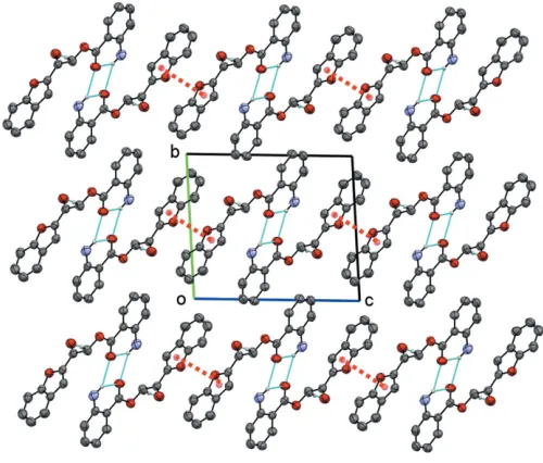
graph-set motif (Fig. 5). These dimers are further expanded by C10—H10A O3 hydrogen bonds into infinite chains along the [100] direction (Fig. 6). In addition, neighbouring chains are interconnected by – interactions involving adjacent furan rings [centroid–centroid distance = 3.7982 (15) A˚ ; symmetry code:x,y+ 1,z), forming a sheet parallel to theacplane (Fig. 7).
4. Database survey
A survey of the Cambridge Structural Database (Groomet al., 2016) revealed five benzofuran structures (Kumaret al., 2015) similar to the title compounds: ITAXUY, ITAYAF, ITAYEJ, ITAYIN and ITAYOT. The molecular structures of the studied and previous compounds differ only at their substituted phenyl rings. By comparing their torsion angles at the
C( O)—O—C( O) carbonyl bridges, the title compounds
exhibit asyn-clinal conformation similar to ITAXUY, ITAYEJ and ITAYIN with respect to their torsion angles which range from 75 to 80.
research communications
Acta Cryst.(2017). E73, 1087–1091 Thenet al. C
[image:3.610.45.296.71.299.2] [image:3.610.308.567.75.196.2]17H11NO6and C17H13NO4
1089
Figure 5 [image:3.610.317.565.308.442.2]Intramolecular and intermolecular N1—H1A O4 hydrogen bonds.
Figure 7
The packing of compound (II), showing the hydrogen bonds (cyan dotted lines) and–interactions (red dotted lines).
Figure 3
[image:3.610.314.564.498.711.2]Molecules in compound (I) joined by intermolecular hydrogen bonds, forming a fishbone chain.
Figure 4
Fishbone chains in an up–down manner are shown in different colours.
Figure 6
[image:3.610.44.314.592.728.2]5. Synthesis and crystallization
The synthesis was carried out by reacting
1-(benzofuran-2-yl)-2-bromoethan-1-one (1 mmol) with 2-nitrobenzoic acid
(1 mmol) for compound (I) and 2-aminobenzoic acid
(1 mmol) for compound (II) in 8 ml of N,N -dimethyl-formamide in the presence of a catalytic amount of anhydrous potassium carbonate at room temperature. The reaction solution was stirred for about two h and monitored by
thin-layer chromatography (TLC). After the reaction was
complete, the resultant mixture was then added to a beaker of ice-cooled water to form a precipitate. The precipitate was then filtered, rinsed with distilled water and dried. Crystals suitable for X-ray analysis were obtained by slow evaporation using a suitable solvent.
2-(Benzofuran-2-yl)-2-oxoethyl 2-nitrobenzoate (I):
Solvents used to grow crystal: acetone + methanol 1:1v/v); yield: 80%, m.p. 381–383 K; 1H NMR (500MHz, CDCl3) in
ppm: 8.041–8.025 (d, 1H,J =7.9Hz,14CH), 7.995–7.980 (d, 1H,J =7.9Hz,17CH), 7.796–7.763 (m, 2H,2CH,3CH), 7.726– 7.695 (t, 1H,J =7.9Hz,15CH), 7.673 (s, 1H,7CH), 7.644–7.627 (d, 1H,J =8.4Hz,5CH), 7.578–7.544 (t, 1H,J =8.4Hz,4CH), 7.398–7.366 (t, 1H,J =7.9Hz,16CH), 5.609 (s, 2H,10CH2).
13
C NMR (125 MHz, CDCl3) in ppm: 182.94 (C9), 165.67 (C11),
155.80 (C1), 150.25 (C13), 133.31 (C16), 132.04 (C15), 130.39 (C17), 130.10 (C8), 128.97 (C3), 127.23 (C12), 126.70 (C6), 124.34 (C5), 124.11 (C4), 123.60 (C14), 113.75 (C7), 112.57 (C2), 67.10 (C10). FT–IR (ATR (solid) cm1): 3089 (Ar C—H,
), 2953 (C—H,), 1744, 1686 (C O,), 1612 (C C,), 1554, 1422 (Ar C C,), 1529, 1344 (N O,), 1278, 1123 (C—O,
).
2-(Benzofuran-2-yl)-2-oxoethyl 2-aminobenzoate (II): Solvents used to grow crystal: acetone + acetonitrile (1:1v/
v); yield: 83%; m.p. 432–434 K;1H NMR (500 MHz, DMSO) in ppm: 8.083 (s, 1H,7CH), 7.907–7.891 (d, 1H,J= 8.1Hz, 17
CH), 7.848–7.832 (d, 1H,J =8.1Hz,14CH), 7.787–7.770 (d, 1H, J = 8.5Hz, 2CH), 7.617–7.583 (t, 1H, J = 8.5Hz, 3CH), 7.437–7.405 (t, 1H,J = 8.1Hz,15CH), 7.329–7.295 (t, 1H,J =
8.5Hz,4CH), 6.824–6.807 (d, 1H,J =8.5Hz,5CH), 6.669 (br–s, 2H,1NH2), 6.607–6.574 (t, 1H,J =8.1Hz,
16
CH), 5.591 (s, 2H,
10
CH2). 13
C NMR (125MHz, DMSO) in ppm: 184.08 (C9), 166.60 (C11), 154.96 (C1), 151.62 (C15), 149.63 (C13), 134.49 (C8), 130.78 (C17), 128.88 (C3), 126.49 (C6), 124.28 (C5), 123.84 (C4), 116.63 (C14), 114.84 (C16), 114.66 (C7), 112.31 (C2), 107.87 (C2), 65.63 (C10). FT–IR (ATR (solid) cm1):
3473, 3360 (N—H, ), 3078 (Ar C—H, ), 2942 (C—H,),
1697, 1676 (C O,), 1615 (C C,), 1583, 1487 (Ar C C,), 1244, 1112 (C—O,).
6. Refinement
Crystal data, data collection and structure refinement details for both compounds are summarized in Table 3. All C-bound H atoms were positioned geometrically (C—H = 0.93–0.97 A˚ ) and refined using a riding model withUiso(H) = 1.2Ueq(parent
1090
Thenet al. C17H11NO6and C17H13NO4 Acta Cryst.(2017). E73, 1087–1091
[image:4.610.47.563.92.384.2]research communications
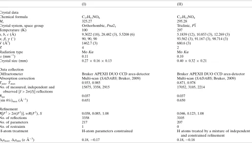
Table 3
Experimental details.
(I) (II)
Crystal data
Chemical formula C17H11NO6 C17H13NO4
Mr 325.27 295.28
Crystal system, space group Orthorhombic,Pna21 Triclinic,P1
Temperature (K) 100 297
a,b,c(A˚ ) 9.3022 (10), 28.482 (3), 5.5208 (6) 5.1839 (12), 10.853 (3), 12.269 (3)
,,(
) 90, 90, 90 93.562 (3), 91.167 (3), 98.714 (3)
V(A˚3) 1462.7 (3) 680.6 (3)
Z 4 2
Radiation type MoK MoK
(mm1) 0.11 0.10
Crystal size (mm) 0.270.160.13 0.400.320.21
Data collection
Diffractometer Bruker APEXII DUO CCD area-detector Bruker APEXII DUO CCD area-detector
Absorption correction Multi-scan (SADABS; Bruker, 2009) Multi-scan (SADABS; Bruker, 2009)
Tmin,Tmax 0.933, 0.985 0.871, 0.978
No. of measured, independent and observed [I> 2(I)] reflections
15875, 3358, 2915 17052, 3105, 2214
Rint 0.037 0.037
(sin/)max(A˚1) 0.651 0.650
Refinement
R[F2> 2(F2)],wR(F2),S 0.038, 0.085, 1.08 0.046, 0.123, 1.08
No. of reflections 3358 3105
No. of parameters 217 207
No. of restraints 1 0
H-atom treatment H-atom parameters constrained H atoms treated by a mixture of independent
and constrained refinement
max,min(e A˚3) 0.18,0.17 0.18,0.18
atom). The N-bound H atoms of compound (II) were located in a difference-Fourier map and refined freely.
Acknowledgements
LYT thanks Universiti Sains Malaysia for the USM Fellowship Scheme and the Malaysian Government for a MyBrain15
(MyMaster) scholarship. HCK thanks the Malaysian
Government for a MyBrain15 (MyPhD) scholarship. The authors extend their appreciation to Vidya Vikas Research & Development Center for the facilities and encouragement.
References
Bruker (2009). APEX2, SAINT and SADABS. Bruker AXS Inc., Madison, Wisconsin, USA.
Groom, C. R., Bruno, I. J., Lightfoot, M. P. & Ward, S. C. (2016).Acta
Cryst.B72, 171–179.
Kumar, C., Then, L., Chia, T., Chandraju, S., Win, Y.-F., Sulaiman, S., Hashim, N., Ooi, K., Quah, C. & Fun, H.-K. (2015).Molecules,20, 16566–16581.
Macrae, C. F., Edgington, P. R., McCabe, P., Pidcock, E., Shields, G. P., Taylor, R., Towler, M. & van de Streek, J. (2006).J. Appl. Cryst.39, 453–457.
Naik, N., Vijay, K., Dias, S. M. & Ranga, S. (2013).Int. J. Pharm.
Pharm. Sci.5, 242–247.
Nevagi, R. J., Dighe, S. N. & Dighe, S. N. (2015).Eur. J. Med. Chem.
97, 561–581.
Sheldrick, G. M. (2015a).Acta Cryst.A71, 3–8. Sheldrick, G. M. (2015b).Acta Cryst.C71, 3–8. Spek, A. L. (2009).Acta Cryst.D65, 148–155.
Swamy, P. M. G., Prasad, Y. R., Ashvini, H. M., Giles, D., Shashidhar, B. V. & Agasimundin, Y. S. (2015). Med. Chem. Res.24, 3437– 3452.
Ugale, V., Patel, H., Patel, B. & Bari, S. (2012).Arab. J. Chem.10, S389–A396.
Zhou, X., Li, M., Wang, X.-B., Wang, T. & Kong, L.-Y. (2010).
Molecules,15, 8593–8601.
research communications
Acta Cryst.(2017). E73, 1087–1091 Thenet al. C
supporting information
sup-1 Acta Cryst. (2017). E73, 1087-1091
supporting information
Acta Cryst. (2017). E73, 1087-1091 [https://doi.org/10.1107/S2056989017009422]
Two closely related 2-(benzofuran-2-yl)-2-oxoethyl benzoates: structural
differences and C
—
H
···
O hydrogen-bonded supramolecular assemblies
Li Yee Then, C. S. Chidan Kumar, Huey Chong Kwong, Yip-Foo Win, Siau Hui Mah, Ching Kheng
Quah, S. Naveen and Ismail Warad
Computing details
For both compounds, data collection: APEX2 (Bruker, 2009); cell refinement: SAINT (Bruker, 2009); data reduction:
SAINT (Bruker, 2009); program(s) used to solve structure: SHELXT2013 (Sheldrick, 2015a); program(s) used to refine
structure: SHELXL2013 (Sheldrick, 2015b). Molecular graphics: SHELXL2013 (Sheldrick, 2015b) and Mercury (Macrae
et al., 2006) for (I); SHELXL2013 (Sheldrick, 2015b) for (II). For both compounds, software used to prepare material for
publication: SHELXL2013 (Sheldrick, 2015b) and PLATON (Spek, 2009).
(I) 2-(1H-1-Benzofuran-2-yl)-2-oxoethyl 2-nitrobenzoate
Crystal data
C17H11NO6
Mr = 325.27
Orthorhombic, Pna21
a = 9.3022 (10) Å
b = 28.482 (3) Å
c = 5.5208 (6) Å
V = 1462.7 (3) Å3
Z = 4
F(000) = 672
Dx = 1.477 Mg m−3
Mo Kα radiation, λ = 0.71073 Å
Cell parameters from 3687 reflections
θ = 2.3–25.3°
µ = 0.11 mm−1
T = 100 K
Block, colourless 0.27 × 0.16 × 0.13 mm
Data collection
Bruker APEXII DUO CCD area-detector diffractometer
Radiation source: fine-focus sealed tube Graphite monochromator
φ and ω scans
Absorption correction: multi-scan (SADABS; Bruker, 2009)
Tmin = 0.933, Tmax = 0.985
15875 measured reflections 3358 independent reflections 2915 reflections with I > 2σ(I)
Rint = 0.037
θmax = 27.6°, θmin = 1.4°
h = −12→12
k = −36→37
l = −7→7
Refinement
Refinement on F2
Least-squares matrix: full
R[F2 > 2σ(F2)] = 0.038
wR(F2) = 0.085
S = 1.08
3358 reflections
217 parameters 1 restraint
Hydrogen site location: inferred from neighbouring sites
supporting information
sup-2 Acta Cryst. (2017). E73, 1087-1091
w = 1/[σ2(F
o2) + (0.0345P)2 + 0.279P], where P = (Fo2 + 2Fc2)/3
(Δ/σ)max < 0.001
Δρmax = 0.18 e Å−3
Δρmin = −0.17 e Å−3
Special details
Geometry. All esds (except the esd in the dihedral angle between two l.s. planes) are estimated using the full covariance matrix. The cell esds are taken into account individually in the estimation of esds in distances, angles and torsion angles; correlations between esds in cell parameters are only used when they are defined by crystal symmetry. An approximate (isotropic) treatment of cell esds is used for estimating esds involving l.s. planes.
Fractional atomic coordinates and isotropic or equivalent isotropic displacement parameters (Å2)
x y z Uiso*/Ueq
N1 0.5075 (2) 0.27391 (8) 0.1493 (4) 0.0290 (5)
O1 0.72961 (19) 0.53112 (6) 0.6220 (3) 0.0309 (4)
O2 0.51143 (18) 0.39614 (6) 0.2506 (3) 0.0289 (4)
O3 0.5376 (2) 0.45933 (6) 0.6044 (4) 0.0328 (4)
O4 0.6811 (2) 0.36342 (6) 0.4887 (4) 0.0350 (5)
O5 0.5946 (2) 0.30243 (6) 0.0721 (4) 0.0343 (4)
O6 0.4877 (2) 0.23524 (7) 0.0592 (4) 0.0460 (6)
C1 0.8415 (3) 0.56187 (8) 0.5803 (5) 0.0270 (6)
C2 0.8830 (3) 0.59794 (10) 0.7294 (6) 0.0362 (6)
H2A 0.8343 0.6045 0.8768 0.043*
C3 0.9982 (3) 0.62384 (10) 0.6543 (6) 0.0412 (7)
H3A 1.0313 0.6488 0.7539 0.049*
C4 1.0686 (3) 0.61491 (10) 0.4372 (6) 0.0413 (8)
H4A 1.1475 0.6341 0.3910 0.050*
C5 1.0259 (3) 0.57859 (11) 0.2866 (6) 0.0380 (7)
H5A 1.0743 0.5725 0.1385 0.046*
C6 0.9074 (3) 0.55093 (9) 0.3613 (5) 0.0278 (6)
C7 0.8303 (3) 0.51167 (9) 0.2669 (5) 0.0284 (6)
H7A 0.8489 0.4958 0.1187 0.034*
C8 0.7272 (3) 0.50142 (9) 0.4259 (5) 0.0312 (6)
C9 0.6161 (3) 0.46488 (9) 0.4317 (5) 0.0286 (6)
C10 0.6080 (3) 0.43440 (9) 0.2082 (5) 0.0299 (6)
H10A 0.5737 0.4533 0.0691 0.036*
H10B 0.7047 0.4221 0.1685 0.036*
C11 0.5621 (3) 0.36359 (9) 0.4045 (5) 0.0272 (6)
C12 0.4463 (3) 0.32984 (8) 0.4742 (5) 0.0250 (5)
C13 0.4219 (3) 0.28707 (9) 0.3614 (4) 0.0247 (5)
C14 0.3186 (3) 0.25567 (9) 0.4415 (5) 0.0307 (6)
H14A 0.3052 0.2264 0.3622 0.037*
C15 0.2354 (3) 0.26790 (10) 0.6397 (5) 0.0345 (6)
H15A 0.1640 0.2469 0.6979 0.041*
C16 0.2562 (3) 0.31066 (10) 0.7531 (5) 0.0348 (6)
H16A 0.1978 0.3191 0.8874 0.042*
C17 0.3614 (3) 0.34129 (10) 0.6725 (5) 0.0319 (6)
supporting information
sup-3 Acta Cryst. (2017). E73, 1087-1091
Atomic displacement parameters (Å2)
U11 U22 U33 U12 U13 U23
N1 0.0311 (11) 0.0268 (11) 0.0292 (12) 0.0017 (9) 0.0055 (10) −0.0006 (10)
O1 0.0304 (10) 0.0307 (9) 0.0316 (9) 0.0006 (8) 0.0030 (8) 0.0025 (8)
O2 0.0337 (10) 0.0216 (9) 0.0314 (9) 0.0024 (7) −0.0008 (9) 0.0025 (8)
O3 0.0371 (10) 0.0267 (9) 0.0346 (10) 0.0017 (8) 0.0038 (9) −0.0001 (8)
O4 0.0278 (10) 0.0305 (10) 0.0467 (12) 0.0005 (8) −0.0025 (9) 0.0064 (9)
O5 0.0374 (10) 0.0291 (9) 0.0364 (11) 0.0010 (8) 0.0133 (9) 0.0021 (9)
O6 0.0516 (13) 0.0338 (11) 0.0527 (13) −0.0047 (10) 0.0184 (11) −0.0161 (10)
C1 0.0267 (13) 0.0262 (12) 0.0280 (13) 0.0043 (10) −0.0017 (11) 0.0067 (11)
C2 0.0419 (16) 0.0360 (16) 0.0307 (14) 0.0065 (13) −0.0075 (13) −0.0020 (12)
C3 0.0426 (17) 0.0341 (15) 0.0470 (18) 0.0033 (13) −0.0196 (15) 0.0017 (14)
C4 0.0293 (15) 0.0395 (17) 0.055 (2) −0.0040 (13) −0.0109 (15) 0.0193 (15)
C5 0.0326 (15) 0.0502 (18) 0.0312 (15) 0.0123 (13) 0.0035 (13) 0.0142 (13)
C6 0.0281 (13) 0.0299 (14) 0.0256 (13) 0.0072 (11) −0.0027 (11) 0.0043 (11)
C7 0.0316 (14) 0.0247 (13) 0.0289 (13) 0.0081 (11) −0.0035 (12) −0.0022 (11)
C8 0.0349 (14) 0.0230 (12) 0.0357 (15) 0.0058 (11) −0.0062 (13) −0.0002 (11)
C9 0.0293 (13) 0.0222 (12) 0.0342 (14) 0.0065 (11) −0.0007 (12) 0.0042 (11)
C10 0.0343 (14) 0.0208 (12) 0.0345 (15) 0.0017 (11) 0.0044 (12) 0.0032 (11)
C11 0.0299 (14) 0.0217 (12) 0.0299 (14) 0.0059 (11) 0.0028 (12) −0.0010 (11)
C12 0.0234 (12) 0.0244 (12) 0.0273 (13) 0.0056 (10) 0.0004 (11) 0.0021 (11)
C13 0.0233 (12) 0.0281 (13) 0.0227 (12) 0.0052 (10) 0.0021 (10) 0.0008 (11)
C14 0.0301 (13) 0.0308 (14) 0.0313 (13) −0.0033 (11) −0.0001 (12) −0.0007 (12)
C15 0.0275 (13) 0.0436 (16) 0.0324 (14) −0.0054 (12) 0.0042 (12) 0.0049 (13)
C16 0.0289 (14) 0.0471 (17) 0.0286 (13) 0.0011 (13) 0.0069 (12) −0.0014 (14)
C17 0.0320 (14) 0.0343 (15) 0.0295 (14) 0.0050 (12) 0.0012 (12) −0.0051 (12)
Geometric parameters (Å, º)
N1—O6 1.223 (3) C6—C7 1.427 (4)
N1—O5 1.224 (3) C7—C8 1.333 (4)
N1—C13 1.465 (3) C7—H7A 0.9500
O1—C8 1.374 (3) C8—C9 1.467 (4)
O1—C1 1.380 (3) C9—C10 1.511 (4)
O2—C11 1.343 (3) C10—H10A 0.9900
O2—C10 1.431 (3) C10—H10B 0.9900
O3—C9 1.211 (3) C11—C12 1.494 (4)
O4—C11 1.201 (3) C12—C13 1.387 (3)
C1—C2 1.372 (4) C12—C17 1.389 (4)
C1—C6 1.391 (4) C13—C14 1.385 (4)
C2—C3 1.365 (4) C14—C15 1.385 (4)
C2—H2A 0.9500 C14—H14A 0.9500
C3—C4 1.389 (5) C15—C16 1.383 (4)
C3—H3A 0.9500 C15—H15A 0.9500
C4—C5 1.385 (4) C16—C17 1.384 (4)
C4—H4A 0.9500 C16—H16A 0.9500
supporting information
sup-4 Acta Cryst. (2017). E73, 1087-1091
C5—H5A 0.9500
O6—N1—O5 123.8 (2) O3—C9—C10 122.6 (2)
O6—N1—C13 118.3 (2) C8—C9—C10 115.1 (2)
O5—N1—C13 117.9 (2) O2—C10—C9 109.6 (2)
C8—O1—C1 105.8 (2) O2—C10—H10A 109.8
C11—O2—C10 114.1 (2) C9—C10—H10A 109.8
C2—C1—O1 126.0 (3) O2—C10—H10B 109.8
C2—C1—C6 124.4 (3) C9—C10—H10B 109.8
O1—C1—C6 109.6 (2) H10A—C10—H10B 108.2
C3—C2—C1 116.3 (3) O4—C11—O2 124.9 (2)
C3—C2—H2A 121.8 O4—C11—C12 124.2 (2)
C1—C2—H2A 121.8 O2—C11—C12 110.7 (2)
C2—C3—C4 122.2 (3) C13—C12—C17 117.8 (2)
C2—C3—H3A 118.9 C13—C12—C11 124.6 (2)
C4—C3—H3A 118.9 C17—C12—C11 117.5 (2)
C5—C4—C3 121.3 (3) C14—C13—C12 122.5 (2)
C5—C4—H4A 119.4 C14—C13—N1 117.8 (2)
C3—C4—H4A 119.4 C12—C13—N1 119.7 (2)
C4—C5—C6 117.6 (3) C13—C14—C15 118.5 (3)
C4—C5—H5A 121.2 C13—C14—H14A 120.7
C6—C5—H5A 121.2 C15—C14—H14A 120.7
C1—C6—C5 118.1 (3) C16—C15—C14 120.1 (3)
C1—C6—C7 105.7 (2) C16—C15—H15A 120.0
C5—C6—C7 136.1 (3) C14—C15—H15A 120.0
C8—C7—C6 107.0 (2) C15—C16—C17 120.6 (3)
C8—C7—H7A 126.5 C15—C16—H16A 119.7
C6—C7—H7A 126.5 C17—C16—H16A 119.7
C7—C8—O1 111.9 (2) C16—C17—C12 120.5 (3)
C7—C8—C9 132.6 (3) C16—C17—H17A 119.8
O1—C8—C9 115.5 (2) C12—C17—H17A 119.8
O3—C9—C8 122.3 (3)
C8—O1—C1—C2 179.6 (2) O3—C9—C10—O2 −7.9 (3)
C8—O1—C1—C6 −0.3 (3) C8—C9—C10—O2 171.4 (2)
O1—C1—C2—C3 179.2 (2) C10—O2—C11—O4 −5.4 (4)
C6—C1—C2—C3 −0.8 (4) C10—O2—C11—C12 169.6 (2)
C1—C2—C3—C4 1.1 (4) O4—C11—C12—C13 −90.2 (4)
C2—C3—C4—C5 −0.8 (4) O2—C11—C12—C13 94.7 (3)
C3—C4—C5—C6 0.3 (4) O4—C11—C12—C17 86.9 (3)
C2—C1—C6—C5 0.3 (4) O2—C11—C12—C17 −88.2 (3)
O1—C1—C6—C5 −179.7 (2) C17—C12—C13—C14 −1.0 (4)
C2—C1—C6—C7 −179.7 (2) C11—C12—C13—C14 176.1 (2)
O1—C1—C6—C7 0.3 (3) C17—C12—C13—N1 179.1 (2)
C4—C5—C6—C1 0.0 (4) C11—C12—C13—N1 −3.8 (4)
C4—C5—C6—C7 −180.0 (3) O6—N1—C13—C14 −2.8 (3)
C1—C6—C7—C8 −0.1 (3) O5—N1—C13—C14 177.0 (2)
supporting information
sup-5 Acta Cryst. (2017). E73, 1087-1091
C6—C7—C8—O1 −0.1 (3) O5—N1—C13—C12 −3.1 (3)
C6—C7—C8—C9 −179.1 (3) C12—C13—C14—C15 1.0 (4)
C1—O1—C8—C7 0.3 (3) N1—C13—C14—C15 −179.1 (2)
C1—O1—C8—C9 179.4 (2) C13—C14—C15—C16 0.0 (4)
C7—C8—C9—O3 174.5 (3) C14—C15—C16—C17 −1.0 (4)
O1—C8—C9—O3 −4.5 (3) C15—C16—C17—C12 1.0 (4)
C7—C8—C9—C10 −4.9 (4) C13—C12—C17—C16 0.1 (4)
O1—C8—C9—C10 176.2 (2) C11—C12—C17—C16 −177.3 (2)
C11—O2—C10—C9 −71.4 (3)
Hydrogen-bond geometry (Å, º)
D—H···A D—H H···A D···A D—H···A
C10—H10A···O3i 0.99 2.59 3.471 (4) 148
C15—H15A···O5ii 0.95 2.58 3.380 (3) 142
Symmetry codes: (i) x, y, z−1; (ii) x−1/2, −y+1/2, z+1.
(II) 2-(1H-1-Benzofuran-2-yl)-2-oxoethyl 2-aminobenzoate
Crystal data
C17H13NO4
Mr = 295.28
Triclinic, P1
a = 5.1839 (12) Å
b = 10.853 (3) Å
c = 12.269 (3) Å
α = 93.562 (3)°
β = 91.167 (3)°
γ = 98.714 (3)°
V = 680.6 (3) Å3
Z = 2
F(000) = 308
Dx = 1.441 Mg m−3
Mo Kα radiation, λ = 0.71073 Å
Cell parameters from 5796 reflections
θ = 2.4–27.9°
µ = 0.10 mm−1
T = 297 K
Block, orange
0.40 × 0.32 × 0.21 mm
Data collection
Bruker APEXII DUO CCD area-detector diffractometer
Radiation source: fine-focus sealed tube Graphite monochromator
φ and ω scans
Absorption correction: multi-scan (SADABS; Bruker, 2009)
Tmin = 0.871, Tmax = 0.978
17052 measured reflections 3105 independent reflections 2214 reflections with I > 2σ(I)
Rint = 0.037
θmax = 27.5°, θmin = 1.7°
h = −6→6
k = −14→14
l = −15→15
Refinement
Refinement on F2
Least-squares matrix: full
R[F2 > 2σ(F2)] = 0.046
wR(F2) = 0.123
S = 1.08
3105 reflections 207 parameters 0 restraints
Hydrogen site location: mixed
H atoms treated by a mixture of independent and constrained refinement
w = 1/[σ2(F
o2) + (0.0462P)2 + 0.1899P]
where P = (Fo2 + 2Fc2)/3 (Δ/σ)max < 0.001
Δρmax = 0.18 e Å−3
supporting information
sup-6 Acta Cryst. (2017). E73, 1087-1091
Special details
Geometry. All esds (except the esd in the dihedral angle between two l.s. planes) are estimated using the full covariance matrix. The cell esds are taken into account individually in the estimation of esds in distances, angles and torsion angles; correlations between esds in cell parameters are only used when they are defined by crystal symmetry. An approximate (isotropic) treatment of cell esds is used for estimating esds involving l.s. planes.
Fractional atomic coordinates and isotropic or equivalent isotropic displacement parameters (Å2)
x y z Uiso*/Ueq
N1 0.6563 (4) 0.67887 (18) 0.63733 (16) 0.0583 (4)
H1A 0.611 (4) 0.617 (2) 0.5840 (19) 0.069 (7)*
H1B 0.797 (5) 0.682 (2) 0.6702 (19) 0.074 (7)*
O1 0.2314 (2) 0.47254 (11) 0.10178 (9) 0.0478 (3)
O2 0.0514 (2) 0.72867 (11) 0.41702 (9) 0.0500 (3)
O3 0.3365 (3) 0.67869 (12) 0.24247 (11) 0.0587 (4)
O4 0.2949 (3) 0.59366 (11) 0.47797 (11) 0.0579 (4)
C1 0.1297 (3) 0.35882 (16) 0.05120 (14) 0.0448 (4)
C2 0.2360 (4) 0.3001 (2) −0.03521 (16) 0.0581 (5)
H2A 0.3890 0.3356 −0.0667 0.070*
C3 0.1032 (5) 0.1866 (2) −0.07204 (17) 0.0677 (6)
H3A 0.1669 0.1434 −0.1309 0.081*
C4 −0.1240 (5) 0.1334 (2) −0.02449 (18) 0.0672 (6)
H4A −0.2072 0.0551 −0.0514 0.081*
C5 −0.2283 (4) 0.19380 (18) 0.06135 (17) 0.0585 (5)
H5A −0.3814 0.1581 0.0926 0.070*
C6 −0.0976 (3) 0.31045 (16) 0.10033 (14) 0.0452 (4)
C7 −0.1376 (3) 0.40069 (16) 0.18454 (14) 0.0453 (4)
H7A −0.2762 0.3957 0.2318 0.054*
C8 0.0625 (3) 0.49452 (16) 0.18298 (13) 0.0429 (4)
C9 0.1355 (3) 0.60838 (16) 0.25240 (14) 0.0439 (4)
C10 −0.0582 (3) 0.63287 (18) 0.33799 (14) 0.0507 (4)
H10A −0.2107 0.6571 0.3031 0.061*
H10B −0.1141 0.5567 0.3741 0.061*
C11 0.2338 (3) 0.69697 (16) 0.48521 (13) 0.0431 (4)
C12 0.3415 (3) 0.79867 (15) 0.56403 (13) 0.0407 (4)
C13 0.5539 (3) 0.78708 (16) 0.63365 (13) 0.0441 (4)
C14 0.6573 (4) 0.89064 (19) 0.70265 (15) 0.0567 (5)
H14A 0.7996 0.8855 0.7486 0.068*
C15 0.5549 (4) 0.9990 (2) 0.70424 (16) 0.0611 (5)
H15A 0.6289 1.0667 0.7506 0.073*
C16 0.3431 (4) 1.00947 (18) 0.63798 (16) 0.0603 (5)
H16A 0.2715 1.0831 0.6403 0.072*
C17 0.2401 (4) 0.91065 (17) 0.56910 (15) 0.0515 (4)
supporting information
sup-7 Acta Cryst. (2017). E73, 1087-1091
Atomic displacement parameters (Å2)
U11 U22 U33 U12 U13 U23
N1 0.0538 (10) 0.0643 (11) 0.0604 (11) 0.0199 (9) −0.0078 (8) 0.0097 (9)
O1 0.0432 (7) 0.0505 (7) 0.0489 (7) 0.0035 (5) 0.0037 (5) 0.0054 (5)
O2 0.0511 (7) 0.0514 (7) 0.0492 (7) 0.0158 (6) −0.0066 (6) −0.0011 (6)
O3 0.0505 (8) 0.0579 (8) 0.0635 (8) −0.0041 (6) 0.0051 (6) −0.0002 (6)
O4 0.0647 (8) 0.0426 (7) 0.0680 (8) 0.0158 (6) −0.0080 (7) 0.0023 (6)
C1 0.0456 (9) 0.0457 (10) 0.0441 (9) 0.0097 (8) −0.0051 (7) 0.0072 (8)
C2 0.0588 (12) 0.0676 (13) 0.0508 (11) 0.0174 (10) 0.0053 (9) 0.0058 (9)
C3 0.0846 (16) 0.0697 (14) 0.0532 (12) 0.0292 (12) −0.0042 (11) −0.0027 (10)
C4 0.0846 (16) 0.0519 (12) 0.0630 (13) 0.0095 (11) −0.0195 (12) −0.0043 (10)
C5 0.0565 (11) 0.0540 (11) 0.0622 (12) −0.0018 (9) −0.0073 (9) 0.0083 (10)
C6 0.0457 (9) 0.0462 (10) 0.0442 (9) 0.0075 (8) −0.0060 (7) 0.0081 (8)
C7 0.0399 (9) 0.0523 (10) 0.0438 (9) 0.0047 (8) 0.0017 (7) 0.0087 (8)
C8 0.0408 (9) 0.0489 (10) 0.0409 (9) 0.0105 (7) −0.0014 (7) 0.0093 (7)
C9 0.0405 (9) 0.0458 (10) 0.0458 (9) 0.0065 (8) −0.0051 (7) 0.0080 (7)
C10 0.0432 (10) 0.0576 (11) 0.0508 (10) 0.0083 (8) −0.0033 (8) −0.0008 (8)
C11 0.0422 (9) 0.0447 (10) 0.0444 (9) 0.0098 (7) 0.0042 (7) 0.0093 (7)
C12 0.0416 (9) 0.0414 (9) 0.0403 (8) 0.0078 (7) 0.0058 (7) 0.0064 (7)
C13 0.0417 (9) 0.0511 (10) 0.0404 (9) 0.0064 (8) 0.0069 (7) 0.0104 (7)
C14 0.0538 (11) 0.0684 (13) 0.0461 (10) 0.0039 (10) −0.0026 (8) 0.0032 (9)
C15 0.0724 (13) 0.0573 (12) 0.0488 (11) −0.0023 (10) 0.0052 (9) −0.0049 (9)
C16 0.0776 (14) 0.0471 (11) 0.0579 (11) 0.0156 (10) 0.0074 (10) −0.0006 (9)
C17 0.0565 (11) 0.0482 (10) 0.0520 (10) 0.0152 (9) 0.0008 (8) 0.0034 (8)
Geometric parameters (Å, º)
N1—C13 1.363 (2) C6—C7 1.419 (2)
N1—H1A 0.91 (2) C7—C8 1.340 (2)
N1—H1B 0.82 (2) C7—H7A 0.9300
O1—C1 1.371 (2) C8—C9 1.453 (2)
O1—C8 1.373 (2) C9—C10 1.507 (3)
O2—C11 1.3469 (19) C10—H10A 0.9700
O2—C10 1.420 (2) C10—H10B 0.9700
O3—C9 1.208 (2) C11—C12 1.457 (2)
O4—C11 1.209 (2) C12—C17 1.394 (2)
C1—C2 1.371 (3) C12—C13 1.405 (2)
C1—C6 1.382 (2) C13—C14 1.396 (3)
C2—C3 1.363 (3) C14—C15 1.361 (3)
C2—H2A 0.9300 C14—H14A 0.9300
C3—C4 1.387 (3) C15—C16 1.376 (3)
C3—H3A 0.9300 C15—H15A 0.9300
C4—C5 1.370 (3) C16—C17 1.358 (3)
C4—H4A 0.9300 C16—H16A 0.9300
C5—C6 1.393 (3) C17—H17A 0.9300
supporting information
sup-8 Acta Cryst. (2017). E73, 1087-1091
C13—N1—H1A 119.3 (14) O3—C9—C10 122.21 (16)
C13—N1—H1B 117.8 (16) C8—C9—C10 115.14 (15)
H1A—N1—H1B 118 (2) O2—C10—C9 111.46 (14)
C1—O1—C8 105.72 (13) O2—C10—H10A 109.3
C11—O2—C10 115.04 (13) C9—C10—H10A 109.3
C2—C1—O1 125.63 (17) O2—C10—H10B 109.3
C2—C1—C6 124.29 (18) C9—C10—H10B 109.3
O1—C1—C6 110.08 (15) H10A—C10—H10B 108.0
C3—C2—C1 115.8 (2) O4—C11—O2 121.09 (16)
C3—C2—H2A 122.1 O4—C11—C12 126.02 (15)
C1—C2—H2A 122.1 O2—C11—C12 112.89 (14)
C2—C3—C4 122.1 (2) C17—C12—C13 118.93 (16)
C2—C3—H3A 119.0 C17—C12—C11 120.32 (15)
C4—C3—H3A 119.0 C13—C12—C11 120.72 (15)
C5—C4—C3 121.3 (2) N1—C13—C14 119.72 (17)
C5—C4—H4A 119.3 N1—C13—C12 122.51 (17)
C3—C4—H4A 119.3 C14—C13—C12 117.76 (16)
C4—C5—C6 117.9 (2) C15—C14—C13 121.56 (18)
C4—C5—H5A 121.1 C15—C14—H14A 119.2
C6—C5—H5A 121.1 C13—C14—H14A 119.2
C1—C6—C5 118.65 (17) C14—C15—C16 120.74 (19)
C1—C6—C7 105.84 (15) C14—C15—H15A 119.6
C5—C6—C7 135.50 (18) C16—C15—H15A 119.6
C8—C7—C6 106.96 (16) C17—C16—C15 119.00 (18)
C8—C7—H7A 126.5 C17—C16—H16A 120.5
C6—C7—H7A 126.5 C15—C16—H16A 120.5
C7—C8—O1 111.38 (15) C16—C17—C12 121.99 (18)
C7—C8—C9 132.36 (17) C16—C17—H17A 119.0
O1—C8—C9 116.19 (14) C12—C17—H17A 119.0
O3—C9—C8 122.65 (17)
C8—O1—C1—C2 179.91 (16) O1—C8—C9—C10 177.70 (13)
C8—O1—C1—C6 −0.32 (16) C11—O2—C10—C9 −70.85 (18)
O1—C1—C2—C3 179.33 (15) O3—C9—C10—O2 −13.3 (2)
C6—C1—C2—C3 −0.4 (3) C8—C9—C10—O2 166.80 (13)
C1—C2—C3—C4 −0.5 (3) C10—O2—C11—O4 −0.3 (2)
C2—C3—C4—C5 1.0 (3) C10—O2—C11—C12 179.52 (14)
C3—C4—C5—C6 −0.5 (3) O4—C11—C12—C17 −175.17 (17)
C2—C1—C6—C5 0.8 (3) O2—C11—C12—C17 5.1 (2)
O1—C1—C6—C5 −178.95 (14) O4—C11—C12—C13 6.8 (3)
C2—C1—C6—C7 −179.49 (16) O2—C11—C12—C13 −172.97 (14)
O1—C1—C6—C7 0.74 (17) C17—C12—C13—N1 176.84 (17)
C4—C5—C6—C1 −0.3 (2) C11—C12—C13—N1 −5.1 (2)
C4—C5—C6—C7 −179.90 (18) C17—C12—C13—C14 −1.8 (2)
C1—C6—C7—C8 −0.87 (18) C11—C12—C13—C14 176.29 (15)
C5—C6—C7—C8 178.74 (18) N1—C13—C14—C15 −177.67 (18)
C6—C7—C8—O1 0.71 (18) C12—C13—C14—C15 1.0 (3)
supporting information
sup-9 Acta Cryst. (2017). E73, 1087-1091
C1—O1—C8—C7 −0.26 (17) C14—C15—C16—C17 −1.3 (3)
C1—O1—C8—C9 177.21 (13) C15—C16—C17—C12 0.4 (3)
C7—C8—C9—O3 174.59 (17) C13—C12—C17—C16 1.1 (3)
O1—C8—C9—O3 −2.2 (2) C11—C12—C17—C16 −176.94 (17)
C7—C8—C9—C10 −5.5 (3)
Hydrogen-bond geometry (Å, º)
D—H···A D—H H···A D···A D—H···A
N1—H1A···O4 0.91 (2) 2.05 (2) 2.700 (3) 127.7 (18)
N1—H1A···O4i 0.91 (2) 2.49 (2) 3.246 (2) 141.4 (18)
C10—H10A···O3ii 0.97 2.50 3.444 (2) 165
C17—H17A···O2 0.93 2.35 2.687 (2) 101