Received 17 June 2017 Accepted 24 July 2017
Edited by W. T. A. Harrison, University of Aberdeen, Scotland
Keywords:crystal structure; Schiff base; hydrogen bonding; tautomerism.
CCDC reference:1564085
Supporting information:this article has supporting information at journals.iucr.org/e
Crystal structure of a
1,1,2,2-tetrachloroethane-solvated hydrazinecarbothioamide compound
Sayed Riyadh,aDavid L. Hughesb* and Musa A Saida*
aChemistry Department, Taibah University, PO Box 30002, Code 14177, Al-Madinah Al-Munawarah, Kingdom of Saudi Arabia, andbSchool of Chemistry, University of East Anglia, Norwich NR4 7TJ, UK. *Correspondence e-mail: [email protected], [email protected]
The title compound, [(1-{4-[2-(2,4-dihydroxyphenyl)diazen-1-yl]phenyl}ethyl-idene)amino]thiourea, 1,1,2,2-tetrachloroethane monosolvate, C15H15N5O2S
-C2H2Cl4, was prepared from 4-(4-acetylphenyldiazendiyl)resorcinol and
thio-semicarbazide and recrystallized from mixed solvents of tetrachloroethane and
n-hexane. 1H NMR and X-ray diffraction data are in support of the thione tautomeric form. The X-ray analysis shows the molecule crystallizes as a zwitterion, with proton transfer from the nominal phenol to the azide group; the N—N bond length is 1.291 (5) A˚ , and an intramolecular N—H O hydrogen bond is formed. In the crystal, N—H O, N—H N and O—H S hydrogen bonds connect the molecules into a three-dimensional network. The tetra-chloroethane solvent molecules are linked to this network through weak C— H O linkages.
1. Chemical context
Ethylidenethiosemicarbazides are polyfunctional compounds with several nucleophilic centers (NH, SH, NH2). These
compounds exist in both thione and thiol tautomeric forms, Fig. 1. 1-(1-Arylethylidene)thiosemicarbazides have been found to exhibit potent inhibitory activities against mush-room-tyrosinase (a multifunctional copper-containing enzyme that causes dermatological disorders) (Liuet al., 2008, 2009). Also, 1-[1-(heterocyclic)ethylidene]thiosemicarbazides and their metal complexes have been investigated as potential anticancer agents (Finchet al., 2000; Soareset al., 2012; Serda
et al., 2012). On the other hand, ethylidenethiosemicarbazides are reactive building blocks for the construction of bioactive heterocycles, such as: [1,2,3]-thiadiazoles (El-Sadek et al., 2012], imidazolinones (Thanusu et al., 2010), thiazoles (Chimentiet al., 2010; Abdel-Gawadet al., 2010; Vazzanaet al., 2004; Vigato & Tamburini, 2004), and thiazolidin-4-ones (Abdel-Gawadet al., 2010). It has been demonstrated that the azomethine group is accountable for biological activities shown by various types of Schiff bases (Vazzanaet al., 2004; Vigato & Tamburini, 2004). As part of our studies in this area, we now report the synthesis and crystal structure of the
[image:1.610.208.458.667.728.2]ISSN 2056-9890
Figure 1
solvated title compound, (I), containing azomethine groups
and we investigate its keto and enol tautomeric forms. 2. Structural commentary
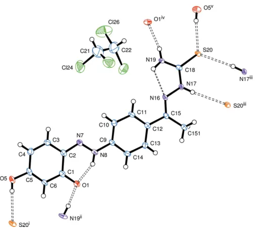
The main molecule comprises two essentially planar groups, which share the C12—C15 bond; the angle between the normals to the two planes is 13.77 (8), Fig. 2.
This molecule is a zwitterion, with a negative charge on O1 and a positive charge on N8: this nitrogen atom is bonded to a
1272
Riyadhet al. C15H15N5O2SC2H2Cl4 Acta Cryst.(2017). E73, 1271–1274
[image:2.610.45.296.71.328.2] [image:2.610.311.566.77.196.2]research communications
Figure 2
The molecular structure of (I) and the hydrogen-bond interactions (not including the ‘weak’ hydrogen bonds). Displacement ellipsoids are drawn at the 50% probability level. Symmetry operations (in the text and all figures): (i) 2 +x,1
2y,z 1
2; (ii)x+ 1, 1 2y,
1
2z; (iii)x1, 1y,
1z; (iv)x1,1 2y,
1
2+z; (v)x2, 1 2y,
1
2+z; (vi)x+ 1,y,z; (vii)x,
1y, 1z; (viii)x1,y,z.
Figure 3
The 1H NMR spectrum for compound
[image:2.610.315.566.260.366.2]3, showing one form of the product.
Table 1
Hydrogen-bond geometry (A˚ ,).
D—H A D—H H A D A D—H A
C6—H6 S20i 0.93 2.89 3.634 (4) 137
C10—H10 Cl25ii 0.93 2.88 3.807 (5) 172 C151—H15C S20iii 0.96 2.87 3.458 (4) 121
N17—H17 S20iii 0.86 2.63 3.483 (4) 173 C22—H22 O1iv 0.98 2.37 3.345 (8) 175
O5—H5O S20i 0.80 (2) 2.43 (2) 3.227 (4) 173 (5) N8—H8N O1 0.85 (2) 1.78 (3) 2.528 (4) 147 (4) N19—H19A O1iv 0.84 (2) 2.05 (2) 2.862 (5) 163 (4)
N19—H19B N16 0.84 (2) 2.16 (5) 2.578 (5) 111 (4)
Symmetry codes: (i)xþ2;yþ1
2;z12; (ii)xþ1;y;z; (iii)x1;yþ1;zþ1; (iv)
[image:2.610.46.299.447.677.2]x1;yþ1 2;zþ12.
Figure 4
[image:2.610.313.564.494.717.2]hydrogen atom (clearly identified in the X-ray analysis) and forms an intramolecular hydrogen bond N8—H8N O1. There is delocalized bonding throughout the O1—C1—C2— N7—N8—C9 chain, with bond dimensions very similar to those found in a series of 1-(2-phenyldiazen-2-ium-1-yl)-naphthalen-2-olate compounds studied by Benosmane et al.
(2013), Bougueria et al.(2013a,b) and Chetioui et al.(2013), showing a structure midway between the keto and phenolate forms of compound3in the Scheme; in particular, the C1—O1 bond length is 1.296 (5) A˚ and N7—N8 is 1.291 (5) A˚.
There is a more pronounced arrangement of double and single bonds for the C N—N group further along the mol-ecule, with C15 N16 at 1.280 (5) A˚ , and N16—N17 at 1.377 (5) A˚ ; N16 is the acceptor of a strained hydrogen bond from H19B.
The structure of the product was substantiated via spec-troscopic data. For example,1H NMR spectra of compounds3
revealed two singlet signals at = 2.35 and 10.28 ppm, attributed to the methyl group adjacent to hydrazone (CH3–
C N–NH) (de Oliveiraet al., 2014) and NH of the hydrazone group (C N–NH), respectively; there are also two signals (= 8.11 and 8.43 ppm) for the NH2group. A singlet signal at = 10.70 ppm is due to the OH group whereas the C O HN appears at= 12.53 ppm, see Fig. 3.
3. Supramolecular features
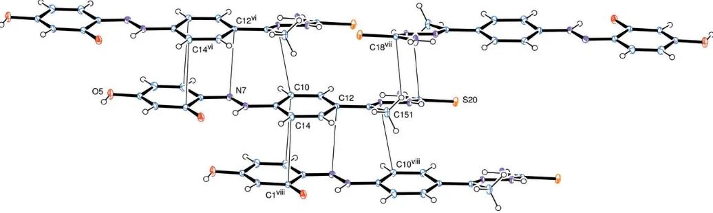
Intermolecular hydrogen bonds are shown in Table 1 and Fig. 4, and connect the molecules into a three-dimensional network. The solvent tetrachloroethane molecules are linked to this network through weak hydrogen bonds C10— H10 Cl25vi and C22—H22 O1iv. Other short inter-molecular contacts connect molecules by –stacking along theaaxis, Fig. 5; the phenyl ring of C1–C6 lies over the almost parallel ring of C9–C14 in the adjacent molecule, with C1 C14vi= 3.309 A˚ , C3 C10vi= 3.360 A˚ , and C5 N8vi= 3.263 A˚ . The hydrazone group of C15 N17 is sandwiched between the C15 C18 section of an inverted molecule [with closest contacts of N17 N17vii= 3.405 A˚ and C18 H15Cvii
= 2.83A˚ ] and the chain of N7 C11 of the stacked contact [with closest contacts N16 C10viii = 3.314 A˚ and H15A C14viii= 2.93 A˚ ].
4. Synthesis and crystallization
4-Acetylphenylazoresorcinol (Torrey & MacPherson, 1909) (1) (12.8 g, 50 mmol) was dissolved in 100ml of ethanol and stirred with an equimolar quantity of thiosemicarbazide (2) (4.55 g, 50 mmol) for 24 h at room temperature using catalytic amounts of HCl. The product, precipitated from the reaction mixture, was filtered, washed with ethanol and recrystallized from hot ethanol solution to give compound 3 as dark-red microcrystals (12.34g, 75%). Dark-red prisms of (I) were obtained by recrystallization from mixed solvents of tetra-chloromethane andn-hexane 1:1; m.p. 521–523 K; IR (KBr):
(cm1) 3456–3257 (OH+NH+NH2), 1596 (C N); 1
H NMR (DMSO-d6): 2.35 (s, 3H, CH3-C=N-NH), 6.37 (s, 1H,
CH—CO), 6.56 (d, 1H,J= 4Hz, CH—C N), 7.69 (d, 1H,J= 4Hz, CH—C—OH), 7.90 (d, 2H,J= 7Hz, Ar-H), 8.25 (d, 2H,J= 7Hz, Ar-H), 8.11 & 8.50 (2s, 2H, NH2), 10.28
(s, 1H, NH—C S), 10.75 (s, 1H, OH), 12.50 (s, 1H, NH— O C) ppm; 13C-NMR (DMSO-d6): 13.86 CH3—C N—
NH), 102.99 (C6), 109.33 (C3), 121.39 (C10 & C14), 127.64 (C12), 132.54 (C11 & C13), 134.35 (C2), 138.86 (C4), 146.86 (C9), 150.79 (C15), 156.64 (C5), 163.28 (C O), 179.02 (C S) ppm; MSm/z(%): 329 (M+, 38), 227 (35), 171 (70), 146 (70). Analysis calculated for C15H15N5O2S (329.09): C, 54.70; H,
4.59; N, 21.26; S, 9.74. Found: C, 54.61; H, 4.66; N, 21.33; S, 9.51%.
5. Refinement
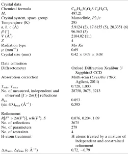
[image:3.610.47.565.571.726.2]Crystal data, data collection and structure refinement details are summarized in Table 2. The non-hydrogen atoms were refined with anisotropic thermal parameters. Hydrogen atoms on the O and N atoms were located in difference maps and were refined with distance constraints viz O—H distances were set to 0.82 (2) A˚ and N—H distances to 0.86 A˚; their
Figure 5
isotropic thermal parameters were refined freely. The remaining H atoms were included in idealized positions with their Uisovalues set to ride on the Ueqvalues of the parent
carbon atoms.
References
Abdel-Gawad, H., Mohamed, H. A., Dawood, K. M. & Badria, F. A.-R. (2010).Chem. Pharm. Bull.58, 1529–1531.
Agilent (2014).CrysAlis PRO. Agilent Technologies Ltd, Yarnton, Oxfordshire, England.
Benosmane, A., Mili, A., Bouguerria, H. & Bouchoul, A. (2013).Acta
Cryst.E69, o1021.
Bougueria, H., Benaouida, M. A., Bouacida, S. & Bouchoul, A. El K. (2013a).Acta Cryst.E69, o1175–o1176.
Bougueria, H., Benosmane, A., Benaouida, M. A., Bouchoul, A. El K. & Bouaoud, S. E. (2013b).Acta Cryst.E69, o1052.
Cardoso, M. V. de O., de Siqueira, L. R. P., da Silva, E. B., Costa, L. B., Hernandes, M. Z., Rabello, M. M., Ferreira, R. S., da Cruz, L. F., Magalha˜es Moreira, D. R., Pereira, V. R. A., de Castro, M. C. A. B., Bernhardt, P. V. & Leite, A. C. L. (2014).Eur. J. Med. Chem.86, 48– 59.
Chetioui, S., Boudraa, I., Bouacida, S., Bouchoul, A. & Bouaoud, S. E. (2013).Acta Cryst.E69, o1322–o1323.
Chimenti, F., Bolasco, A., Secci, D., Chimenti, P., Granese, A., Carradori, S., Ya´n˜ez, M., Orallo, F., Ortuso, F. & Alcaro, S. (2010).
Bioorg. Med. Chem.18, 5715–5723.
El-Sadek, M. M., Hassan, S. Y., Abdelwahab, H. E. & Yacout, G. A. (2012).Molecules,17, 8378–8396.
Farrugia, L. J. (2012).J. Appl. Cryst.45, 849–854.
Finch, R. A., Liu, M.-C., Grill, S. P., Rose, W. C., Loomis, R., Vasquez, K. M., Cheng, Y.-C. & Sartorelli, A. C. (2000). Biochem.
Pharmacol.59, 983–991.
Johnson, C. K. (1976).ORTEPII. Report ORNL-5138. Oak Ridge National Laboratory, Tennessee, USA.
Liu, J., Cao, R., Yi, W., Ma, C., Wan, Y., Zhou, B., Ma, L. & Song, H. (2009).Eur. J. Med. Chem.44, 1773–1778.
Liu, J., Yi, W., Wan, Y., Ma, L. & Song, H. (2008).Bioorg. Med. Chem.
16, 1096–1102.
Serda, M., Kalinowski, D. S., Mrozek-Wilczkiewicz, A., Musiol, R., Szurko, A., Ratuszna, A., Pantarat, N., Kovacevic, Z., Merlot, A. M., Richardson, D. R. & Polanski, J. (2012).Bioorg. Med. Chem.
Lett.22, 5527–5531.
Sheldrick, G. M. (2008).Acta Cryst.A64, 112–122. Sheldrick, G. M. (2015).Acta Cryst.C71, 3–8.
Soares, M. A., Lessa, J. A., Mendes, I. C., Da Silva, J. G., Dos Santos, R. G., Salum, L. B., Daghestani, H., Andricopulo, A. D., Day, B. W., Vogt, A., Pesquero, J. L., Rocha, W. R. & Beraldo, H. (2012).
Bioorg. Med. Chem.20, 3396–3409.
Thanusu, J., Kanagarajan, V. & Gopalakrishnan, M. (2010).Bioorg.
Med. Chem. Lett.20, 713–717.
Torrey, H. A. & MacPherson, W. (1909).J. Am. Chem. Soc.31, 579– 583.
Vazzana, I., Terranova, E., Mattioli, F. & Sparatore, F. (2004).
Arkivoc, (v), 364–374.
Vigato, P. A. & Tamburini, S. (2004).Coord. Chem. Rev.248, 1717– 2128.
1274
Riyadhet al. C15H15N5O2SC2H2Cl4 Acta Cryst.(2017). E73, 1271–1274
[image:4.610.43.294.92.395.2]research communications
Table 2
Experimental details.
Crystal data
Chemical formula C15H15N5O2SC2H2Cl4
Mr 497.21
Crystal system, space group Monoclinic,P21/c
Temperature (K) 295
a,b,c(A˚ ) 5.9124 (2), 17.6155 (5), 20.3351 (6)
() 96.563 (3)
V(A˚3) 2104.02 (11)
Z 4
Radiation type MoK
(mm1) 0.69
Crystal size (mm) 0.420.090.08
Data collection
Diffractometer Oxford Diffraction Xcalibur 3/ Sapphire3 CCD
Absorption correction Multi-scan (CrysAlis PRO; Agilent, 2014) Tmin,Tmax 0.728, 1.000
No. of measured, independent and observed [I> 2(I)] reflections
28750, 3675, 3213
Rint 0.053
(sin /)max(A˚ 1
) 0.595
Refinement
R[F2> 2(F2)],wR(F2),S 0.076, 0.204, 1.09
No. of reflections 3675 No. of parameters 279 No. of restraints 4
H-atom treatment H atoms treated by a mixture of independent and constrained refinement
max,min(e A˚3) 0.72,0.79
Computer programs: CrysAlis PRO(Agilent, 2014), SHELXS97 (Sheldrick, 2008),
ORTEP(Johnson, 1976; Farrugia, 2012),SHELXL2014(Sheldrick, 2015) andWinGX
sup-1
Acta Cryst. (2017). E73, 1271-1274
supporting information
Acta Cryst. (2017). E73, 1271-1274 [https://doi.org/10.1107/S2056989017010830]
Crystal structure of a 1,1,2,2-tetrachloroethane-solvated
hydrazinecarbothio-amide compound
Sayed Riyadh, David L. Hughes and Musa A Said
Computing details
Data collection: CrysAlis PRO (Agilent, 2014); cell refinement: CrysAlis PRO (Agilent, 2014); data reduction: CrysAlis
PRO (Agilent, 2014); program(s) used to solve structure: SHELXS97 (Sheldrick, 2008); program(s) used to refine
structure: SHELXL2014 (Sheldrick, 2015); molecular graphics: ORTEP (Johnson, 1976; Farrugia, 2012); software used
to prepare material for publication: SHELXL2014 (Sheldrick, 2015) and WinGX (Farrugia, 2012).
[(1-{4-[2-(2,4-Dihydroxyphenyl)diazen-1-yl]phenyl}ethylidene)amino]thiourea 1,1,2,2-tetrachloroethane
monosolvate
Crystal data
C15H15N5O2S·C2H2Cl4 Mr = 497.21
Monoclinic, P21/c a = 5.9124 (2) Å
b = 17.6155 (5) Å
c = 20.3351 (6) Å
β = 96.563 (3)°
V = 2104.02 (11) Å3 Z = 4
F(000) = 1016
Dx = 1.570 Mg m−3
Mo Kα radiation, λ = 0.71073 Å Cell parameters from 5694 reflections
θ = 3.5–28.7°
µ = 0.69 mm−1 T = 295 K Prism, dark red 0.42 × 0.09 × 0.08 mm
Data collection
Oxford Diffraction Xcalibur 3/Sapphire3 CCD diffractometer
Radiation source: Enhance (Mo) X-ray Source Graphite monochromator
Detector resolution: 16.0050 pixels mm-1
Thin slice φ and ω scans
Absorption correction: multi-scan (CrysAlis PRO; Agilent, 2014)
Tmin = 0.728, Tmax = 1.000
28750 measured reflections 3675 independent reflections 3213 reflections with I > 2σ(I)
Rint = 0.053
θmax = 25.0°, θmin = 3.6°
h = −7→7
k = −20→20
l = −24→24
Refinement
Refinement on F2
Least-squares matrix: full
R[F2 > 2σ(F2)] = 0.076 wR(F2) = 0.204 S = 1.09 3675 reflections 279 parameters 4 restraints
Primary atom site location: structure-invariant direct methods
Secondary atom site location: difference Fourier map
Hydrogen site location: mixed
supporting information
sup-2
Acta Cryst. (2017). E73, 1271-1274
w = 1/[σ2(Fo2) + (0.0938P)2 + 8.2654P]
where P = (Fo2 + 2Fc2)/3
(Δ/σ)max < 0.001
Δρmax = 0.72 e Å−3
Δρmin = −0.79 e Å−3
Special details
Geometry. All esds (except the esd in the dihedral angle between two l.s. planes) are estimated using the full covariance matrix. The cell esds are taken into account individually in the estimation of esds in distances, angles and torsion angles; correlations between esds in cell parameters are only used when they are defined by crystal symmetry. An approximate (isotropic) treatment of cell esds is used for estimating esds involving l.s. planes.
Fractional atomic coordinates and isotropic or equivalent isotropic displacement parameters (Å2)
x y z Uiso*/Ueq
O1 0.9543 (5) 0.22114 (17) 0.20187 (14) 0.0225 (7)
C1 1.0971 (7) 0.1827 (2) 0.2420 (2) 0.0183 (9)
C2 1.0684 (7) 0.1769 (2) 0.3120 (2) 0.0181 (9)
C3 1.2286 (7) 0.1342 (2) 0.3548 (2) 0.0217 (9)
H3 1.2112 0.1308 0.3996 0.026*
C4 1.4060 (7) 0.0984 (3) 0.3313 (2) 0.0239 (10)
H4 1.5092 0.0706 0.3596 0.029*
C5 1.4325 (7) 0.1039 (2) 0.2629 (2) 0.0203 (9)
O5 1.6154 (5) 0.06701 (19) 0.24376 (17) 0.0270 (7)
C6 1.2855 (7) 0.1452 (2) 0.2200 (2) 0.0187 (9)
H6 1.3101 0.1486 0.1757 0.022*
N7 0.8994 (6) 0.2111 (2) 0.34044 (17) 0.0189 (8)
N8 0.7553 (6) 0.2510 (2) 0.30244 (17) 0.0184 (8)
C9 0.5772 (7) 0.2898 (2) 0.3284 (2) 0.0170 (8)
C10 0.5488 (7) 0.2886 (2) 0.3949 (2) 0.0189 (9)
H10 0.6486 0.2612 0.4246 0.023*
C11 0.3698 (7) 0.3286 (2) 0.4168 (2) 0.0194 (9)
H11 0.3503 0.3277 0.4615 0.023*
C12 0.2182 (7) 0.3702 (2) 0.3729 (2) 0.0159 (8)
C13 0.2511 (7) 0.3713 (2) 0.3063 (2) 0.0205 (9)
H13 0.1541 0.3996 0.2765 0.025*
C14 0.4281 (7) 0.3304 (3) 0.2839 (2) 0.0221 (9)
H14 0.4468 0.3303 0.2391 0.027*
C15 0.0244 (7) 0.4110 (2) 0.3983 (2) 0.0158 (8)
C151 −0.1144 (7) 0.4677 (3) 0.3562 (2) 0.0235 (10)
H15A −0.2643 0.4477 0.3439 0.035*
H15B −0.0428 0.4779 0.3171 0.035*
H15C −0.1251 0.5139 0.3807 0.035*
N16 −0.0111 (5) 0.39265 (19) 0.45721 (17) 0.0156 (7)
N17 −0.1850 (5) 0.42611 (19) 0.48636 (17) 0.0165 (7)
H17 −0.2660 0.4619 0.4670 0.020*
C18 −0.2233 (7) 0.4003 (2) 0.5467 (2) 0.0166 (9)
N19 −0.0835 (7) 0.3482 (2) 0.57381 (19) 0.0249 (9)
S20 −0.44392 (18) 0.43497 (6) 0.58404 (5) 0.0214 (3)
C21 0.2745 (18) 0.1205 (5) 0.5978 (4) 0.091 (3)
sup-3
Acta Cryst. (2017). E73, 1271-1274
C22 0.0344 (16) 0.1387 (4) 0.5960 (4) 0.076 (2)
H22 0.0155 0.1820 0.6251 0.091*
Cl23 0.4115 (4) 0.21230 (11) 0.58528 (10) 0.0710 (6)
Cl24 0.3333 (5) 0.05129 (12) 0.53859 (10) 0.0888 (8)
Cl25 −0.0877 (4) 0.16005 (11) 0.51483 (9) 0.0729 (6)
Cl26 −0.0903 (5) 0.05355 (12) 0.62934 (11) 0.0894 (8)
H5O 1.612 (8) 0.068 (3) 0.2043 (10) 0.019 (13)*
H8N 0.776 (8) 0.249 (3) 0.2620 (11) 0.021 (12)*
H19A −0.101 (7) 0.330 (2) 0.6111 (13) 0.006 (10)*
H19B 0.023 (6) 0.336 (3) 0.552 (2) 0.025 (13)*
Atomic displacement parameters (Å2)
U11 U22 U33 U12 U13 U23
O1 0.0237 (16) 0.0269 (17) 0.0174 (15) 0.0069 (13) 0.0040 (12) −0.0021 (13)
C1 0.018 (2) 0.016 (2) 0.021 (2) −0.0031 (16) 0.0043 (17) −0.0043 (17)
C2 0.016 (2) 0.016 (2) 0.023 (2) −0.0029 (16) 0.0061 (16) −0.0041 (17)
C3 0.024 (2) 0.022 (2) 0.021 (2) 0.0003 (17) 0.0072 (18) 0.0012 (17)
C4 0.022 (2) 0.024 (2) 0.026 (2) 0.0029 (18) 0.0021 (18) 0.0023 (18)
C5 0.016 (2) 0.016 (2) 0.029 (2) −0.0015 (16) 0.0068 (17) −0.0071 (18)
O5 0.0237 (16) 0.0311 (18) 0.0277 (19) 0.0112 (14) 0.0090 (14) −0.0021 (15)
C6 0.020 (2) 0.019 (2) 0.018 (2) 0.0012 (17) 0.0079 (17) −0.0043 (17)
N7 0.0172 (17) 0.0191 (18) 0.0212 (18) −0.0015 (14) 0.0056 (14) −0.0021 (15)
N8 0.0173 (18) 0.0221 (19) 0.0167 (19) 0.0001 (14) 0.0065 (14) −0.0041 (15)
C9 0.0158 (19) 0.017 (2) 0.020 (2) −0.0013 (16) 0.0074 (16) −0.0032 (16)
C10 0.017 (2) 0.019 (2) 0.021 (2) 0.0003 (16) 0.0023 (16) −0.0008 (17)
C11 0.020 (2) 0.024 (2) 0.015 (2) 0.0007 (17) 0.0057 (16) −0.0029 (17)
C12 0.0152 (19) 0.013 (2) 0.020 (2) −0.0036 (15) 0.0046 (16) −0.0029 (16)
C13 0.020 (2) 0.020 (2) 0.022 (2) 0.0050 (17) 0.0049 (17) 0.0043 (17)
C14 0.026 (2) 0.025 (2) 0.017 (2) 0.0006 (18) 0.0104 (18) −0.0005 (17)
C15 0.0140 (19) 0.014 (2) 0.020 (2) −0.0013 (15) 0.0043 (16) −0.0029 (16)
C151 0.024 (2) 0.025 (2) 0.024 (2) 0.0067 (18) 0.0113 (18) 0.0018 (18)
N16 0.0137 (16) 0.0143 (17) 0.0198 (18) 0.0004 (13) 0.0061 (13) −0.0023 (14)
N17 0.0137 (16) 0.0159 (17) 0.0205 (18) 0.0026 (13) 0.0054 (13) 0.0006 (14)
C18 0.017 (2) 0.016 (2) 0.017 (2) −0.0047 (16) 0.0062 (16) −0.0034 (16)
N19 0.028 (2) 0.031 (2) 0.019 (2) 0.0117 (17) 0.0143 (16) 0.0073 (16)
S20 0.0186 (5) 0.0267 (6) 0.0208 (6) 0.0054 (4) 0.0112 (4) 0.0042 (4)
C21 0.134 (8) 0.088 (6) 0.049 (4) 0.040 (6) −0.003 (5) −0.025 (4)
C22 0.122 (7) 0.052 (4) 0.053 (4) 0.027 (4) 0.004 (4) −0.005 (3)
Cl23 0.0926 (14) 0.0562 (11) 0.0691 (12) −0.0062 (10) 0.0310 (10) −0.0010 (9)
Cl24 0.136 (2) 0.0733 (13) 0.0533 (11) 0.0549 (13) −0.0076 (11) −0.0175 (9)
Cl25 0.1051 (15) 0.0583 (11) 0.0539 (10) 0.0306 (10) 0.0031 (10) 0.0093 (8)
Cl26 0.136 (2) 0.0649 (13) 0.0658 (13) −0.0453 (13) 0.0050 (12) 0.0031 (10)
Geometric parameters (Å, º)
O1—C1 1.296 (5) C12—C15 1.494 (5)
supporting information
sup-4
Acta Cryst. (2017). E73, 1271-1274
C1—C2 1.456 (6) C13—H13 0.9300
C2—N7 1.351 (5) C14—H14 0.9300
C2—C3 1.426 (6) C15—N16 1.280 (5)
C3—C4 1.356 (6) C15—C151 1.499 (6)
C3—H3 0.9300 C151—H15A 0.9600
C4—C5 1.420 (6) C151—H15B 0.9600
C4—H4 0.9300 C151—H15C 0.9600
C5—O5 1.356 (5) N16—N17 1.377 (5)
C5—C6 1.369 (6) N17—C18 1.352 (5)
O5—H5O 0.801 (19) N17—H17 0.8600
C6—H6 0.9300 C18—N19 1.314 (6)
N7—N8 1.291 (5) C18—S20 1.696 (4)
N8—C9 1.408 (5) N19—H19A 0.841 (19)
N8—H8N 0.846 (19) N19—H19B 0.84 (2)
C9—C10 1.381 (6) C21—C22 1.451 (13)
C9—C14 1.388 (6) C21—Cl24 1.776 (8)
C10—C11 1.387 (6) C21—Cl23 1.840 (11)
C10—H10 0.9300 C21—H21 0.9800
C11—C12 1.398 (6) C22—Cl25 1.766 (8)
C11—H11 0.9300 C22—Cl26 1.834 (9)
C12—C13 1.392 (6) C22—H22 0.9800
O1—C1—C6 121.7 (4) C14—C13—H13 119.8
O1—C1—C2 120.8 (4) C12—C13—H13 119.8
C6—C1—C2 117.5 (4) C9—C14—C13 120.0 (4)
N7—C2—C3 116.5 (4) C9—C14—H14 120.0
N7—C2—C1 124.2 (4) C13—C14—H14 120.0
C3—C2—C1 119.3 (4) N16—C15—C12 114.6 (4)
C4—C3—C2 121.1 (4) N16—C15—C151 124.4 (4)
C4—C3—H3 119.4 C12—C15—C151 121.0 (4)
C2—C3—H3 119.4 C15—C151—H15A 109.5
C3—C4—C5 119.3 (4) C15—C151—H15B 109.5
C3—C4—H4 120.3 H15A—C151—H15B 109.5
C5—C4—H4 120.3 C15—C151—H15C 109.5
O5—C5—C6 122.7 (4) H15A—C151—H15C 109.5
O5—C5—C4 115.4 (4) H15B—C151—H15C 109.5
C6—C5—C4 121.8 (4) C15—N16—N17 120.3 (3)
C5—O5—H5O 110 (4) C18—N17—N16 117.3 (3)
C5—C6—C1 120.9 (4) C18—N17—H17 121.4
C5—C6—H6 119.6 N16—N17—H17 121.4
C1—C6—H6 119.6 N19—C18—N17 116.9 (4)
N8—N7—C2 117.1 (4) N19—C18—S20 122.9 (3)
N7—N8—C9 120.7 (3) N17—C18—S20 120.2 (3)
N7—N8—H8N 114 (3) C18—N19—H19A 121 (3)
C9—N8—H8N 125 (3) C18—N19—H19B 116 (3)
C10—C9—C14 120.5 (4) H19A—N19—H19B 124 (5)
C10—C9—N8 122.7 (4) C22—C21—Cl24 113.7 (6)
sup-5
Acta Cryst. (2017). E73, 1271-1274
C9—C10—C11 119.3 (4) Cl24—C21—Cl23 112.7 (5)
C9—C10—H10 120.4 C22—C21—H21 108.7
C11—C10—H10 120.4 Cl24—C21—H21 108.7
C10—C11—C12 121.2 (4) Cl23—C21—H21 108.7
C10—C11—H11 119.4 C21—C22—Cl25 111.4 (6)
C12—C11—H11 119.4 C21—C22—Cl26 104.1 (6)
C13—C12—C11 118.6 (4) Cl25—C22—Cl26 112.4 (5)
C13—C12—C15 121.9 (4) C21—C22—H22 109.6
C11—C12—C15 119.5 (4) Cl25—C22—H22 109.6
C14—C13—C12 120.4 (4) Cl26—C22—H22 109.6
O1—C1—C2—N7 −1.3 (6) C10—C11—C12—C13 −0.5 (6)
C6—C1—C2—N7 178.5 (4) C10—C11—C12—C15 178.5 (4)
O1—C1—C2—C3 180.0 (4) C11—C12—C13—C14 1.5 (6)
C6—C1—C2—C3 −0.2 (6) C15—C12—C13—C14 −177.6 (4)
N7—C2—C3—C4 −179.3 (4) C10—C9—C14—C13 1.1 (6)
C1—C2—C3—C4 −0.5 (6) N8—C9—C14—C13 −178.7 (4)
C2—C3—C4—C5 0.2 (7) C12—C13—C14—C9 −1.7 (6)
C3—C4—C5—O5 179.5 (4) C13—C12—C15—N16 166.6 (4)
C3—C4—C5—C6 0.9 (7) C11—C12—C15—N16 −12.5 (5)
O5—C5—C6—C1 180.0 (4) C13—C12—C15—C151 −12.8 (6)
C4—C5—C6—C1 −1.6 (6) C11—C12—C15—C151 168.2 (4)
O1—C1—C6—C5 −178.9 (4) C12—C15—N16—N17 −179.9 (3)
C2—C1—C6—C5 1.2 (6) C151—C15—N16—N17 −0.6 (6)
C3—C2—N7—N8 178.9 (4) C15—N16—N17—C18 175.4 (4)
C1—C2—N7—N8 0.2 (6) N16—N17—C18—N19 3.4 (5)
C2—N7—N8—C9 −178.6 (4) N16—N17—C18—S20 −176.5 (3)
N7—N8—C9—C10 1.1 (6) Cl24—C21—C22—Cl25 −50.4 (9)
N7—N8—C9—C14 −179.1 (4) Cl23—C21—C22—Cl25 72.7 (6)
C14—C9—C10—C11 −0.1 (6) Cl24—C21—C22—Cl26 71.0 (7)
N8—C9—C10—C11 179.6 (4) Cl23—C21—C22—Cl26 −165.9 (4)
C9—C10—C11—C12 −0.1 (6)
Hydrogen-bond geometry (Å, º)
D—H···A D—H H···A D···A D—H···A
C6—H6···S20i 0.93 2.89 3.634 (4) 137
C10—H10···Cl25ii 0.93 2.88 3.807 (5) 172
C151—H15C···S20iii 0.96 2.87 3.458 (4) 121
N17—H17···S20iii 0.86 2.63 3.483 (4) 173
C22—H22···O1iv 0.98 2.37 3.345 (8) 175
O5—H5O···S20i 0.80 (2) 2.43 (2) 3.227 (4) 173 (5)
N8—H8N···O1 0.85 (2) 1.78 (3) 2.528 (4) 147 (4)
N19—H19A···O1iv 0.84 (2) 2.05 (2) 2.862 (5) 163 (4)
N19—H19B···N16 0.84 (2) 2.16 (5) 2.578 (5) 111 (4)