research communications
Acta Cryst.(2017). E73, 1417–1420 https://doi.org/10.1107/S2056989017012488
1417
Received 4 August 2017 Accepted 29 August 2017
Edited by G. Smith, Queensland University of Technology, Australia
Keywords:1,3-thiazin-4-one; twisted half-chair pucker; N—H O and C—H O interactions; crystal structure.
CCDC reference:1571357
Supporting information:this article has supporting information at journals.iucr.org/e
Crystal structure of (1
S
,2
S
,5
R
)-5-acetylamino-4-oxo-2,3-diphenyl-1,3-thiazinan-1-ium-1-olate
Hemant P. Yennawar,aDuncan J. Nobleband Lee J. Silverbergb*
aDepartment of Biochemistry and Molecular Biology, Pennsylvania State University, University Park, PA 16802, USA, and b
Pennsylvania State University, Schuylkill Campus, 200 University Drive, Schuylkill Haven, PA 17972, USA. *Correspondence e-mail: [email protected]
The asymmetric unit of the enantiomerically pure title compound, C18H18N2O3S,
comprises two independent molecules (A and B) having almost identical conformations. When overlayed, the alignment–r.m.s. deviation value is 0.30 A˚ . The six-membered heterocycle has a twisted half-chair conformation in both molecules. The O atom on the S atom of the ring is pseudo-axial on the thiazine ring andtransto both a phenyl group substituent and the acetamide group in each case. The two benzene rings in each molecule are almost orthogonal to each other, with interplanar dihedral angles of 83.79 (17) and 86.95 (16). The acetamide group is pseudo-equatorial and a phenyl ring is pseudo-axial on the thiazine ring. Both molecules show a weak intramolecular C—H O interaction between H-atom donors of one of the phenyl rings and the acetamide group. In the crystal, an intermolecular N—H O(thiazine) hydrogen bond links B
molecules along the 21(b) screw axis and, in addition, an N—H O(acetamide)
hydrogen bond links AandBmolecules acrossa. A two-dimensional layered structure lying parallel to (001) is generated, also involving weak intermolecular C—H O interactions.
1. Chemical context
The 1,3-thiazin-4-ones are a group of six-membered hetero-cycles with a wide range of biological activity (Ryabukhin et al., 1996). Surrey’s research (Surrey et al., 1958; Surrey, 1963a,b) resulted in the discovery of two drugs, the anti-anxiety and muscle relaxant chlormezanone [2-(4-chloro-phenyl)-3-methyl-2,3,5,6-tetrahydro-4H-1,3-thiazin-4-one 1,1-dioxide] (O’Neil, 2006; Tanaka & Horayama, 2005) and muscle relaxant dichloromezanone [2-(3,4-dichlorophenyl)-3-methyl-2,3,5,6-tetrahydro-4H-1,3-thiazin-4-one 1,1-dioxide] (Elks & Ganellin, 1990). These sulfones showed greater activity than the sulfides from which they were synthesized (Surrey et al., 1958). Surrey also prepared a variety of other sulfoxides and sulfones of 3-alkyl-2-aryl-2,3,5,6-tetrahydro-4H-1,3-thiazin-4-ones (Surrey, 1963a,b). We have reported previously the crystal structure of the firstN-aryl sulfoxide in this family, racemic 2,3-diphenyl-2,3,5,6-tetrahydro-4H -1,3-thiazin-4-one 1-oxide (Yennawaret al., 2016).
A sulfoxide typically has an S—O bond that is between a double bond and a single bond, with one of the lone pairs that was on the sulfide coordinating to the O atom, while O atom contributes electrons from a lone pair to a dorbital of the S atom. The geometry of a sulfoxide is pyramidal, with a high energy barrier for inversion, making it possible to isolate stable enantiomers (Bentley, 2005). Herein, we report the crystal structure of the sulfoxide of N-[(2S,5R )-4-oxo-2,3-diphenyl-1,3-thiazinan-5-yl]acetamide (Yennawar, Singh & Silverberg, 2015), C18H18N2O3S, prepared using the method
we have reported previously for the oxidation of other 2,3-diphenyl-1,3-thiazin-4-ones (Yennawaret al., 2016; Yennawar, Noble et al., 2017) and 1,3-thiazolidinones (Yennawar, Hullihenet al., 2015; Cannonet al., 2015). The oxidation of the confirmed enantiopure sulfideN-[(2S,5R )-4-oxo-2,3-diphenyl-1,3-thiazinan-5-yl]acetamide 0.375-hydrate (Yennawar, Singh & Silverberg, 2015), derived fromN-acetyl-l-cysteine, yielded a single stereoisomer as the only product.
2. Structural commentary
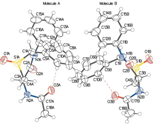
The crystal structure of the title compound has two indepen-dent homochiral molecules (AandB) in the asymmetric unit (Fig. 1), which have almost identical conformational features, having an alignment–r.m.s. deviation value of 0.3 A˚ . Both have
the thiazine rings in a twisted half-chair configuration, with puckering amplitudes = 0.6753 (19)/0.653 (2) A˚ and = 131.05 (17)/135.66 (18) in molecules A/B, respectively
(Cremer & Pople, 1975). The O atom on the S atom of the ring is pseudo-axial on the thiazine ring and trans to both the 2-phenyl group and the acetamide group in each case. The two phenyl rings in each molecule are almost orthogonal to one another, with dihedral angles of 83.79 (17) and 86.95 (16) in molecules A and B, respectively. The acetamide group is pseudo-equatorial and the 2-phenyl group is pseudo-axial on the thiazine ring. A weak intramolecular C—H O hydrogen bond between the 2-phenyl ring and the O atom of the acet-amide group is seen in both molecules (C10A—H O3Aand C10B—H O3B), as detailed in Table 1.
We reported previously the crystal structure of the starting sulfide, N-[(2S,5R )-4-oxo-2,3-diphenyl-1,3-thiazinan-5-yl]ace-tamide 0.375-hydrate (Yennawar, Singh & Silverberg, 2015), which also had two independent homochiral molecules in the asymmetric unit. However, they were not identical: in one molecule, the thiazine ring was in a half-chair conformation in which the 2-phenyl ring was nearly pseudo-axial and the acetamide group was nearly pseudo-equatorial. The other molecule had the thiazine ring in a boat conformation in which both substituents were pseudo-equatorial.
1418
Yennawaret al. C18H18N2O3S Acta Cryst.(2017). E73, 1417–1420
research communications
Table 1
Hydrogen-bond geometry (A˚ ,).
D—H A D—H H A D A D—H A
N2A—H2A O2Bi 0.91 (3) 2.25 (3) 3.137 (3) 164 (2)
N2B—H2B O1Bii 0.79 (3) 2.14 (3) 2.916 (3) 168 (3) C10A—H10A O3A 0.93 2.42 3.259 (3) 149 C10B—H10B O3B 0.93 2.44 3.232 (4) 143 C4B—H4BB O2Aiii 0.97 2.25 3.116 (3) 148
Symmetry codes: (i)xþ1;y;z; (ii)x;yþ1
[image:2.610.108.234.296.389.2]2;z; (iii)xþ1;yþ 1 2;z.
Figure 2
Crystal packing diagram with red dotted lines for intermolecular N— H O contacts between 21-related molecules, forming helical chains
[image:2.610.312.563.389.691.2]along theb-axis direction, as well as the interaction with an independent molecule. Blue dotted lines represent the intramolecular C—H O contacts.
Figure 1
[image:2.610.46.295.498.700.2]3. Supramolecular features
In the crystal, the B molecule and its 21-related symmetry
neighbours form a continuous hydrogen-bonded chain along theb-cell direction through N—H O interactions involving the acetamide N atom and the thiazin-1-ium-1-olate O atoms [N2B—H O1Bii; symmetry code: (ii)x,y+12,z; Table 1]
(Fig. 2). MoleculesAandBinteract, wherein the O atom in the 4-position of moleculeBaccepts a proton from the acet-amide N atom of molecule A [N2A—H O1Bi; symmetry code: (i)x+ 1,y,z]. The sulfoxide O atom of moleculeAdoes not participate in any hydrogen bonding. A two-dimensional sheet structure lying parallel to (001) is generated. No benzene ring in either of the molecules participates in face-to-face–
stacking interactions.
4. Database survey
Crystal structures of a number of 1,3-thiazolidin-4-one 1-oxides have been reported (Wanget al., 2010; Johnsonet al., 1983; Chen et al., 2011; Colombo et al., 2008; Yennawar, Hullihen et al., 2015) and the structure of chlormezanone [2-(4-chlorophenyl)-3-methyl-2,3,5,6-tetrahydro-4H -1,3-thiazin-4-one 1,1-dioxide] has also been reported (Tanaka & Horayama, 2005). We have reported previously the crystal
structure of 2,3-diphenyl-2,3,5,6-tetrahydro-4H -1,3-thiazin-4-one 1-oxide (Yennawar et al., 2016). We have also reported recently the crystal structures of 2,3-diphenyl-2,3-dihydro-4H -1,3-benzothiazin-4-one 1-oxide (Yennawar, Fox et al., 2017) and 2,3-diphenyl-2,3-dihydro-4H-pyrido[3,2-e ][1,3]thiazin-4-one 1-oxide (Yennawar, Nobleet al., 2017).
5. Synthesis and crystallization
A 5 ml round-bottomed flask was charged with 53.9 mg of
N-[(2S,5R)-4-oxo-2,3-diphenyl-1,3-thiazinan-5-yl]acetamide 0.375-hydrate, whose configuration was established previously (Yennawar, Singh & Silverberg, 2015), and 1.4 ml of methanol and stirred. A solution of 79.5 mg of Oxone1 and 1 ml of distilled water was added dropwise and the mixture was stirred until the reaction was complete, as determined by thin-layer chromatography (TLC). The solids were dissolved by the addition of 5 ml of distilled water. The solution was extracted with 10 ml of dichloromethane. The organic layer was washed with 5 ml of distilled water and then with 5 ml of saturated sodium chloride. The solution was dried over Na2SO4 and
concentrated under vacuum giving a crude solid. This was chromatographed on flash silica gel, eluting with a gradient of 0–60% acetone in ethyl acetate, giving 55.8 mg of product [98.6% yield; m.p. 449–452 K; RF= 0.20 (30% acetone/70%
ethyl acetate)]. Crystals suitable for X-ray crystallography were grown by slow evaporation from propan-2-ol.
6. Refinement
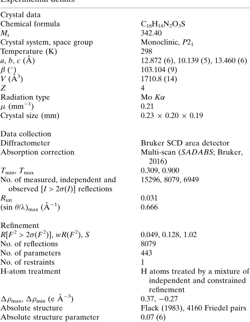
Crystal data, data collection and structure refinement details are summarized in Table 2. The H atoms, excepting those on N atoms, were placed geometrically and allowed to ride on their parent C atoms during refinement, with C—H distances of 0.93 (aromatic), 0.96 (methyl), 0.97 or (methylene) and 0.98 A˚ (methyl), and withUiso(H) = 1.2Ueq(aromatic or methylene C)
or 1.5Ueq(methyl C). H atoms on N atoms were located in a
difference Fourier map and were refined isotropically. The absolute configuration for the chiral centres in the molecule was determined as (1S,2S,5R) (for the arbitrarily numbered atoms C1A/B,C3A/B), with a Flack absolute structure para-meter (Flack, 1983) of 0.07 (6) for 4160 Friedel pairs.
Funding information
Funding for this research was provided by: National Science Foundation (grant No. CHEM-0131112 for the X-ray diffractometer); Penn State Schuylkill.
References
Bentley, R. (2005).Chem. Soc. Rev.34, 609–624.
Bruker (2016).SMART,SAINT and SADABS. Bruker AXS Inc., Madison, Wisconsin, USA.
Cannon, K., Gandla, D., Lauro, S., Silverberg, L., Tierney, J. & Lagalante, A. (2015).Int. J. Chem.7(2), 73–84.
Chen, H., Zai-Hong, G., Qing-Mei, Y. & Xiao-Liu, L. (2011).Chin. J. Org. Chem.31, 249–255.
research communications
Acta Cryst.(2017). E73, 1417–1420 Yennawaret al. C
[image:3.610.45.290.86.401.2]18H18N2O3S
1419
Table 2Experimental details.
Crystal data
Chemical formula C18H18N2O3S
Mr 342.40
Crystal system, space group Monoclinic,P21
Temperature (K) 298
a,b,c(A˚ ) 12.872 (6), 10.139 (5), 13.460 (6)
() 103.104 (9)
V(A˚3) 1710.8 (14)
Z 4
Radiation type MoK
(mm1) 0.21
Crystal size (mm) 0.230.200.19
Data collection
Diffractometer Bruker SCD area detector Absorption correction Multi-scan (SADABS; Bruker,
2016)
Tmin,Tmax 0.309, 0.900
No. of measured, independent and observed [I> 2(I)] reflections
15296, 8079, 6949
Rint 0.031
(sin/)max(A˚
1) 0.666
Refinement
R[F2> 2(F2)],wR(F2),S 0.049, 0.128, 1.02
No. of reflections 8079
No. of parameters 443
No. of restraints 1
H-atom treatment H atoms treated by a mixture of independent and constrained refinement
max, min(e A˚
3
) 0.37,0.27
Absolute structure Flack (1983), 4160 Friedel pairs Absolute structure parameter 0.07 (6)
Computer programs: SMART (Bruker, 2016), SAINT (Bruker, 2016), SHELXS97
Colombo, A., Ferna`ndez, J. C., Ferna´ndez-Forner, D., de la Figuera, N., Albericio, F. & Forns, P. (2008).Tetrahedron Lett.49, 1569–1572. Cremer, D. & Pople, J. A. (1975).J. Am. Chem. Soc.97, 1354–1358. Dolomanov, O. V., Bourhis, L. J., Gildea, R. J., Howard, J. A. K. &
Puschmann, H. (2009).J. Appl. Cryst.42, 339–341.
Elks, J. & Ganellin, C. R. (1990). Editors.Dictionary of Drugs, p. 382. Cambridge, UK: Chapman and Hall.
Flack, H. D. (1983).Acta Cryst.A39, 876–881.
Johnson, M. R., Fazio, M. J., Ward, D. L. & Sousa, L. R. (1983).J. Org. Chem.48, 494–499.
O’Neil, M. J. (2006). Editor. The Merck Index, 14th ed., p. 349, Whitehouse Station, NJ: Merck & Co. Inc.
Ryabukhin, Y. I., Korzhavina, O. B. & Suzdalev, K. F. (1996).Adv. Heterocycl. Chem.66, 131–190.
Sheldrick, G. M. (2008).Acta Cryst.A64, 112–122. Surrey, A. R. (1963a). US Patent 3082209.
Surrey, A. R. (1963b). US Patent 3093639.
Surrey, A. R., Webb, W. G. & Gesler, R. M. (1958).J. Am. Chem. Soc. 80, 3469–3471.
Tanaka, R. & Horayama, N. (2005).X-Ray Struct. Anal. Online,21, x57–x58.
Wang, Q., Xu, Z. & Sun, Y. (2010).Acta Cryst.E66, o1422. Yennawar, H. P., Fox, R., Moyer, Q. J., Yang, Z. & Silverberg, L. J.
(2017).Acta Cryst.E73, 1189–1191.
Yennawar, H. P., Noble, D. J., Yang, Z. & Silverberg, L. J. (2017). IUCrData,2, x171112.
Yennawar, H. P., Singh, H. & Silverberg, L. J. (2015).Acta Cryst.E71, 62–64.
Yennawar, H. P., Tierney, J., Hullihen, P. D. & Silverberg, L. J. (2015). Acta Cryst.E71, 264–267.
Yennawar, H. P., Yang, Z. & Silverberg, L. J. (2016).Acta Cryst.E72, 1541–1543.
1420
Yennawaret al. C18H18N2O3S Acta Cryst.(2017). E73, 1417–1420
supporting information
sup-1
Acta Cryst. (2017). E73, 1417-1420
supporting information
Acta Cryst. (2017). E73, 1417-1420 [https://doi.org/10.1107/S2056989017012488]
Crystal structure of (1
S
,2
S
,5
R
)-5-acetylamino-4-oxo-2,3-diphenyl-1,3-thia-zinan-1-ium-1-olate
Hemant P. Yennawar, Duncan J. Noble and Lee J. Silverberg
Computing details
Data collection: SMART (Bruker, 2016); cell refinement: SAINT (Bruker, 2016); data reduction: SAINT (Bruker, 2016);
program(s) used to solve structure: SHELXS97 (Sheldrick, 2008); program(s) used to refine structure: SHELXL97
(Sheldrick, 2008); molecular graphics: OLEX2 (Dolomanov et al., 2009); software used to prepare material for
publication: OLEX2 (Dolomanov et al., 2009).
(1S,2S,5R)-5-Acetylamino-4-oxo-2,3-diphenyl-1,3-thiazinan-1-ium-1-olate
Crystal data
C18H18N2O3S
Mr = 342.40
Monoclinic, P21
a = 12.872 (6) Å
b = 10.139 (5) Å
c = 13.460 (6) Å
β = 103.104 (9)°
V = 1710.8 (14) Å3
Z = 4
F(000) = 720
Dx = 1.329 Mg m−3
Melting point = 449–452 K Mo Kα radiation, λ = 0.71073 Å Cell parameters from 7433 reflections
θ = 2.5–28.2°
µ = 0.21 mm−1
T = 298 K Block, colorless 0.23 × 0.20 × 0.19 mm
Data collection
Bruker SCD area detector diffractometer
Radiation source: fine-focus sealed tube Graphite monochromator
φ and ω scans
Absorption correction: multi-scan (SADABS; Bruker, 2016)
Tmin = 0.309, Tmax = 0.900
15296 measured reflections 8079 independent reflections 6949 reflections with I > 2σ(I)
Rint = 0.031
θmax = 28.2°, θmin = 2.0°
h = −16→17
k = −13→13
l = −17→17
Refinement
Refinement on F2 Least-squares matrix: full
R[F2 > 2σ(F2)] = 0.049
wR(F2) = 0.128
S = 1.02 8079 reflections 443 parameters 1 restraint
Primary atom site location: structure-invariant direct methods
Secondary atom site location: difference Fourier map
Hydrogen site location: inferred from neighbouring sites
supporting information
sup-2
Acta Cryst. (2017). E73, 1417-1420
w = 1/[σ2(F
o2) + (0.0745P)2] where P = (Fo2 + 2Fc2)/3 (Δ/σ)max = 0.001
Δρmax = 0.37 e Å−3
Δρmin = −0.27 e Å−3
Absolute structure: Flack (1983), 4160 Friedel pairs
Absolute structure parameter: 0.07 (6)
Special details
Experimental. The data collection nominally covered a full sphere of reciprocal space by a combination of 4 sets of ω
scans each set at different φ and/or 2θ angles and each scan (10 s exposure) covering -0.300° degrees in ω. The crystal to detector distance was 5.82 cm.
Geometry. All esds (except the esd in the dihedral angle between two l.s. planes) are estimated using the full covariance matrix. The cell esds are taken into account individually in the estimation of esds in distances, angles and torsion angles; correlations between esds in cell parameters are only used when they are defined by crystal symmetry. An approximate (isotropic) treatment of cell esds is used for estimating esds involving l.s. planes.
Refinement. Refinement of F2 against ALL reflections. The weighted R-factor wR and goodness of fit S are based on F2, conventional R-factors R are based on F, with F set to zero for negative F2. The threshold expression of F2 > 2sigma(F2) is used only for calculating R-factors(gt) etc. and is not relevant to the choice of reflections for refinement. R-factors based on F2 are statistically about twice as large as those based on F, and R- factors based on ALL data will be even larger.
Fractional atomic coordinates and isotropic or equivalent isotropic displacement parameters (Å2)
x y z Uiso*/Ueq
C1A 0.79355 (19) 0.2939 (3) 0.48969 (16) 0.0397 (5)
H1A 0.7893 0.2095 0.5241 0.048*
C2A 0.86711 (19) 0.3246 (3) 0.33058 (17) 0.0393 (5)
C3A 0.93950 (18) 0.4386 (2) 0.37719 (15) 0.0344 (5)
H3A 1.0109 0.4009 0.4010 0.041*
C4A 0.91189 (19) 0.5047 (2) 0.46957 (15) 0.0356 (5)
H4AA 0.8426 0.5468 0.4499 0.043*
H4AB 0.9645 0.5718 0.4964 0.043*
C5A 0.69426 (19) 0.3706 (3) 0.49564 (17) 0.0431 (5)
C6A 0.6572 (2) 0.3596 (4) 0.5857 (2) 0.0598 (8)
H6A 0.6895 0.3011 0.6365 0.072*
C7A 0.5723 (3) 0.4365 (5) 0.5981 (2) 0.0759 (11)
H7A 0.5479 0.4297 0.6579 0.091*
C8A 0.5243 (3) 0.5217 (5) 0.5243 (3) 0.0763 (10)
H8A 0.4674 0.5728 0.5340 0.092*
C9A 0.5592 (2) 0.5335 (4) 0.4343 (2) 0.0636 (8)
H9A 0.5262 0.5926 0.3841 0.076*
C10A 0.6433 (2) 0.4567 (3) 0.4200 (2) 0.0497 (6)
H10A 0.6658 0.4627 0.3592 0.060*
C11A 0.7454 (2) 0.1476 (2) 0.34101 (17) 0.0398 (5)
C16A 0.7794 (2) 0.0238 (3) 0.3750 (2) 0.0563 (7)
H16A 0.8400 0.0136 0.4273 0.068*
C15A 0.7227 (3) −0.0862 (3) 0.3309 (3) 0.0719 (10)
H15A 0.7447 −0.1701 0.3546 0.086*
C14A 0.6350 (3) −0.0716 (4) 0.2531 (3) 0.0709 (10)
H14A 0.5983 −0.1455 0.2228 0.085*
C13A 0.6014 (3) 0.0507 (4) 0.2197 (3) 0.0734 (10)
supporting information
sup-3
Acta Cryst. (2017). E73, 1417-1420
C12A 0.6561 (2) 0.1616 (4) 0.2634 (2) 0.0588 (7)
H12A 0.6325 0.2453 0.2403 0.071*
C17A 0.8618 (2) 0.5983 (3) 0.2465 (2) 0.0487 (6)
C18A 0.8817 (3) 0.7020 (4) 0.1734 (3) 0.0784 (11)
H18A 0.9497 0.7427 0.1999 0.118*
H18B 0.8266 0.7676 0.1648 0.118*
H18C 0.8814 0.6621 0.1087 0.118*
N1A 0.80481 (17) 0.26229 (19) 0.38641 (14) 0.0384 (4)
N2A 0.94797 (16) 0.5352 (2) 0.29910 (14) 0.0392 (4)
H2A 1.014 (2) 0.561 (3) 0.2912 (19) 0.036 (7)*
O1A 1.00529 (16) 0.2968 (2) 0.56800 (15) 0.0581 (5)
O2A 0.87065 (18) 0.2840 (2) 0.24629 (13) 0.0615 (6)
O3A 0.77158 (16) 0.5727 (3) 0.25622 (17) 0.0702 (7)
S1A 0.91010 (5) 0.38151 (6) 0.56562 (4) 0.04051 (15)
C1B 0.22990 (19) 0.4488 (2) 0.06076 (17) 0.0389 (5)
H1B 0.2075 0.3562 0.0584 0.047*
C2B 0.16558 (17) 0.6316 (2) 0.16162 (17) 0.0357 (5)
C3B 0.13964 (19) 0.7241 (3) 0.06778 (18) 0.0410 (5)
H3B 0.0625 0.7164 0.0407 0.049*
C4B 0.1904 (2) 0.6890 (3) −0.02038 (17) 0.0418 (5)
H4BA 0.2672 0.6983 0.0009 0.050*
H4BB 0.1649 0.7488 −0.0769 0.050*
C5B 0.34898 (18) 0.4469 (3) 0.06755 (16) 0.0377 (5)
C6B 0.3913 (2) 0.3500 (3) 0.0150 (2) 0.0528 (7)
H6B 0.3468 0.2870 −0.0229 0.063*
C7B 0.4994 (3) 0.3473 (3) 0.0189 (2) 0.0615 (8)
H7B 0.5269 0.2836 −0.0177 0.074*
C8B 0.5669 (2) 0.4381 (4) 0.0764 (2) 0.0578 (7)
H8B 0.6397 0.4350 0.0791 0.069*
C9B 0.5264 (2) 0.5332 (3) 0.1298 (2) 0.0499 (6)
H9B 0.5721 0.5941 0.1692 0.060*
C10B 0.4174 (2) 0.5390 (3) 0.12537 (18) 0.0424 (5)
H10B 0.3903 0.6043 0.1609 0.051*
C11B 0.2058 (2) 0.4150 (2) 0.23392 (18) 0.0419 (5)
C12B 0.2934 (3) 0.4162 (4) 0.3126 (2) 0.0722 (10)
H12B 0.3466 0.4787 0.3143 0.087*
C13B 0.3032 (4) 0.3236 (5) 0.3903 (3) 0.0893 (13)
H13B 0.3635 0.3232 0.4436 0.107*
C14B 0.2239 (3) 0.2330 (4) 0.3880 (3) 0.0775 (11)
H14B 0.2307 0.1705 0.4397 0.093*
C15B 0.1358 (3) 0.2339 (3) 0.3110 (3) 0.0669 (9)
H15B 0.0818 0.1729 0.3107 0.080*
C16B 0.1249 (2) 0.3260 (3) 0.2317 (2) 0.0505 (6)
H16B 0.0644 0.3268 0.1787 0.061*
C17B 0.2575 (2) 0.9072 (3) 0.13509 (19) 0.0458 (6)
C18B 0.2671 (4) 1.0542 (3) 0.1506 (3) 0.0763 (10)
H18D 0.3007 1.0727 0.2204 0.114*
supporting information
sup-4
Acta Cryst. (2017). E73, 1417-1420
H18F 0.3095 1.0903 0.1069 0.114*
N1B 0.19754 (16) 0.5070 (2) 0.14898 (14) 0.0381 (4)
N2B 0.15836 (19) 0.8610 (2) 0.09612 (17) 0.0469 (5)
H2B 0.110 (2) 0.910 (3) 0.089 (2) 0.042 (8)*
O1B 0.04145 (15) 0.5076 (3) −0.06247 (17) 0.0666 (6)
O2B 0.14719 (15) 0.66808 (19) 0.24289 (14) 0.0484 (4)
O3B 0.33503 (15) 0.83467 (19) 0.15418 (15) 0.0530 (5)
S1B 0.15756 (5) 0.52300 (7) −0.06050 (4) 0.04663 (17)
Atomic displacement parameters (Å2)
U11 U22 U33 U12 U13 U23
C1A 0.0472 (13) 0.0435 (13) 0.0292 (10) −0.0069 (11) 0.0101 (9) −0.0013 (9)
C2A 0.0442 (12) 0.0432 (13) 0.0316 (10) −0.0011 (10) 0.0108 (9) −0.0049 (9)
C3A 0.0338 (11) 0.0389 (12) 0.0300 (10) −0.0003 (9) 0.0065 (8) −0.0027 (9)
C4A 0.0397 (11) 0.0343 (12) 0.0319 (9) 0.0014 (9) 0.0062 (9) −0.0056 (8)
C5A 0.0441 (12) 0.0518 (14) 0.0355 (11) −0.0110 (12) 0.0135 (10) −0.0064 (11)
C6A 0.0595 (16) 0.083 (2) 0.0409 (13) −0.0101 (16) 0.0195 (12) −0.0040 (14)
C7A 0.0571 (18) 0.125 (3) 0.0529 (16) −0.0064 (19) 0.0280 (15) −0.0207 (19)
C8A 0.0464 (17) 0.110 (3) 0.073 (2) 0.0079 (19) 0.0156 (15) −0.025 (2)
C9A 0.0417 (14) 0.084 (2) 0.0621 (16) 0.0076 (16) 0.0050 (12) −0.0074 (17)
C10A 0.0429 (13) 0.0638 (18) 0.0428 (12) −0.0026 (13) 0.0106 (10) −0.0038 (12)
C11A 0.0426 (13) 0.0425 (13) 0.0367 (11) −0.0081 (11) 0.0137 (9) −0.0063 (10)
C16A 0.0598 (17) 0.0445 (15) 0.0607 (16) −0.0018 (14) 0.0055 (13) −0.0077 (13)
C15A 0.100 (3) 0.0442 (18) 0.076 (2) −0.0166 (17) 0.029 (2) −0.0106 (14)
C14A 0.084 (2) 0.070 (2) 0.0665 (19) −0.0399 (19) 0.0320 (19) −0.0292 (17)
C13A 0.062 (2) 0.094 (3) 0.0601 (18) −0.0213 (19) 0.0051 (15) −0.0221 (18)
C12A 0.0604 (18) 0.0622 (19) 0.0468 (14) −0.0062 (14) −0.0028 (13) −0.0074 (13)
C17A 0.0390 (13) 0.0625 (18) 0.0442 (13) −0.0003 (12) 0.0085 (11) 0.0123 (12)
C18A 0.0573 (18) 0.097 (3) 0.081 (2) 0.0043 (19) 0.0149 (17) 0.050 (2)
N1A 0.0482 (11) 0.0372 (11) 0.0311 (9) −0.0086 (9) 0.0115 (8) −0.0076 (7)
N2A 0.0349 (10) 0.0446 (12) 0.0388 (9) −0.0033 (9) 0.0102 (8) 0.0027 (9)
O1A 0.0572 (11) 0.0548 (12) 0.0561 (11) 0.0131 (10) −0.0005 (9) 0.0087 (9)
O2A 0.0785 (14) 0.0743 (14) 0.0388 (9) −0.0276 (12) 0.0280 (10) −0.0233 (9)
O3A 0.0353 (10) 0.103 (2) 0.0692 (13) −0.0017 (10) 0.0059 (9) 0.0341 (13)
S1A 0.0493 (3) 0.0405 (3) 0.0289 (2) 0.0010 (3) 0.0029 (2) −0.0018 (2)
C1B 0.0428 (12) 0.0363 (12) 0.0386 (11) −0.0073 (10) 0.0114 (10) −0.0046 (9)
C2B 0.0284 (10) 0.0408 (12) 0.0396 (11) −0.0031 (9) 0.0111 (9) 0.0040 (9)
C3B 0.0320 (11) 0.0487 (14) 0.0423 (12) 0.0031 (10) 0.0083 (9) 0.0088 (10)
C4B 0.0400 (12) 0.0535 (15) 0.0296 (10) −0.0044 (11) 0.0030 (9) 0.0073 (10)
C5B 0.0390 (12) 0.0417 (13) 0.0334 (10) 0.0012 (10) 0.0099 (9) 0.0018 (9)
C6B 0.0530 (15) 0.0572 (18) 0.0474 (14) 0.0036 (13) 0.0099 (11) −0.0136 (12)
C7B 0.0560 (16) 0.070 (2) 0.0626 (17) 0.0151 (15) 0.0223 (14) −0.0136 (15)
C8B 0.0397 (14) 0.074 (2) 0.0604 (16) 0.0124 (14) 0.0134 (12) 0.0068 (15)
C9B 0.0410 (13) 0.0488 (15) 0.0568 (14) −0.0004 (12) 0.0047 (11) 0.0037 (13)
C10B 0.0423 (12) 0.0381 (13) 0.0465 (12) 0.0000 (10) 0.0093 (10) −0.0022 (10)
C11B 0.0459 (13) 0.0422 (14) 0.0414 (12) 0.0020 (10) 0.0178 (10) 0.0077 (9)
supporting information
sup-5
Acta Cryst. (2017). E73, 1417-1420
C13B 0.096 (3) 0.104 (3) 0.061 (2) −0.010 (2) 0.0021 (19) 0.040 (2)
C14B 0.090 (3) 0.075 (2) 0.075 (2) 0.011 (2) 0.034 (2) 0.0384 (19)
C15B 0.067 (2) 0.0498 (18) 0.097 (2) 0.0029 (15) 0.046 (2) 0.0232 (17)
C16B 0.0485 (14) 0.0436 (15) 0.0639 (17) 0.0020 (12) 0.0224 (13) 0.0070 (12)
C17B 0.0572 (16) 0.0417 (15) 0.0410 (12) 0.0062 (12) 0.0160 (11) 0.0010 (10)
C18B 0.098 (3) 0.0469 (19) 0.084 (2) 0.0031 (17) 0.021 (2) −0.0066 (16)
N1B 0.0428 (10) 0.0384 (11) 0.0360 (9) −0.0021 (8) 0.0148 (8) 0.0055 (8)
N2B 0.0459 (12) 0.0432 (13) 0.0541 (12) 0.0160 (11) 0.0163 (10) 0.0089 (10)
O1B 0.0388 (10) 0.0814 (16) 0.0722 (13) −0.0158 (11) −0.0029 (9) −0.0075 (12)
O2B 0.0558 (11) 0.0522 (11) 0.0435 (9) 0.0023 (8) 0.0246 (8) 0.0015 (8)
O3B 0.0478 (10) 0.0490 (11) 0.0607 (11) 0.0041 (8) 0.0093 (9) −0.0076 (8)
S1B 0.0416 (3) 0.0590 (4) 0.0362 (3) −0.0103 (3) 0.0023 (2) −0.0070 (3)
Geometric parameters (Å, º)
C1A—H1A 0.9800 C1B—H1B 0.9800
C1A—C5A 1.514 (4) C1B—C5B 1.515 (3)
C1A—N1A 1.465 (3) C1B—N1B 1.468 (3)
C1A—S1A 1.841 (3) C1B—S1B 1.846 (3)
C2A—C3A 1.527 (3) C2B—C3B 1.547 (3)
C2A—N1A 1.371 (3) C2B—N1B 1.351 (3)
C2A—O2A 1.217 (3) C2B—O2B 1.227 (3)
C3A—H3A 0.9800 C3B—H3B 0.9800
C3A—C4A 1.524 (3) C3B—C4B 1.521 (3)
C3A—N2A 1.459 (3) C3B—N2B 1.445 (4)
C4A—H4AA 0.9700 C4B—H4BA 0.9700
C4A—H4AB 0.9700 C4B—H4BB 0.9700
C4A—S1A 1.801 (2) C4B—S1B 1.788 (3)
C5A—C6A 1.404 (3) C5B—C6B 1.392 (4)
C5A—C10A 1.388 (4) C5B—C10B 1.394 (4)
C6A—H6A 0.9300 C6B—H6B 0.9300
C6A—C7A 1.383 (5) C6B—C7B 1.380 (4)
C7A—H7A 0.9300 C7B—H7B 0.9300
C7A—C8A 1.355 (6) C7B—C8B 1.377 (5)
C8A—H8A 0.9300 C8B—H8B 0.9300
C8A—C9A 1.389 (5) C8B—C9B 1.374 (4)
C9A—H9A 0.9300 C9B—H9B 0.9300
C9A—C10A 1.382 (4) C9B—C10B 1.392 (4)
C10A—H10A 0.9300 C10B—H10B 0.9300
C11A—C16A 1.372 (4) C11B—C12B 1.361 (4)
C11A—C12A 1.375 (4) C11B—C16B 1.372 (4)
C11A—N1A 1.449 (3) C11B—N1B 1.460 (3)
C16A—H16A 0.9300 C12B—H12B 0.9300
C16A—C15A 1.390 (5) C12B—C13B 1.389 (5)
C15A—H15A 0.9300 C13B—H13B 0.9300
C15A—C14A 1.362 (6) C13B—C14B 1.369 (6)
C14A—H14A 0.9300 C14B—H14B 0.9300
supporting information
sup-6
Acta Cryst. (2017). E73, 1417-1420
C13A—H13A 0.9300 C15B—H15B 0.9300
C13A—C12A 1.385 (5) C15B—C16B 1.401 (4)
C12A—H12A 0.9300 C16B—H16B 0.9300
C17A—C18A 1.502 (4) C17B—C18B 1.505 (4)
C17A—N2A 1.336 (3) C17B—N2B 1.350 (4)
C17A—O3A 1.226 (3) C17B—O3B 1.219 (3)
C18A—H18A 0.9600 C18B—H18D 0.9600
C18A—H18B 0.9600 C18B—H18E 0.9600
C18A—H18C 0.9600 C18B—H18F 0.9600
N2A—H2A 0.91 (3) N2B—H2B 0.79 (3)
O1A—S1A 1.491 (2) O1B—S1B 1.497 (2)
C5A—C1A—H1A 106.6 C5B—C1B—H1B 106.0
C5A—C1A—S1A 108.30 (17) C5B—C1B—S1B 111.05 (15)
N1A—C1A—H1A 106.6 N1B—C1B—H1B 106.0
N1A—C1A—C5A 115.4 (2) N1B—C1B—C5B 115.16 (19)
N1A—C1A—S1A 112.87 (15) N1B—C1B—S1B 111.87 (17)
S1A—C1A—H1A 106.6 S1B—C1B—H1B 106.0
N1A—C2A—C3A 120.12 (19) N1B—C2B—C3B 118.6 (2)
O2A—C2A—C3A 119.4 (2) O2B—C2B—C3B 119.7 (2)
O2A—C2A—N1A 120.3 (2) O2B—C2B—N1B 121.3 (2)
C2A—C3A—H3A 106.2 C2B—C3B—H3B 105.6
C4A—C3A—C2A 115.76 (18) C4B—C3B—C2B 116.3 (2)
C4A—C3A—H3A 106.2 C4B—C3B—H3B 105.6
N2A—C3A—C2A 110.52 (18) N2B—C3B—C2B 112.0 (2)
N2A—C3A—H3A 106.2 N2B—C3B—H3B 105.6
N2A—C3A—C4A 111.2 (2) N2B—C3B—C4B 110.8 (2)
C3A—C4A—H4AA 109.9 C3B—C4B—H4BA 109.7
C3A—C4A—H4AB 109.9 C3B—C4B—H4BB 109.7
C3A—C4A—S1A 108.90 (16) C3B—C4B—S1B 109.99 (17)
H4AA—C4A—H4AB 108.3 H4BA—C4B—H4BB 108.2
S1A—C4A—H4AA 109.9 S1B—C4B—H4BA 109.7
S1A—C4A—H4AB 109.9 S1B—C4B—H4BB 109.7
C6A—C5A—C1A 117.5 (3) C6B—C5B—C1B 119.1 (2)
C10A—C5A—C1A 123.3 (2) C6B—C5B—C10B 119.1 (2)
C10A—C5A—C6A 119.1 (3) C10B—C5B—C1B 121.8 (2)
C5A—C6A—H6A 120.2 C5B—C6B—H6B 119.9
C7A—C6A—C5A 119.5 (3) C7B—C6B—C5B 120.1 (3)
C7A—C6A—H6A 120.2 C7B—C6B—H6B 119.9
C6A—C7A—H7A 119.6 C6B—C7B—H7B 119.6
C8A—C7A—C6A 120.8 (3) C8B—C7B—C6B 120.7 (3)
C8A—C7A—H7A 119.6 C8B—C7B—H7B 119.6
C7A—C8A—H8A 119.6 C7B—C8B—H8B 120.1
C7A—C8A—C9A 120.7 (3) C9B—C8B—C7B 119.8 (3)
C9A—C8A—H8A 119.6 C9B—C8B—H8B 120.1
C8A—C9A—H9A 120.3 C8B—C9B—H9B 119.8
C10A—C9A—C8A 119.4 (3) C8B—C9B—C10B 120.4 (3)
supporting information
sup-7
Acta Cryst. (2017). E73, 1417-1420
C5A—C10A—H10A 119.8 C5B—C10B—H10B 120.0
C9A—C10A—C5A 120.5 (3) C9B—C10B—C5B 119.9 (2)
C9A—C10A—H10A 119.8 C9B—C10B—H10B 120.0
C16A—C11A—C12A 119.8 (3) C12B—C11B—C16B 120.9 (3)
C16A—C11A—N1A 119.7 (2) C12B—C11B—N1B 120.3 (2)
C12A—C11A—N1A 120.5 (3) C16B—C11B—N1B 118.8 (2)
C11A—C16A—H16A 120.2 C11B—C12B—H12B 120.1
C11A—C16A—C15A 119.6 (3) C11B—C12B—C13B 119.9 (3)
C15A—C16A—H16A 120.2 C13B—C12B—H12B 120.1
C16A—C15A—H15A 119.9 C12B—C13B—H13B 120.1
C14A—C15A—C16A 120.3 (3) C14B—C13B—C12B 119.8 (4)
C14A—C15A—H15A 119.9 C14B—C13B—H13B 120.1
C15A—C14A—H14A 120.0 C13B—C14B—H14B 119.9
C13A—C14A—C15A 120.0 (3) C15B—C14B—C13B 120.3 (3)
C13A—C14A—H14A 120.0 C15B—C14B—H14B 119.9
C14A—C13A—H13A 119.7 C14B—C15B—H15B 119.7
C14A—C13A—C12A 120.6 (3) C14B—C15B—C16B 120.7 (3)
C12A—C13A—H13A 119.7 C16B—C15B—H15B 119.7
C11A—C12A—C13A 119.7 (3) C11B—C16B—C15B 118.5 (3)
C11A—C12A—H12A 120.2 C11B—C16B—H16B 120.7
C13A—C12A—H12A 120.2 C15B—C16B—H16B 120.7
N2A—C17A—C18A 116.0 (2) N2B—C17B—C18B 116.0 (3)
O3A—C17A—C18A 121.6 (3) O3B—C17B—C18B 122.0 (3)
O3A—C17A—N2A 122.4 (2) O3B—C17B—N2B 121.9 (2)
C17A—C18A—H18A 109.5 C17B—C18B—H18D 109.5
C17A—C18A—H18B 109.5 C17B—C18B—H18E 109.5
C17A—C18A—H18C 109.5 C17B—C18B—H18F 109.5
H18A—C18A—H18B 109.5 H18D—C18B—H18E 109.5
H18A—C18A—H18C 109.5 H18D—C18B—H18F 109.5
H18B—C18A—H18C 109.5 H18E—C18B—H18F 109.5
C2A—N1A—C1A 127.96 (19) C2B—N1B—C1B 128.89 (19)
C2A—N1A—C11A 117.19 (18) C2B—N1B—C11B 117.93 (19)
C11A—N1A—C1A 114.79 (18) C11B—N1B—C1B 113.1 (2)
C3A—N2A—H2A 120.0 (16) C3B—N2B—H2B 120 (2)
C17A—N2A—C3A 121.0 (2) C17B—N2B—C3B 121.5 (2)
C17A—N2A—H2A 118.6 (16) C17B—N2B—H2B 119 (2)
C4A—S1A—C1A 94.46 (11) C4B—S1B—C1B 94.54 (11)
O1A—S1A—C1A 107.22 (13) O1B—S1B—C1B 105.93 (12)
O1A—S1A—C4A 105.71 (11) O1B—S1B—C4B 105.70 (14)
Hydrogen-bond geometry (Å, º)
D—H···A D—H H···A D···A D—H···A
N2A—H2A···O2Bi 0.91 (3) 2.25 (3) 3.137 (3) 164 (2)
N2B—H2B···O1Bii 0.79 (3) 2.14 (3) 2.916 (3) 168 (3)
supporting information
sup-8
Acta Cryst. (2017). E73, 1417-1420
C10B—H10B···O3B 0.93 2.44 3.232 (4) 143
C4B—H4BB···O2Aiii 0.97 2.25 3.116 (3) 148