Received 21 September 2016 Accepted 18 October 2016
Edited by A. Van der Lee, Universite´ de Montpellier II, France
Keywords:crystal structure; cafenstrole; coor-dination polymer; silver; triazole; herbicide.
CCDC reference:1510357
Supporting information:this article has supporting information at journals.iucr.org/e
Crystal structure of
catena-poly[[(N,N-diethyl-3-
mesitylsulfonyl-1H-1,2,4-triazole-1-carboxamide-j
N
1)silver(I)]-
l
-nitrato-
j
3O,O
000:O]
Hyunjin Park,aEunjin Kwon,aIl Yoonb* and Jineun Kima*
a
Department of Chemistry (BK21 plus) and Research Institute of Natural Sciences, Gyeongsang National University, Jinju 52828, Republic of Korea, andbPhotodynamic Therapy Research Institute, School of Nanoscience and Engineering, Inje University, 197 Injero, Gimhae, Gyeongnam 50834, Republic of Korea. *Correspondence e-mail: yoonil71@inje.ac.kr, jekim@gnu.ac.kr
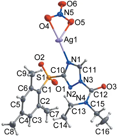
The reaction of silver nitrate and cafenstrole (N,N-diethyl-3-mesitylsulfonyl-1H -1,2,4-triazole-1-carboxamide), a triazole herbicide, leads to the title coordina-tion polymer, [Ag(NO3)(C16H22N4O3S)]n, whose asymmetric unit comprises one
cafenstrole ligand molecule, one AgIatom and one nitrate ion. The AgIatom, with a distorted trigonal–pyramidal environment, is coordinated by one nitrogen atom of a triazole ring, two oxygen atoms of a nitrate ion and one oxygen atom of a neighboring nitrate ion. The coordination bonds between silver and oxygen atoms give rise to a one-dimensional (1D) coordination polymer structure along [001]. The dihedral angle between the planes of the triazole and benzene rings is 87.13 (11). In the crystal, the coordination polymer is stabilized by C—H O
hydrogen bonds and C—H interactions, resulting in a three-dimensional architecture.
1. Chemical context
Recently, we have reported the crystal structure of the ligand cafenstrole (L; Kang et al., 2015). Cafenstrole is a triazole herbicide and has been used for rice cultivation as an inhibitor of the germination of grass weeds (Takahashi et al., 2001). Triazole derivatives have been investigated intensively over the years for pharmaceutical and agricultural purposes (Kumar et al., 2013; Zhanget al., 2014). It is very likely that triazole–metal interactions play a major role in the biological actions of triazole-containing drugs and agricultural chemicals. 1,2,4-Triazole and its derivatives have gained great attention as ligands to transition metals (Haasnoot, 2000). To under-stand the interactions of triazoles with metals, further research on the structures of triazole–metal compounds is of great necessity. Thus, our attention will be focused on the diversity of the coordination geometries of 1,2,4-triazole complexes with transition metal ions. Herein, we report the reaction of silver nitrate and cafenstrole to produce the title compound, which is a 1D silver(I) coordination polymer.
2. Structural commentary
The asymmetric unit of the title compound is shown in Fig. 1. It contains one Lligand and one silver nitrate ion. Reaction between silver nitrate and L afforded a 1D coordination polymer, in which the AgI atom has a distorted trigonal– pyramidal environment with one nitrogen atom (N1) [Ag1— N1 = 2.250 (3) A˚ ] and three oxygen atoms (O4, O5, O5i)
[Ag1—O4 = 2.708 (3), Ag1—O5 = 2.450 (3) and Ag1—O5i= 2.396 (3) A˚ ; symmetry code: (i) x + 1, y + 1, z 1
2], as
shown in Fig. 2.
Atom Ag1 lies almost in the plane constituted by atoms O5, N1, and O5i[deviation = 0.0436 (12) A˚ ]. The Ag1, O5, N1, and O5iatoms form a slightly distorted triangular basal plane with bond angles O5—Ag1—O5i = 106.52 (5), O5—Ag1—N1 = 118.75 (11) and O5i—Ag1—N1 = 134.63 (11). The apex atom,
O4, deviates considerably from the normal to the basal plane, as indicated by the O4—Ag1—N1 bond angle of 149.66 (10).
Other bond angles are 48.93 (10) and 67.18 (10) for O4—
Ag1—O5 and O4—Ag1—O5i, respectively. One oxygen atom of the nitrate ion (O6) is not bound to the AgIion, whereas the other two oxygen atoms of the nitrate ion (O4 and O5) are bound to the AgIion. One of the bound O atoms (O5) links
neighbouring AgIion ions, thus forming a 1D polymer along [001]. The triazole plane is rotated about the S1–C10 axis in the opposite direction in comparison with free cafenstrol (Kanget al., 2015). Thus, the diethyl amino group is located above the phenyl ring in the title compound, while that of free cafenstrol is placed outside the phenyl ring.
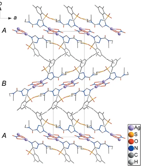
3. Supramolecular features
The O5 atom is bound to both Ag1 and neighboring Ag1ii [symmetry code: (ii)x+ 1,y+ 1,z+1
2], where the
neigh-bouring asymmetric unit is related to the asymmetric unit by 21symmetry, resulting in a 1D chain along [001] (Fig. 3). C—
H O hydrogen bonds between the 1D chains (yellow dashed lines) lead to the formation of layers parallel to (100). The layers are packed in an ABABpattern along [010] (Fig. 4). Weak intermolecular C—H interactions (black dashed lines) between the A andB layers generate a three-dimen-sional network structure (Fig. 4). Thus the structure of the AgI coordination polymer is stabilized by C13—H13B O2 and
research communications
Acta Cryst.(2016). E72, 1610–1613 Parket al. [Ag(NO
[image:2.610.311.563.70.264.2]3)(C16H22N4O3S)]
1611
Figure 2The coordination environment of the AgIatom in the title compound.
[Symmetry codes: (i)x+ 1,y+ 1,z1
[image:2.610.54.287.101.291.2]2; (ii)x+ 1,y+ 1,z+ 1 2.]
Figure 3
[image:2.610.66.273.466.704.2]The packing of the title compound showing chains along [001].
Figure 1
[image:2.610.316.566.570.726.2]C16—H16B O2 hydrogen bonds and weak intermolecular C8—H8C Cg1 (Cg1 is the centroid of the C1–C6 ring) interactions (Fig. 4 and Table 1).
4. Database survey
The crystal structure of cafenstrole has been reported (Kanget al., 2015). The crystal structure of a 1,2,3-thiadiazole compound containing a 1,2,4-triazole moiety, C15H14FN5O2S2,
has been determined by Min et al.(2014) whereas the struc-ture of a similar triazole herbicide, methyl 2-(1-diethylcarb-amoyl-1,2,4-triazole-3-ylsulfonyl)acetate, has been reported by Ohkata et al. (2002). The structure of 5-{4-cyclopropyl-5-[(3-fluorobenzyl)sulfinyl]-4H -1,2,4-triazol-3-yl}-4-methyl-1,2,3-thiadiazole (C15H14FN5OS2), was determined by Minet
al.(2015) and the crystal structure of
1-(mesityl-2-sulfonyl)-3-nitro-1,2,4-triazole has been determined by Kuroda et al.
(1982). The complex, [Pr(C7H5O3)2(NO3)(C12H8N2)]
-2C12H8N2, has a polymeric chain structure, where nitrate ions
show similar coordination bonds compared to those in the title compound, but with AgI ions replaced by with PrIII atoms (Wanget al., 2012).
5. Synthesis and crystallization
The title compound was prepared from a mixed solution of the cafenstrole ligand (0.05 g, 0.14 mmol) in acetone (5 mL) and Ag(NO3) (0.06 g, 0.35 mmol) in methanol (5 mL). The ligand
was purchased from the Dr Ehrenstorfer GmbH Company. Single crystals suitable for X-ray crystallography were obtained by slow evaporation of the solvent at room temperature after one week.
6. Refinement
Crystal data, data collection and structure refinement details are summarized in Table 2. All H atoms were positioned geometrically and refined using a riding model withd(C—H) = 0.98 A˚ , Uiso(H) = 1.5Ueq(C) for methyl group,d(C—H) =
0.99 A˚ , Uiso(H) = 1.2Ueq(C) for Csp 3
[image:3.610.314.561.92.395.2]—H and d(C—H) = 0.95 A˚ ,Uiso(H) = 1.2Ueq(C) for aromatic C—H.
Table 1
Hydrogen-bond geometry (A˚ ,).
Cg1 is the centroid of the C1–C6 ring.
D—H A D—H H A D A D—H A
C8—H8C Cg1i 0.98 2.66 3.614 (5) 166
C13—H13B O2ii 0.99 2.58 3.395 (4) 140
C16—H16B O2iii 0.98 2.52 3.412 (6) 152
Symmetry codes: (i)xþ1 2;yþ
3
2;z; (ii)xþ1;y;z; (iii)xþ1;y;zþ1.
Table 2
Experimental details.
Crystal data
Chemical formula [Ag(NO3)(C16H22N4O3S)]
Mr 520.31
Crystal system, space group Orthorhombic,Pna21
Temperature (K) 173
a,b,c(A˚ ) 9.0947 (2), 31.0133 (6), 7.1934 (1)
V(A˚3) 2028.95 (7)
Z 4
Radiation type MoK
(mm1) 1.14
Crystal size (mm) 0.480.100.02
Data collection
Diffractometer Bruker APEXII CCD
Absorption correction Multi-scan (SADABS; Bruker, 2014)
Tmin,Tmax 0.579, 0.746
No. of measured, independent and observed [I> 2(I)] reflections
17454, 4760, 4259
Rint 0.042
(sin/)max(A˚ 1
) 0.667
Refinement
R[F2> 2(F2)],wR(F2),S 0.030, 0.053, 0.98
No. of reflections 4760
No. of parameters 267
No. of restraints 1
H-atom treatment H-atom parameters constrained
max,min(e A˚
3) 0.65,0.39
Absolute structure Flackxdetermined using 1577 quotients [(I+)(I
)]/[(I+)+(I )] (Parsons et al., 2013)
Absolute structure parameter 0.003 (14)
[image:3.610.49.296.415.707.2]Computer programs:APEX2andSAINT(Bruker, 2014),SHELXS97andSHELXTL (Sheldrick, 2008),SHELXL2014(Sheldrick, 2015),DIAMOND(Brandenburg, 2010) andpublCIF(Westrip, 2010).
Figure 4
Acknowledgements
This work was supported by the National Research Founda-tion (NRF) of Korea, Grant funded by the Ministry of Education, Science and Technology (2014R1A1A4A01008346 and 2014R1A1A4A01009105).
References
Brandenburg, K. (2010).DIAMOND. Crystal Impact GbR, Bonn,
Germany.
Bruker (2014). APEX2, SAINT and SADABS. Bruker AXS Inc.,
Madison, Wisconsin, USA.
Haasnoot, J. G. (2000).Coord. Chem. Rev.200–202, 131–185. Kang, G., Kim, J., Park, H. & Kim, T. H. (2015).Acta Cryst.E71, o614. Kumar, R., Yar, M. S., Chaturvedi, S. & Srivastava, A. (2013).Int. J.
PharmTech Res.5, 1844–1869.
Kuroda, R., Sanderson, M. R., Neidle, S. & Reese, C. B. (1982).J. Chem. Soc. Perkin Trans. 2. pp. 617–620.
Min, L.-J., Tan, C.-X., Weng, J.-Q. & Liu, X.-H. (2014).Phosphorus Sulfur Silicon,189, 379–386.
Min, L.-J., Yang, M.-Y., Mu, J.-X., Sun, Z.-H., Tan, C.-X., Weng, J.-Q., Liu, X.-H. & Zhang, Y.-G. (2015).Phosphorus Sulfur Silicon,190, 1884–1892.
Ohkata, K., Yano, T., Kojima, S., Hiraga, Y., Yoshii, T. & Hori, M. (2002).Bull. Chem. Soc. Jpn,75, 567–574.
Parsons, S., Flack, H. D. & Wagner, T. (2013).Acta Cryst.B69, 249– 259.
Sheldrick, G. M. (2008).Acta Cryst. A64, 112–122. Sheldrick, G. M. (2015).Acta Cryst. C71, 3–8.
Takahashi, H., Ohki, A., Kanzaki, M., Tanaka, A., Sato, Y., Matthes, B., Bo¨ger, P. & Wakabayashi, K. (2001).J. Biosci.56, 781–786. Wang, P., Xu, D. & Wang, X. (2012).Acta Cryst.E68, m1148. Westrip, S. P. (2010).J. Appl. Cryst.43, 920–925.
Zhang, H.-Z., Damu, G. L. V., Cai, G.-X. & Zhou, C.-H. (2014).Curr. Org. Chem.18, 359–406.
research communications
Acta Cryst.(2016). E72, 1610–1613 Parket al. [Ag(NO
sup-1
Acta Cryst. (2016). E72, 1610-1613
supporting information
Acta Cryst. (2016). E72, 1610-1613 [https://doi.org/10.1107/S2056989016016662]
Crystal structure of
catena
-poly[[(
N
,
N
-diethyl-3-mesitylsulfonyl-1
H
-1,2,4-triazole-1-carboxamide-
κ
N
1)silver(I)]-
µ
-nitrato-
κ
3O
,
O
′
:
O
]
Hyunjin Park, Eunjin Kwon, Il Yoon and Jineun Kim
Computing details
Data collection: APEX2 (Bruker, 2014); cell refinement: SAINT (Bruker, 2014); data reduction: SAINT (Bruker, 2014); program(s) used to solve structure: SHELXS97 (Sheldrick, 2008); program(s) used to refine structure: SHELXL2014
(Sheldrick, 2015); molecular graphics: DIAMOND (Brandenburg, 2010); software used to prepare material for publication: SHELXTL (Sheldrick, 2008) and publCIF (Westrip, 2010).
catena-Poly[[(N,N-diethyl-3-mesitylsulfonyl-1H-1,2,4-triazole-1-carboxamide-κN1)silver(I)]-µ-nitrato-κ3O,O′:O]
Crystal data
[Ag(C16H22N4O3S)(NO3)]
Mr = 520.31
Orthorhombic, Pna21
a = 9.0947 (2) Å
b = 31.0133 (6) Å
c = 7.1934 (1) Å
V = 2028.95 (7) Å3
Z = 4
F(000) = 1056
Dx = 1.703 Mg m−3
Mo Kα radiation, λ = 0.71073 Å Cell parameters from 7087 reflections
θ = 2.3–27.1°
µ = 1.14 mm−1
T = 173 K Plate, colourless 0.48 × 0.10 × 0.02 mm
Data collection
Bruker APEXII CCD diffractometer
φ and ω scans
Absorption correction: multi-scan (SADABS; Bruker, 2014)
Tmin = 0.579, Tmax = 0.746
17454 measured reflections
4760 independent reflections 4259 reflections with I > 2σ(I)
Rint = 0.042
θmax = 28.3°, θmin = 2.3°
h = −12→11
k = −39→41
l = −9→9
Refinement
Refinement on F2
Least-squares matrix: full
R[F2 > 2σ(F2)] = 0.030
wR(F2) = 0.053
S = 0.98 4760 reflections 267 parameters 1 restraint
Hydrogen site location: inferred from neighbouring sites
H-atom parameters constrained
w = 1/[σ2(F
o2) + (0.0179P)2]
where P = (Fo2 + 2Fc2)/3
(Δ/σ)max = 0.002
Δρmax = 0.65 e Å−3
Δρmin = −0.39 e Å−3
Absolute structure: Flack x determined using 1577 quotients [(I+)-(I-)]/[(I+)+(I-)] (Parsons et
al., 2013)
supporting information
sup-2
Acta Cryst. (2016). E72, 1610-1613
Special details
Geometry. All esds (except the esd in the dihedral angle between two l.s. planes) are estimated using the full covariance matrix. The cell esds are taken into account individually in the estimation of esds in distances, angles and torsion angles; correlations between esds in cell parameters are only used when they are defined by crystal symmetry. An approximate (isotropic) treatment of cell esds is used for estimating esds involving l.s. planes.
Fractional atomic coordinates and isotropic or equivalent isotropic displacement parameters (Å2)
x y z Uiso*/Ueq
Ag1 0.62392 (3) 0.52477 (2) 0.76161 (6) 0.03314 (9)
S1 0.73402 (9) 0.64406 (3) 0.83512 (13) 0.01964 (19)
O1 0.6829 (3) 0.66757 (8) 0.9943 (4) 0.0300 (7)
O2 0.6265 (2) 0.62494 (9) 0.7166 (4) 0.0309 (8)
O3 1.1685 (2) 0.53847 (7) 1.2570 (5) 0.0290 (5)
O4 0.3336 (4) 0.51667 (11) 0.6854 (5) 0.0486 (9)
O5 0.4227 (3) 0.49348 (10) 0.9434 (4) 0.0382 (7)
O6 0.1895 (3) 0.48605 (10) 0.8835 (5) 0.0424 (8)
N1 0.8072 (3) 0.55871 (9) 0.9126 (4) 0.0206 (7)
N2 0.9749 (3) 0.60970 (9) 0.9892 (4) 0.0187 (6)
N3 1.0242 (3) 0.56942 (9) 1.0338 (4) 0.0183 (6)
N4 1.2736 (3) 0.58767 (9) 1.0626 (4) 0.0206 (7)
N5 0.3120 (4) 0.49853 (10) 0.8351 (5) 0.0266 (8)
C1 0.8614 (3) 0.67468 (11) 0.7068 (5) 0.0205 (9)
C2 0.9148 (3) 0.71461 (10) 0.7730 (7) 0.0218 (7)
C3 1.0110 (4) 0.73701 (13) 0.6569 (6) 0.0286 (9)
H3 1.0465 0.7642 0.6973 0.034*
C4 1.0571 (4) 0.72185 (14) 0.4873 (6) 0.0300 (9)
C5 1.0073 (4) 0.68155 (14) 0.4304 (6) 0.0303 (10)
H5 1.0412 0.6703 0.3152 0.036*
C6 0.9102 (4) 0.65722 (12) 0.5352 (5) 0.0232 (8)
C7 0.8791 (4) 0.73432 (14) 0.9579 (6) 0.0353 (10)
H7A 0.9450 0.7587 0.9811 0.053*
H7B 0.8919 0.7127 1.0559 0.053*
H7C 0.7769 0.7444 0.9576 0.053*
C8 1.1603 (5) 0.74778 (18) 0.3669 (7) 0.0526 (14)
H8A 1.1032 0.7677 0.2896 0.079*
H8B 1.2166 0.7283 0.2868 0.079*
H8C 1.2279 0.7642 0.4459 0.079*
C9 0.8679 (5) 0.61345 (14) 0.4610 (6) 0.0391 (11)
H9A 0.7697 0.6150 0.4054 0.059*
H9B 0.8673 0.5925 0.5630 0.059*
H9C 0.9391 0.6044 0.3665 0.059*
C10 0.8450 (3) 0.60100 (11) 0.9190 (5) 0.0177 (8)
C11 0.9224 (4) 0.53946 (12) 0.9906 (5) 0.0216 (8)
H11 0.9315 0.5094 1.0124 0.026*
C12 1.1648 (4) 0.56330 (12) 1.1294 (5) 0.0193 (8)
C13 1.2803 (4) 0.60769 (12) 0.8765 (6) 0.0254 (9)
sup-3
Acta Cryst. (2016). E72, 1610-1613
H13B 1.3697 0.5974 0.8116 0.030*
C14 1.2828 (4) 0.65612 (13) 0.8847 (7) 0.0365 (11)
H14A 1.1910 0.6666 0.9400 0.055*
H14B 1.2929 0.6678 0.7587 0.055*
H14C 1.3661 0.6656 0.9608 0.055*
C15 1.4092 (4) 0.58914 (14) 1.1768 (6) 0.0319 (10)
H15A 1.4890 0.6026 1.1035 0.038*
H15B 1.4398 0.5593 1.2073 0.038*
C16 1.3886 (5) 0.6139 (2) 1.3531 (8) 0.0642 (17)
H16A 1.3571 0.6433 1.3238 0.096*
H16B 1.4816 0.6148 1.4217 0.096*
H16C 1.3134 0.5998 1.4294 0.096*
Atomic displacement parameters (Å2)
U11 U22 U33 U12 U13 U23
Ag1 0.01833 (12) 0.03174 (15) 0.04934 (19) −0.00517 (11) −0.00399 (18) −0.01117 (19)
S1 0.0136 (4) 0.0221 (4) 0.0232 (5) 0.0013 (3) 0.0017 (4) 0.0027 (4)
O1 0.0276 (14) 0.0292 (15) 0.0333 (18) 0.0050 (11) 0.0131 (13) 0.0002 (13)
O2 0.0178 (11) 0.0376 (15) 0.037 (2) −0.0045 (10) −0.0073 (12) 0.0054 (13)
O3 0.0248 (12) 0.0308 (13) 0.0315 (14) −0.0015 (9) −0.0036 (18) 0.0114 (18)
O4 0.064 (2) 0.044 (2) 0.037 (2) −0.0106 (17) 0.0084 (16) 0.0004 (17)
O5 0.0227 (13) 0.057 (2) 0.0346 (19) 0.0013 (14) −0.0001 (14) −0.0022 (16)
O6 0.0200 (14) 0.053 (2) 0.054 (2) −0.0094 (13) 0.0092 (14) −0.0063 (16)
N1 0.0154 (14) 0.0192 (16) 0.0272 (19) −0.0024 (12) 0.0000 (13) −0.0001 (14)
N2 0.0189 (14) 0.0181 (15) 0.0192 (17) 0.0011 (11) −0.0022 (13) 0.0017 (13)
N3 0.0164 (14) 0.0183 (16) 0.0203 (17) −0.0009 (11) −0.0002 (13) 0.0040 (14)
N4 0.0152 (14) 0.0212 (16) 0.0253 (19) −0.0034 (12) −0.0062 (13) 0.0060 (14)
N5 0.0290 (18) 0.0226 (18) 0.028 (2) −0.0009 (14) 0.0059 (16) −0.0059 (16)
C1 0.0175 (16) 0.0221 (18) 0.022 (2) 0.0025 (13) 0.0002 (15) 0.0049 (14)
C2 0.0220 (15) 0.0231 (16) 0.0204 (19) 0.0025 (12) −0.006 (2) 0.003 (2)
C3 0.029 (2) 0.028 (2) 0.029 (2) −0.0089 (17) −0.0079 (18) 0.0088 (19)
C4 0.0210 (19) 0.045 (3) 0.024 (2) −0.0072 (17) −0.0035 (18) 0.013 (2)
C5 0.026 (2) 0.045 (3) 0.019 (2) 0.0022 (18) 0.0054 (18) 0.0061 (19)
C6 0.0224 (18) 0.026 (2) 0.021 (2) 0.0042 (15) 0.0016 (16) 0.0023 (17)
C7 0.044 (2) 0.025 (2) 0.037 (3) −0.0028 (18) −0.001 (2) −0.0045 (19)
C8 0.043 (3) 0.078 (4) 0.037 (3) −0.026 (3) 0.002 (2) 0.019 (3)
C9 0.059 (3) 0.031 (2) 0.027 (3) −0.004 (2) 0.008 (2) −0.008 (2)
C10 0.0126 (16) 0.0185 (18) 0.022 (2) 0.0003 (13) 0.0030 (15) 0.0006 (16)
C11 0.0222 (18) 0.0211 (18) 0.021 (2) −0.0047 (14) 0.0032 (17) 0.0029 (16)
C12 0.0166 (17) 0.0185 (19) 0.023 (2) 0.0010 (13) 0.0000 (16) 0.0005 (16)
C13 0.0181 (17) 0.031 (2) 0.027 (2) −0.0019 (15) 0.0039 (16) 0.0053 (17)
C14 0.030 (2) 0.027 (2) 0.052 (3) −0.0036 (17) 0.000 (2) 0.016 (2)
C15 0.0199 (19) 0.033 (2) 0.042 (3) −0.0039 (17) −0.0091 (18) 0.007 (2)
supporting information
sup-4
Acta Cryst. (2016). E72, 1610-1613
Geometric parameters (Å, º)
Ag1—N1 2.250 (3) C4—C5 1.391 (6)
Ag1—O5i 2.396 (3) C4—C8 1.509 (5)
Ag1—O5 2.450 (3) C5—C6 1.385 (5)
S1—O2 1.426 (3) C5—H5 0.9500
S1—O1 1.435 (3) C6—C9 1.509 (5)
S1—C1 1.760 (4) C7—H7A 0.9800
S1—C10 1.779 (3) C7—H7B 0.9800
O3—C12 1.198 (5) C7—H7C 0.9800
O4—N5 1.231 (4) C8—H8A 0.9800
O5—N5 1.282 (4) C8—H8B 0.9800
O5—Ag1ii 2.396 (3) C8—H8C 0.9800
O6—N5 1.230 (4) C9—H9A 0.9800
N1—C11 1.330 (5) C9—H9B 0.9800
N1—C10 1.357 (4) C9—H9C 0.9800
N2—C10 1.313 (4) C11—H11 0.9500
N2—N3 1.365 (4) C13—C14 1.503 (5)
N3—C11 1.348 (4) C13—H13A 0.9900
N3—C12 1.465 (4) C13—H13B 0.9900
N4—C12 1.334 (4) C14—H14A 0.9800
N4—C13 1.477 (5) C14—H14B 0.9800
N4—C15 1.483 (5) C14—H14C 0.9800
C1—C2 1.413 (5) C15—C16 1.494 (6)
C1—C6 1.419 (5) C15—H15A 0.9900
C2—C3 1.395 (5) C15—H15B 0.9900
C2—C7 1.499 (7) C16—H16A 0.9800
C3—C4 1.373 (6) C16—H16B 0.9800
C3—H3 0.9500 C16—H16C 0.9800
N1—Ag1—O5i 134.63 (11) H7A—C7—H7C 109.5
N1—Ag1—O5 118.75 (11) H7B—C7—H7C 109.5
O5i—Ag1—O5 106.52 (5) C4—C8—H8A 109.5
O2—S1—O1 117.78 (16) C4—C8—H8B 109.5
O2—S1—C1 111.23 (17) H8A—C8—H8B 109.5
O1—S1—C1 110.96 (16) C4—C8—H8C 109.5
O2—S1—C10 106.22 (17) H8A—C8—H8C 109.5
O1—S1—C10 107.14 (17) H8B—C8—H8C 109.5
C1—S1—C10 102.10 (16) C6—C9—H9A 109.5
N5—O5—Ag1ii 118.1 (2) C6—C9—H9B 109.5
N5—O5—Ag1 102.3 (2) H9A—C9—H9B 109.5
Ag1ii—O5—Ag1 137.37 (12) C6—C9—H9C 109.5
C11—N1—C10 102.7 (3) H9A—C9—H9C 109.5
C11—N1—Ag1 125.2 (2) H9B—C9—H9C 109.5
C10—N1—Ag1 131.0 (2) N2—C10—N1 116.1 (3)
C10—N2—N3 101.4 (3) N2—C10—S1 119.1 (3)
C11—N3—N2 110.5 (3) N1—C10—S1 124.8 (3)
sup-5
Acta Cryst. (2016). E72, 1610-1613
N2—N3—C12 121.0 (3) N1—C11—H11 125.4
C12—N4—C13 126.5 (3) N3—C11—H11 125.4
C12—N4—C15 115.7 (3) O3—C12—N4 128.3 (3)
C13—N4—C15 117.1 (3) O3—C12—N3 117.9 (3)
O6—N5—O4 122.4 (4) N4—C12—N3 113.9 (3)
O6—N5—O5 120.0 (4) N4—C13—C14 112.6 (3)
O4—N5—O5 117.6 (4) N4—C13—H13A 109.1
C2—C1—C6 121.3 (3) C14—C13—H13A 109.1
C2—C1—S1 121.5 (3) N4—C13—H13B 109.1
C6—C1—S1 117.2 (3) C14—C13—H13B 109.1
C3—C2—C1 116.8 (4) H13A—C13—H13B 107.8
C3—C2—C7 117.6 (3) C13—C14—H14A 109.5
C1—C2—C7 125.6 (3) C13—C14—H14B 109.5
C4—C3—C2 123.6 (4) H14A—C14—H14B 109.5
C4—C3—H3 118.2 C13—C14—H14C 109.5
C2—C3—H3 118.2 H14A—C14—H14C 109.5
C3—C4—C5 118.0 (4) H14B—C14—H14C 109.5
C3—C4—C8 121.2 (4) N4—C15—C16 112.4 (4)
C5—C4—C8 120.8 (4) N4—C15—H15A 109.1
C6—C5—C4 122.5 (4) C16—C15—H15A 109.1
C6—C5—H5 118.8 N4—C15—H15B 109.1
C4—C5—H5 118.8 C16—C15—H15B 109.1
C5—C6—C1 117.7 (4) H15A—C15—H15B 107.9
C5—C6—C9 117.4 (4) C15—C16—H16A 109.5
C1—C6—C9 124.8 (3) C15—C16—H16B 109.5
C2—C7—H7A 109.5 H16A—C16—H16B 109.5
C2—C7—H7B 109.5 C15—C16—H16C 109.5
H7A—C7—H7B 109.5 H16A—C16—H16C 109.5
C2—C7—H7C 109.5 H16B—C16—H16C 109.5
C10—N2—N3—C11 −0.7 (4) N3—N2—C10—N1 −0.7 (4)
C10—N2—N3—C12 −176.0 (3) N3—N2—C10—S1 −179.2 (2)
Ag1ii—O5—N5—O6 −14.1 (4) C11—N1—C10—N2 1.8 (4)
Ag1—O5—N5—O6 179.9 (3) Ag1—N1—C10—N2 −166.7 (3)
Ag1ii—O5—N5—O4 164.7 (3) C11—N1—C10—S1 −179.8 (3)
Ag1—O5—N5—O4 −1.3 (4) Ag1—N1—C10—S1 11.7 (5)
O2—S1—C1—C2 140.4 (3) O2—S1—C10—N2 161.7 (3)
O1—S1—C1—C2 7.2 (3) O1—S1—C10—N2 −71.6 (3)
C10—S1—C1—C2 −106.7 (3) C1—S1—C10—N2 45.1 (3)
O2—S1—C1—C6 −40.6 (3) O2—S1—C10—N1 −16.7 (4)
O1—S1—C1—C6 −173.8 (3) O1—S1—C10—N1 110.0 (3)
C10—S1—C1—C6 72.3 (3) C1—S1—C10—N1 −133.3 (3)
C6—C1—C2—C3 3.5 (5) C10—N1—C11—N3 −2.1 (4)
S1—C1—C2—C3 −177.6 (3) Ag1—N1—C11—N3 167.3 (2)
C6—C1—C2—C7 −175.3 (3) N2—N3—C11—N1 1.9 (4)
S1—C1—C2—C7 3.6 (5) C12—N3—C11—N1 176.8 (3)
C1—C2—C3—C4 −1.4 (5) C13—N4—C12—O3 160.4 (4)
supporting information
sup-6
Acta Cryst. (2016). E72, 1610-1613
C2—C3—C4—C5 −1.3 (6) C13—N4—C12—N3 −21.2 (5)
C2—C3—C4—C8 179.4 (4) C15—N4—C12—N3 168.4 (3)
C3—C4—C5—C6 2.1 (6) C11—N3—C12—O3 −39.4 (5)
C8—C4—C5—C6 −178.6 (4) N2—N3—C12—O3 135.0 (4)
C4—C5—C6—C1 −0.1 (6) C11—N3—C12—N4 142.0 (4)
C4—C5—C6—C9 −178.3 (4) N2—N3—C12—N4 −43.6 (4)
C2—C1—C6—C5 −2.8 (5) C12—N4—C13—C14 116.8 (4)
S1—C1—C6—C5 178.2 (3) C15—N4—C13—C14 −73.0 (4)
C2—C1—C6—C9 175.2 (4) C12—N4—C15—C16 −70.8 (5)
S1—C1—C6—C9 −3.8 (5) C13—N4—C15—C16 117.9 (4)
Symmetry codes: (i) −x+1, −y+1, z−1/2; (ii) −x+1, −y+1, z+1/2.
Hydrogen-bond geometry (Å, º)
Cg1 is the centroid of the C1–C6 ring.
D—H···A D—H H···A D···A D—H···A
C8—H8C···Cg1iii 0.98 2.66 3.614 (5) 166
C13—H13B···O2iv 0.99 2.58 3.395 (4) 140
C16—H16B···O2v 0.98 2.52 3.412 (6) 152