Original Article
Downregulation of lncRNA NEAT1 inhibits mouse
mesangial cell proliferation, fibrosis, and inflammation
but promotes apoptosis in diabetic nephropathy
Jian Ma, Na Zhao, Likun Du, Yang Wang
Department of Endocrinology, First Affiliated Hospital, Heilongjiang University of Chinese Medicine, Harbin, China
Received December 5, 2018; Accepted January 18, 2019; Epub April 1, 2019; Published April 15, 2019
Abstract: This study aimed to investigate the effect of long non-coding RNA nuclear enriched abundant transcript 1
(lnc-NEAT1) on mouse mesangial cells (MMCs) proliferation, apoptosis, fibrosis as well as inflammation in diabetic
nephropathy (DN). MMCs (SV40 MES13 cells) were cultured under 30 mM glucose to construct DN cellular model (high glucose (HG) group); meanwhile, MMCs cultured under 5.6 mM glucose (normal glucose (NG) group) and 5.6 mM glucose plus 24.4 mM 3-O-methyl-D-glucose (osmotic control (OC) group) served as controls, and lnc-NEAT1 expression was determined by qPCR assay. Lnc-NEAT1 interference plasmids and control interference plasmids
were transfected into DN cellular model as Sh-NEAT1 group and Sh-NC group. Cell proliferation, apoptosis, fibrosis, and inflammation were detected using Counting Kit-8, Annexin V/propidium iodide, western blot and quantitative
polymerase chain reaction assays. Lnc-NEAT1 expression was elevated in HG group compared to NG group and OC group. Cell proliferation was decreased, and proliferative marker protein Cyclin D1 and proliferating cell nuclear antigen expressions also decreased in Sh-NEAT1 group compared to Sh-NC group. For cell apoptosis, apoptosis rate was increased, and apoptotic protein Cleaved Caspase3 expression enhanced but anti-apoptosis protein Bcl-2
expression decreased in Sh-NEAT1 group compared to Sh-NC group. For fibrosis markers (including fibronectin and collagen I) and inflammatory cytokines (including tumor necrosis factor-α, interleukin-1β and interleukin-6), their
expressions were reduced in Sh-NEAT1 group compared to Sh-NC group. Lnc-NEAT1 is overexpressed, and its
down-regulation inhibits cell proliferation, fibrosis, and inflammation but promotes cell apoptosis in HG-induced MMCs
DN cellular model.
Keywords: Long non-coding RNA, enriched abundant transcript 1, diabetic nephropathy, proliferation, apoptosis,
fibrosis, inflammatory cytokines
Introduction
Diabetic nephropathy (DN), with the functional derangement and structural remodeling of kid-ney triggered by hyperglycemic injury, is one of the most frequent microvascular complications of diabetes patients and the leading cause of end-stage renal disease worldwide, and is char-acterized by a series of pathologic abnormali-ties such as glomerular hypertrophy, mesangial proliferation, thickening of the glomerular base-ment membrane and accumulation of the extracellular matrix [1-6]. According torecent reports, around 30%-40% diabetes patients tend to develop DN, and about 50% of DN patients would further develop end-stage renal disease (ESRD), which has the requirement of dialysis or renal transplantation [7-9]. Prev-
Long non-coding RNA (lncRNA), a subgroup of RNAs with insufficient protein coding capability, is composed of more than 200 nucleotides [12, 13]. Increasing studies have shown that lncRNA plays crucial roles in multiple biologic process-es (including regulation of gene exprprocess-ession, recruitment of chromatin modification, X chro -mosome inactivation, chro-mosome recombina-tion and protein folding). Also, some of them are identified as important regulators in the pathologic processes of several endocrine and metabolic diseases, such as diabetes, osteo-porosis and fatty liver disease [14-16]. LncRNA nuclear enriched abundant transcript 1 (lnc-NEAT1), located on chromosome 11q13.1, works as an important structural component of paraspeckles [17]. Majority of previous studies focus on the role of lnc-NEAT1 in different carci-nomas, and identify its ability to affect cell ac- tivities via targeting miRNAs or regulating sig-naling pathways in cancer progression [18-20]. Recently, some recent studies disclose lnc-NEAT1 is aberrantly expressed in diabetes mice, moreover, there are some studies show that lnc-NEAT1 has proinflammatory influence through affecting inflammatory pathways such as activation of mitogen-activated protein kinase (MAPK) pathways or Toll-like receptor 3 (TLR3)-p38 pathway in some inflammatory dis -eases [21, 22]. Considering inflammation is a key pathogenic mechanism in DN and lnc-NEAT1 is involved in inflammatory processes, we hypothesized that lnc-NEAT1 might partici-pate in the pathology of DN, while the underly-ing mechanism of lnc-NEAT1 in DN was not deeply investigated [23, 24]. In this study, we constructed a DN cellular model with mouse mesangial cells (MMCs) under high glucose (HG) condition, and investigated the effect of lnc-NEAT1 on cell proliferation, apoptosis, fibro -sis as well as inflammation in DN.
Materials and methods
Cells source
MMCs cell line (SV40 MES13 cells) was pur-chased from Cell Bank of Type Culture Collection of Chinese Academy of Sciences (Shanghai, China).
Cells culture
SV40 MES13 cells were cultured in 90% Dulbecco’s modified eagle medium (DMEM)
(Gibco, USA) supplemented with 10% fetal bovine serum (FBS) (Gibco, USA) in a humidified incubator under 95% air and 5% CO2 at 37°C. Measurement of lnc-NEAT1 expression in SV40 MES13 cells under HG condition
30 mM glucose (Sigma, USA), 5.6 mM glucose, 5.6 mM glucose plus 24.4 mM 3-O-methyl-D-glucose (Wako, Japan) were added to treat SV40 MES13 cells for 96 h, and these cells were divided into three groups accordingly as HG group, normal glucose group (NG group) and osmotic control group (OC group). Quantitative polymerase chain reaction (qPCR) was subsequently performed to measure lnc-NEAT1 expression in each group.
Construction and transfection of lnc-NEAT1 interference plasmids
Lnc-NEAT1 interference plasmids and control interference plasmids were constructed by Shanghai GenePharma Bio-Tech Company (Shanghai, China). These plasmids were trans-fected into SV40 MES13 cells under HG condi-tion as Sh-NEAT1 and Sh-NC groups.
Measurement of cell proliferation
Cell proliferation ability was measured using Counting Kit-8 (CCK-8) (Sangon, China) at 0 h, 24 h, 48 h and 72 h after transfection accord-ing to the instructions of manufacturer. In addi-tion, cell proliferative markers (Cyclin D1 and proliferating cell nuclear antigen (PCNA)) were also measured using qPCR and western blot at 48 h after transfection.
Measurement of apoptosis
Cell apoptosis rate was measured using Annexin V (AV) apoptosis detection kit with propidium iodide (PI) (BD, USA) at 24 h after transfection according to the instructions of manufacturer. In addition, apoptotic markers (Cleaved Caspase3 (C-Caspase3) and Bcl-2) were also measured using western blot at 24 h after transfection.
Measurement of cell fibrosis
Measurement of inflammatory cytokines
Inflammatory cytokines including tumor necro -sis factor-α (TNF-α), interleukin (IL)-1β and IL-6 were measured using qPCR and western blot at 24 h after transfection.
Western blot
Total proteins were extracted from each group of cells with 1 mL RIPA (Sigma, USA) on ice and shocked every 5 mins during 30 mins for com-plete pyrolysis. Then, protein concentration was determined by Bicinchoninic Acid Kit (Sigma, USA), followed by thermal denaturation that performed at 98°C for 5 mins. Subse- quently, 20 μg proteins were added to NuPAGE Bis-Tris protein gel (Invitrogen, USA), and after electrophoresis was finished, proteins were transferred to polyvinylidene fluoride mem -branes (Milipore, USA). 5% skim milk was used
3 mins, followed by 40 cycles of 95°C for 5 s, 61°C for 10 s, and then 72°C for 30 s. Finally, the qPCR results were calculated by 2-ΔΔCt for-mula, and glyceraldehyde-3-phosphate dehy-drogenase (GAPDH) was used as the internal references. The primers used in qPCR assay were listed in Table 2.
Statistics
[image:3.612.91.389.85.277.2]Statistics was conducted using SPSS 21.0 Software (IBM, USA) and graphs were drawn using GraphPad 6.01 Software (GraphPad Software, USA). Data were presented as mean ± standard deviation. Comparison among groups was detected using One-way ANOVA fol-lowed by Turkey’s multiple comparison test, and comparison between two groups was detected using t test. P value < 0.05 was con-sidered as significant in this study.
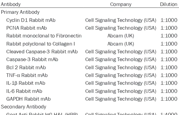
Table 1. Antibodies applied in western blot
Antibody Company Dilution
Primary Antibody
Cyclin D1 Rabbit mAb Cell Signaling Technology (USA) 1:1000 PCNA Rabbit mAb Cell Signaling Technology (USA) 1:1000 Rabbit monoclonal to Fibronectin Abcam (UK) 1:1000 Rabbit polyclonal to Collagen I Abcam (UK) 1:1000 Cleaved Caspase-3 Rabbit mAb Cell Signaling Technology (USA) 1:1000 Caspase-3 Rabbit mAb Cell Signaling Technology (USA) 1:1000 Bcl 2 Rabbit mAb Cell Signaling Technology (USA) 1:1000
TNF-α Rabbit mAb Cell Signaling Technology (USA) 1:1000
IL-1β Rabbit mAb Cell Signaling Technology (USA) 1:1000 IL-6 Rabbit mAb Cell Signaling Technology (USA) 1:1000 GAPDH Rabbit mAb Cell Signaling Technology (USA) 1:1000 Secondary Antibody
Goat Anti-Rabbit IgG H&L (HRP) Cell Signaling Technology (USA) 1:4000
Table 2. Primers applied in qPCR
Gene Forward Primer (5’-3’) Reverse Primer (5’-3’) Lnc-NEAT1 TGAGTAGTGGAAGCAGGAGGAT GGAGGCAAGGACGAGACAGA Cyclin D1 CCAGAGGCGGATGAGAACAAG GCGGTAGCAGGAGAGGAAGT PCNA GCCGAGACCTTAGCCACATTG ATGGTTACCGCCTCCTCTTCTT FN CAGTAGAAGGCAGTAGCACAGA CCAGACACCACACTATCAGGAG Collagen I CTCGTGGATTGCCTGGAACA GCACCAACAGCACCATCGT
TNF-α CGTGGAACTGGCAGAAGAGG TCAGTAGACAGAAGAGCGTGGT
IL-1β TCTCGCAGCAGCACATCAAC TGTTCATCTCGGAGCCTGTAGT IL-6 CTTCCATCCAGTTGCCTTCTTG GTAATTAAGCCTCCGACTTGTGAA GAPDH GAGTCCACTGGCGTCTTCAC ATCTTGAGGCTGTTGTCATACTTCT
to block the membranes at 37°C for 1 h, and mem-branes were incubated with the corresponding pri-mary antibodies overnight at 4°C. Then, membranes were further incubated with secondary antibodies at room temperature for 2 h. Lastly, the bands were visualized using EasyBlot ECL kit (Sangon, China). The antibodies applied in western blot assay are list-ed in Table 1.
qPCR
[image:3.612.90.388.315.451.2]Results
Comparison of lnc-NEAT1 expression among HG group, OC group and NG group
Lnc-NEAT1 expression was remarkably elevat-ed in HG group comparelevat-ed to NG group (P < 0.001), and it was also greatly higher in HG group than that in OC group (P < 0.001), while no difference of lnc-NEAT1 expression was found between OC group and NG group. These
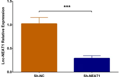
results indicated that lnc-NEAT1 was overex-pressed in DN cellular model (Figure 1). Comparison of lnc-NEAT1 expression after plasmids transfection in DN cellular model
In order to determine the effect of lnc-NEAT1 in DN, we transfected lnc-NEAT1 interference plasmids and control plasmids into DN cellular model. Lnc-NEAT1 expression in Sh-NEAT1 group was dramatically reduced in Sh-NEAT1 group compared to Sh-NC group (P < 0.001) (Figure 2), suggesting the successful transfec-tion of lnc-NEAT1 interference plasmids into DN cellular model.
Comparison of cell proliferation between Sh-NEAT1 group and Sh-NC group
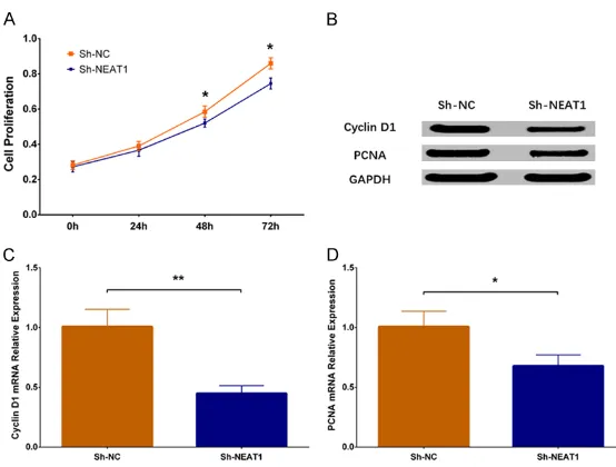
In order to investigate the function of lnc-NEAT1 downregulation on cell proliferation in DN, we performed CCK-8 assay and found that cell pro -liferation was decreased in Sh-NEAT1 group at 48 h (P < 0.05) and 72 h (P < 0.05) compared to Sh-NC group (Figure 3A). Besides, western blot disclosed that expressions of both Cyclin D1 protein and PCNA protein were reduced in Sh-NEAT1 group compared to Sh-NC group (Figure 3B), and qPCR assay displayed that expressions of Cyclin D1 mRNA and PCNA mRNA were lower in Sh-NEAT1 group compared to Sh-NC group (P < 0.01) (Figure 3C, 3D). These results suggested that lnc-NEAT1 down-regulation repressed cell proliferation in the DN cellular model.
Comparison of cell apoptosis between Sh-NEAT1 group and Sh-NC group
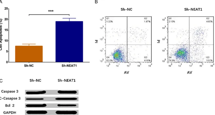
Cell apoptosis rate was increased in Sh-NEAT1 group than that in Sh-NC group (P < 0.001) (Figure 4A, 4B). Western blot revealed that expression of apoptotic protein C-Caspase3 was enhanced in Sh-NEAT1 group compared to Sh-NC group, while expression of anti-apopto-sis protein Bcl 2 was decreased in Sh-NEAT1 group compared to Sh-NC group (Figure 4C). These results indicated that lnc-NEAT1 down-regulation inhibited cell proliferation in a DN cellular model.
Comparison of cell fibrosis between Sh-NEAT1 group and Sh-NC group
[image:4.612.90.286.72.212.2]Expression of cell fibrosis marker mRNAs were measured by qPCR assay, which displayed that Figure 1. Lnc-NEAT1 expression in NG group, OC
group and HG group. Lnc-NEAT1 expression was un-differentiated in OC group compared to NG group, while it was higher in HG group compared to NG group and elevated in HG group compared to OC group. Lnc-NEAT1, long non-coding RNA nuclear en-riched abundant transcript 1; NG, normal glucose; OC, osmotic control; HG, high glucose. Comparison among groups was detected using One-way ANOVA followed by Turkey’s multiple comparison test. P
val-ue < 0.05 was considered significant. ***P < 0.001.
Figure 2. Lnc-NEAT1 expression in DN cellular model after transfection with lnc-NEAT1 interference plas-mids. Lnc-NEAT1 expression was decreased in Sh-NEAT1 group compared to Sh-NC group. Lnc-Sh-NEAT1, long non-coding RNA nuclear enriched abundant transcript 1. Comparison between two groups was determined by t test. P value < 0.05 was considered
[image:4.612.90.286.356.482.2]FN mRNA expression (P < 0.05) (Figure 5A) and collagen I mRNA expression (P < 0.01) (Figure 5B) were decreased in Sh-NEAT1 group com-pared to Sh-NC group. Besides, expressions of fibrotic proteins were assessed by western blot, which showed that FN protein expression and collagen I protein expression were also reduced in the Sh-NEAT1 group compared to the Sh-NC group (Figure 5C), suggesting that lnc-NEAT1 downregulation inhibited fibrosis in a DN cellular model.
Comparison of inflammatory cytokines expres-sions between Sh-NEAT1 group and Sh-NC group
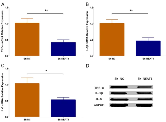
The qPCR assay revealed that expressions of inflammatory cytokines including TNF-α (P < 0.01), IL-1β (P < 0.01) and IL-6 (P < 0.05) were reduced in the Sh-NEAT1 group compared to Sh-NC group (Figure 6A-C). Furthermore, west-ern blot disclosed that protein expressions of TNF-α, IL-1β and IL-6 were attenuated in Sh-NEAT1 group than those in the Sh-NC group (Figure 6D). All these results indicated that lnc-NEAT1 downregulation lowered the inflamma -tion level in a DN cellular model.
Discussion
In this study, we found that: (1) lnc-NEAT1 was
in DN mice and HG-treated MMCs, and it pro-motes glomerular fibrosis through upregulating expression of pro-fibrotic factor transforming growth factor β1 (TGFβ1) [25]. Besides, lnc-LINC00968 represses p21 in DN cellular model (MMCs under HG condition) via binding with zeste homolog 2 (EZH2), thereby facilitates cell proliferation and fibrosis [32]. Furthermore, lnc-Gm4419 knockdown inhibits cell proliferation, represses expressions of pro-inflammatory cytokines (including monocyte chemoattrac-tant protein-1 (mcp-1), IL-1β and TNF-α), and decreases expressions of fibrosis biomarkers (including FN and collagen IV) through reducing activation of nuclear factor kappa light-chain enhancer of activated B cells (NF-κB)/NACHT, leucine-rich repeats (LRR) and (pyrin domain) PYD domain-containing protein 3 (NLRP3) inflammasome signaling pathway in DN cellular model (MMCs under HG condition) [33]. These previous studies reveal that lncRNA may be involved in the pathology of DN and they pres-ent potpres-entials to serve as treatmpres-ent targets. Lnc-NEAT1, which is transcribed by RNA poly-merase II from a common promoter and widely expressed in mammalian cells, functions as scaffolds of nuclear bodies [17, 34-36]. Re- cently, emerging evidence shows lnc-NEAT1 plays a crucial role in regulating gene expres-Figure 3. Cell proliferation in Sh-NEAT1 group and Sh-NC group. Compared
to Sh-NC group, cell proliferation rate was lower in Sh-NEAT1 group (A). Ex-pressions of cell proliferative markers including Cyclin D1 and PCNA were decreased in Sh-NEAT1 group (B-D). NEAT1, nuclear enriched abundant tran-script 1; PCNA, proliferating cell nuclear antigen. Comparison between two groups were determined by t test. P value < 0.05 was considered significant. *P < 0.05; **P < 0.01.
ssed cell proliferation but enhanced apoptosis in a DN cellular model; (2) lnc-NEAT1 downregulation decreased fi-brotic marker expression in a DN cellular model; (3) lnc-NEAT1 downregulation redu- ced inflammatory cytokine expressions in a DN cellular model.
[image:5.612.91.368.69.279.2]logic processes by affecting cell proliferation or apoptosis in several diseases such as diabe-tes, traumatic brain injury, as well as multiple cancers [23, 37-40]. Whereas, limited informa-tion of lnc-NEAT1 in diabetes was found. One previous study conducted microarray analyses on diabetic Akita mice heart and displayed that lnc-NEAT1 expression is downregulated [23]. However, another study disclosed that lnc-NEAT1 is overexpressed and it promotes cell
[image:6.612.95.521.76.305.2]apoptosis as well as autophagy in diabetic rats with myocardial ischemia reperfusion injury [41]. Hence, these previous data reveal that the influence of lnc-NEAT1 in diabetes or diabetic complications is still controversial, and the underlying mechanism of lnc-NEAT1 in DN is rarely disclosed. Since inflammation is the important pathogenetic mechanism in DN and lnc-NEAT1 has potential to serve as a pro-inflammatory factor; and lnc-NEAT1 expression Figure 4. Cell apoptosis in Sh-NEAT1 group and Sh-NC group. As to cell apoptosis, AV/PI assay revealed that cell apoptosis rate was elevated in Sh-NEAT1 group compared to Sh-NC group (A, B). Moreover, western blot displayed that apoptotic protein C-Caspase3 expression was increased but anti-apoptosis protein Bcl 2 expression was de-creased in Sh-NEAT1 group compared to Sh-NC group (C). NEAT1, nuclear enriched abundant transcript 1; AV/PI, Annexin V propidium iodide. Comparison between two groups was determined by t test. P value < 0.05 was
consid-ered significant. ***P < 0.001.
Figure 5. Expression of cell fibrosis markers in Sh-NEAT1 group and Sh-NC group. The qPCR assay disclosed that miRNA expressions of cell fibrosis markers including FN and Collagen Ⅰ were reduced in the Sh-NEAT1 group com-pared to the Sh-NC group (A, B), and western blot revealed that expressions of FN and Collagen Ⅰ proteins were lower in the Sh-NEAT1 group compared to Sh-NC group (C). NEAT1, nuclear enriched abundant transcript 1; qPCR,
quantitative polymerase chain reaction (qPCR); FN, fibronectin. Comparison between two groups was determined by
[image:6.612.95.521.397.512.2]is dysregulated in diabetic rats in the aforemen-tioned previous studies, we suspected that lnc-NEAT1 might act as a key component in the pathology of DN. In the present study, we mea-sured lnc-NEAT1 expression in MMCs (SV40 MES13 cells) with different glucose conditions, and we found that lnc-NEAT1 expression was increased in the HG group compared to the OC group and NG group, indicating that lnc-NEAT1 was overexpressed in the DN cellular model. Moreover, we conducted CCK-8 assay, AV/PI assay and western blot (detection of apoptotic markers) to investigate the influence of lnc-NEAT1 on cell proliferation and apoptosis in DN, and we observed that lnc-NEAT1 downreg-ulation inhibited cell proliferation but enhanced cell apoptosis in DN cellular model. Our data suggested that lnc-NEAT1 might serve as a potential target for preventing or treating the abnormally increase of MMCs in DN.
Some previous studies have proposed that inhi-bition of renal fibrosis is important to alleviate the functional deterioration in DN, while the effect of lnc-NEAT1 on fibrosis is seldom report
[image:7.612.90.372.70.277.2]-ic compl-ications, including DN [45, 46]. In previ-ous studies, one study discloses that lnc- NEAT1 knockdown reduces several inflamma -tory cytokines’ expressions including IL-6 and IL-8 in osteoarthritis synoviocytes [47]. Also, a study observes that lnc-NEAT1 downregulation suppresses the inflammatory response through modulating the intestinal epithelial barrier and regulating exosome-mediated polarization of macrophages in inflammatory cell line models [48]. Another study reveals that silencing of lnc-NEAT1 reduces the expressions of inflammato -ry cytokines including IL-6 and C-X-C motif che -mokine ligand 10 (CXCL10) in monocytes from patients with systemic lupus erythematosus [21]. Furthermore, an interesting study has identified the proinflammatory role of lnc-NEAT1 as a factor of the innate immune response in normal cells through activating the genes transcription by sequestration of tran-scriptional factor splicing factor proline/gluta-mine-rich (SFPQ) [49]. These previous data reveal the pro-inflammatory influence of lnc-NEAT1 in some inflammatory diseases, while evidence of lnc-NEAT1 affecting inflammatory Figure 6. Expressions of inflammatory cytokines in NEAT1 group and
Sh-NC group. TNF-α mRNA expression was lower in the Sh-NEAT1 group com
-pared to the Sh-NC group (A). IL-1β mRNA expression was decreased in the
Sh-NEAT1 group compared to the Sh-NC group (B). IL-6 mRNA expression was reduced in Sh-NEAT1 group compared to Sh-NC group (C). Protein
ex-pressions of inflammatory cytokines including TNF-α, IL-1β and IL-6 were
reduced in the Sh-NEAT1 group compared to the Sh-NC group (D). NEAT1,
nuclear enriched abundant transcript 1; TNF-α, tumor necrosis factor-α; IL-1β, interleukin-1β; IL-6, interleukin-6. Comparison between two groups was
determined by t test. P value < 0.05 was considered significant. *P < 0.05; **P < 0.01.
ed. One related investigation in liver fibrosis mice displays that silencing of lnc-NEAT1 reduces collagen I expression by targeting miR-122 and Kruppel-like factor 6 in liver fibrosis mice, suggesting that lnc-NEAT1 may play a pro-fibrogenic role in liver fibrosis [9, 42-44]. As to the influence of lnc-NEAT1 on fibrosis in DN, no previous studies have been found. In our present study, we found that lnc-NEAT1 downregulation lowered the fibrotic markers’ expressions including FN and collagen I in DN cellular model, which might provide evidence to explore novel preventative or therapeutic measures in inhib-iting fibrosis of DN.
diabet-response in DN is limited. In this present study, we used qPCR and western blot to explore the influence of lnc-NEAT1 downregulation on inflammatory cytokines including TNF-α, IL-1β and IL-6. We found that lnc-NEAT1 downregula-tion reduced the expressions of TNF-α, IL-1β and IL-6 in a DN cellular model, indicating that lnc-NEAT1 aggravated the inflammatory response. This might shed light on the applica-tion of lnc-NEAT1 downregulaapplica-tion in prevenapplica-tion or treatment of DN.
In conclusion, lnc-NEAT1 is overexpressed, and its downregulation inhibits cell proliferation, fibrosis, and inflammation but promotes cell apoptosis in a HG-induced MMCs DN cellular model.
Acknowledgements
This study was supported by New Drug Research Foundation of Heilongjiang Univer- sity of Chinese Medicine (No. 035149), and National Traditional Chinese Medicine Clinical Research Base Foundation (No: 2015D04).
Disclosure of conflict of interest
None.
Address correspondence to: Likun Du, Depart-
ment of Endocrinology, First Affiliated Hospital,
Heilongjiang University of Chinese Medicine, 26 Heping Road, Harbin 150040, China. Tel: +86-451-82111401; Fax: +86-451-+86-451-82111401; E-mail: [email protected]
References
[1] Han Q, Zhu H, Chen X and Liu Z. Non-genetic
mechanisms of diabetic nephropathy. Front Med 2017; 11: 319-332.
[2] Bhattacharjee N, Barma S, Konwar N, Dewan -jee S and Manna P. Mechanistic insight of dia-betic nephropathy and its pharmacotherapeu-tic targets: an update. Eur J Pharmacol 2016; 791: 8-24.
[3] Zhou L, Xu DY, Sha WG, Shen L and Lu GY.
Long non-coding RNA MALAT1 interacts with transcription factor Foxo1 to regulate SIRT1
transcription in high glucose-induced HK-2
cells injury. Biochem Biophys Res Commun 2018; 503: 849-855.
[4] Bjornstad P, Cherney D and Maahs DM. Early diabetic nephropathy in type 1 diabetes: new insights. Curr Opin Endocrinol Diabetes Obes 2014; 21: 279-286.
[5] Mudaliar H, Pollock C and Panchapakesan U. Role of toll-like receptors in diabetic nephropa-thy. Clin Sci (Lond) 2014; 126: 685-694. [6] Sharma D, Bhattacharya P, Kalia K and Tiwari
V. Diabetic nephropathy: new insights into es-tablished therapeutic paradigms and novel molecular targets. Diabetes Res Clin Pract 2017; 128: 91-108.
[7] Feng Y, Chen S, Xu J, Zhu Q, Ye X, Ding D, Yao W
and Lu Y. Dysregulation of lncRNAs GM5524 and GM15645 involved in highglucoseinduced podocyte apoptosis and autophagy in diabetic nephropathy. Mol Med Rep 2018; 18: 3657-3664.
[8] Kanwar YS, Sun L, Xie P, Liu FY and Chen S. A
glimpse of various pathogenetic mechanisms of diabetic nephropathy. Annu Rev Pathol 2011; 6: 395-423.
[9] Magee C, Grieve DJ, Watson CJ and Brazil DP. Diabetic nephropathy: a tangled web to un-weave. Cardiovasc Drugs Ther 2017; 31: 579-592.
[10] Maestroni S and Zerbini G. Glomerular endo-thelial cells versus podocytes as the cellular target in diabetic nephropathy. Acta Diabetol 2018; 55: 1105-1111.
[11] Papadopoulou-Marketou N, Kanaka-Ganten -bein C, Marketos N, Chrousos GP and Papass-otiriou I. Biomarkers of diabetic nephropathy: a 2017 update. Crit Rev Clin Lab Sci 2017; 54: 326-342.
[12] Yang H, Kan QE, Su Y and Man H. Long
non-coding RNA CASC2 improves diabetic
nephrop-athy by inhibiting JNK pathway. Exp Clin Endo -crinol Diabetes 2018.
[13] Cui Z, Ren S, Lu J, Wang F, Xu W, Sun Y, Wei M, Chen J, Gao X, Xu C, Mao JH and Sun Y. The
prostate cancer-up-regulated long noncoding RNA PlncRNA-1 modulates apoptosis and pro-liferation through reciprocal regulation of an-drogen receptor. Urol Oncol 2013; 31: 1117-1123.
[14] Wu QY, Li X, Miao ZN, Ye JX, Wang B, Zhang F, Xu RS, Jiang DL, Zhao MD and Yuan FL. Long
non-coding RNAs: a new regulatory code for os-teoporosis. Front Endocrinol (Lausanne) 2018; 9: 587.
[15] Sun C, Liu X, Yi Z, Xiao X, Yang M, Hu G, Liu H,
Liao L and Huang F. Genome-wide analysis of
long noncoding RNA expression profiles in pa -tients with non-alcoholic fatty liver disease. IUBMB Life 2015; 67: 847-852.
[16] Sathishkumar C, Prabu P, Mohan V and Balas-ubramanyam M. Linking a role of lncRNAs (long non-coding RNAs) with insulin resistance,
accelerated senescence, and inflammation in
[17] Li S, Li J, Chen C, Zhang R and Wang K.
Pan-cancer analysis of long non-coding RNA NEAT1 in various cancers. Genes Dis 2018; 5: 27-35. [18] Yu X, Li Z, Zheng H, Chan MT and Wu WK.
NEAT1: a novel cancer-related long non-coding RNA. Cell Prolif 2017; 50.
[19] Cao J, Zhang Y, Yang J, He S, Li M, Yan S, Chen
Y, Qu C and Xu L. NEAT1 regulates pancreatic
cancer cell growth, invasion and migration though mircroRNA-335-5p/c-met axis. Am J Cancer Res 2016; 6: 2361-2374.
[20] Fang L, Sun J, Pan Z, Song Y, Zhong L, Zhang Y,
Liu Y, Zheng X and Huang P. Long non-coding
RNA NEAT1 promotes hepatocellular carcino-ma cell proliferation through the regulation of miR-129-5p-VCP-IkappaB. Am J Physiol Gastro-intest Liver Physiol 2017; 313: G150-G156. [21] Zhang F, Wu L, Qian J, Qu B, Xia S, La T, Wu Y,
Ma J, Zeng J, Guo Q, Cui Y, Yang W, Huang J,
Zhu W, Yao Y, Shen N and Tang Y. Identification
of the long noncoding RNA NEAT1 as a novel
inflammatory regulator acting through MAPK
pathway in human lupus. J Autoimmun 2016; 75: 96-104.
[22] Santoro M, Nociti V, Lucchini M, De Fino C,
Losavio FA and Mirabella M. Expression profile
of long non-coding RNAs in serum of patients with multiple sclerosis. J Mol Neurosci 2016; 59: 18-23.
[23] Kesherwani V, Shahshahan HR and Mishra PK. Cardiac transcriptome profiling of diabetic aki -ta mice using microarray and next generation sequencing. PLoS One 2017; 12: e0182828. [24] Navarro-González JF, Mora-Fernández C,
Mur-os de Fuentes M, García-Pérez J. Inflammatory
molecules and pathways in the pathogenesis of diabetic nephropathy. Nat Rev Nephrol 2011; 7: 327-340.
[25] Gao Y, Chen ZY, Wang Y, Liu Y, Ma JX and Li YK.
Long non-coding RNA ASncmtRNA-2 is upregu-lated in diabetic kidneys and high glucose-treated mesangial cells. Exp Ther Med 2017; 13: 581-587.
[26] Tang W, Zhang D and Ma X. RNA-sequencing
reveals genome-wide long non-coding RNAs
profiling associated with early development of
diabetic nephropathy. Oncotarget 2017; 8: 105832-105847.
[27] Rinn JL and Chang HY. Genome regulation by long noncoding RNAs. Annu Rev Biochem 2012; 81: 145-166.
[28] Kornienko AE, Guenzl PM, Barlow DP and Paul -er FM. Gene regulation by the act of long non-coding RNA transcription. BMC Biol 2013; 11: 59.
[29] Tripathi V, Ellis JD, Shen Z, Song DY, Pan Q, Watt AT, Freier SM, Bennett CF, Sharma A, Bu-bulya PA, Blencowe BJ, Prasanth SG and
Pras-anth KV. The nuclear-retained noncoding RNA
MALAT1 regulates alternative splicing by mod-ulating SR splicing factor phosphorylation. Mol Cell 2010; 39: 925-938.
[30] Carrieri C, Cimatti L, Biagioli M, Beugnet A, Zucchelli S, Fedele S, Pesce E, Ferrer I, Collavin L, Santoro C, Forrest AR, Carninci P, Biffo S, Stupka E and Gustincich S. Long non-coding antisense RNA controls Uchl1 translation through an embedded SINEB2 repeat. Nature 2012; 491: 454-457.
[31] Wang S, Chen X, Wang M, Yao D, Chen T, Yan Q
and Lu W. Long non-coding RNA
CYP4B1-PS1-001 inhibits proliferation and fibrosis in
diabetic nephropathy by interacting with nucle-olin. Cell Physiol Biochem 2018; 49: 2174-2187.
[32] Li Z, Yu Z, Meng X and Yu P. LncRNA LINC00968 accelerates the proliferation and fibrosis of di -abetic nephropathy by epigenetically repress-ing p21 via recruitrepress-ing EZH2. Biochem Biophys Res Commun 2018; 504: 499-504.
[33] Yi H, Peng R, Zhang LY, Sun Y, Peng HM, Liu HD, Yu LJ, Li AL, Zhang YJ, Jiang WH and Zhang Z. LincRNA-Gm4419 knockdown ameliorates
NF-kappaB/NLRP3 inflammasome-mediated inflammation in diabetic nephropathy. Cell
Death Dis 2017; 8: e2583.
[34] Kawaguchi T and Hirose T. Chromatin remodel -ing complexes in the assembly of long noncod-ing RNA-dependent nuclear bodies. Nucleus 2015; 6: 462-467.
[35] Chujo T, Yamazaki T and Hirose T. Architectural RNAs (arcRNAs): a class of long noncoding RNAs that function as the scaffold of nuclear bodies. Biochim Biophys Acta 2016; 1859: 139-146.
[36] Yu B and Shan G. Functions of long noncoding RNAs in the nucleus. Nucleus 2016; 7: 155-166.
[37] Choudhry H, Albukhari A, Morotti M, Haider S, Moralli D, Smythies J, Schodel J, Green CM, Camps C, Buffa F, Ratcliffe P, Ragoussis J, Har-ris AL and Mole DR. Tumor hypoxia induces nuclear paraspeckle formation through HIF-2alpha dependent transcriptional activation of NEAT1 leading to cancer cell survival. Onco-gene 2015; 34: 4546.
[38] Chakravarty D, Sboner A, Nair SS, Giannopou-lou E, Li R, Hennig S, Mosquera JM, Pauwels J,
Park K, Kossai M, MacDonald TY, Fontugne J,
Erho N, Vergara IA, Ghadessi M, Davicioni E, Jenkins RB, Palanisamy N, Chen Z, Nakagawa S, Hirose T, Bander NH, Beltran H, Fox AH, Ele-mento O and Rubin MA. The oestrogen recep-tor alpha-regulated lncRNA NEAT1 is a critical modulator of prostate cancer. Nat Commun 2014; 5: 5383.
[39] Zhong J, Jiang L, Huang Z, Zhang H, Cheng C,
The long non-coding RNA Neat1 is an impor-tant mediator of the therapeutic effect of bex-arotene on traumatic brain injury in mice. Brain Behav Immun 2017; 65: 183-194. [40] Zhang H, Cai Y, Zheng L, Zhang Z, Lin X and Ji
-ang N. Long noncoding RNA NEAT1 regulate papillary thyroid cancer progression by
modu-lating miR-129-5p/KLK7 expression. J Cell
Physiol 2018; 233: 6638-6648.
[41] Ma M, Hui J, Zhang QY, Zhu Y, He Y and Liu XJ.
Long non-coding RNA nuclear-enriched abun-dant transcript 1 inhibition blunts myocardial
ischemia reperfusion injury via autophagic flux
arrest and apoptosis in streptozotocin-induced diabetic rats. Atherosclerosis 2018; 277: 113-122.
[42] Wang M, Wang S, Yao D, Yan Q and Lu W. A novel long non-coding RNA CYP4B1-PS1-001
regulates proliferation and fibrosis in diabetic
nephropathy. Mol Cell Endocrinol 2016; 426: 136-145.
[43] Alvarez ML, Khosroheidari M, Eddy E and Kief -er J. Role of microRNA 1207-5P and its host gene, the long non-coding RNA Pvt1, as media-tors of extracellular matrix accumulation in the kidney: implications for diabetic nephropathy. PLoS One 2013; 8: e77468.
[44] Yu F, Jiang Z, Chen B, Dong P and Zheng J.
NEAT1 accelerates the progression of liver fi -brosis via regulation of microRNA-122 and
Kruppel-like factor 6. J Mol Med (Berl) 2017;
95: 1191-1202.
[45] Li X, Zeng L, Cao C, Lu C, Lian W, Han J, Zhang X, Zhang J, Tang T and Li M. Long noncoding
RNA MALAT1 regulates renal tubular epithelial pyroptosis by modulated miR-23c targeting of ELAVL1 in diabetic nephropathy. Exp Cell Res 2017; 350: 327-335.
[46] Navarro-Gonzalez JF and Mora-Fernandez C.
The role of inflammatory cytokines in diabetic
nephropathy. J Am Soc Nephrol 2008; 19: 433-442.
[47] Wang Q, Wang W, Zhang F, Deng Y and Long Z. NEAT1/miR-181c regulates osteopontin (OPN)-mediated synoviocyte proliferation in osteoar-thritis. J Cell Biochem 2017; 118: 3775-3784. [48] Liu R, Tang A, Wang X, Chen X, Zhao L, Xiao Z
and Shen S. Inhibition of lncRNA NEAT1
sup-presses the inflammatory response in IBD by
modulating the intestinal epithelial barrier and by exosome-mediated polarization of macro-phages. Int J Mol Med 2018; 42: 2903-2913. [49] Imamura K and Akimitsu N. Long non-coding