Original Article
Neuritin 1 expression in human normal tissues and its
association with various human cancers
Hongchang Dong1*, Xing Luo1*, Yuqin Niu2*, Na Yu1, Rui Gao1, Haiyan Wang1,3, Li Yang1, Jin Huang1
1Department of Biochemistry, The Key Laboratory of Xinjiang Endemic & Ethnic Diseases, School of Medicine,
Shi-hezi University, ShiShi-hezi 832002, Xinjiang, China; 2Medical Center of The Affiliated Hospital of Shihezi University,
Shihezi, Xinjiang, China; 3Hangzhou Normal University, Hangzhou, Zhejiang, China. *Equal contributors. Received February 26, 2018; Accepted March 28, 2018; Epub April 1, 2018; Published April 15, 2018
Abstract: Objective(s): Neuritin (Nrn1) is a glycophosphatidylinositol-linked protein that can be induced by neural activity in the central nervous system. However, its expression outside the nervous system and association with hu-man cancers is unclear. This study investigated the expression of Nrn1 in huhu-man tissues as well as its association with human cancers. Materials and methods: Nrn1 gene expression in human adult tissues was evaluated with the Clontech Multiple Tissue cDNA panel. Nrn1 protein in various tissues was detected by immunohistochemistry. Signal v.4.0 and TMHMM v.2.0 software were used to identify the signal peptide and transmembrane helix of Nrn1. The subcellular localization of Nrn1 in cultured SH-SY5Y cells was assessed by immunocytochemistry and western blotting. The expression of Nrn1 in human cancers were assessed using the online tools GEPIA. Results: Nrn1 mRNA was expressed in various tissues, compared to mRNA levels in the brain tissues, expression was high in the placenta, lungs, skeletal muscle, thymus, pancreas, liver and the heart tissues; lower levels were detected in the small intestine, ovary, spleen, and testes, but there was no detectable expression in the kidneys, colon, prostate or leukocytes. In SY5Y cells, Nrn1 was colocalized with caveolin 1 at the plasma membrane. Nrn1 was downregu-lated in Bladder Urothelial Carcinoma (BLCA); Breast invasive carcinoma (BRCA); Cervical squamous cell carcinoma and endocervical adenocarcinoma (CESC); Colon adenocarcinoma (COAD); Glioblastoma multiforme (GBM); Kidney Chromophobe (KIHC); Kidney renal papillary cell carcinoma (KIRP); Lower Grade GLioma (LGG); Rectum adenocarci-noma (READ); Uterine Corpus Endometrial Carciadenocarci-noma (UCEC); Lung adenocarciadenocarci-noma (LUA), Ovarian serous cystad-enocarcinoma (OV) and upregulated in Lymphoid Neoplasm Diffuse Large B-cell Lymphoma (DLBC). A combination of the overall survival analysis of the 12 kinds of human tumors with Nrn1 downregulation revealed that patients with high levels of Nrn1 present a long term survival. But there is no significant effect on DLBC patients’ survival. Conclusion: Nrn1 is expressed in various human tissues including the nervous system, specifically in the lipid rafts of cell membranes. We also provided the strong evidence that Nrn1 is associated with 13 kinds of human cancers and could function as biomarkers and therapeutic targets for these cancers.
Keywords: Nrn1, expression profile, lipid raft, human cancers, biomarker
Introduction
Neuritin (Nrn1), also known as the candidate plasticity gene (CPG) 15, was identified in a screen of novel genes involved in activity-dependent synaptic plasticity in the neocortex [1, 2]. Nrn1 is involved in neural development, synaptic plasticity, and synapse maturation [3, 4]. In addition, it has been shown to promote retinal ganglion cell survival and axonal regen-eration following optic nerve crush injury [5] and attenuate cognitive impairment in a mou- se model of Alzheimer’s disease [6]. Nrn1 pro -motes neurite outgrowth, dendritic growth, ne-
uronal migration, and synapse maturation in neurons of the visual cortex; it also regulates synaptic plasticity, apoptosis of peripheral neu-rons and spinal axon regeneration and pro-motes recovery following cerebral ischemia. Recombinant Nrn was shown to enhance the structural and functional recovery of injured sci-atic nerves [7], inhibit nerve cell apoptosis, and promote neurite regeneration and recovery of motor function after spinal cord injury in rats [8].
Wide distribution of neuritin and human cancers
and they have also been detected in retinal ganglion cells of the retina and axonal tracts [10], as well as in primary cultured Schwann cells [11]. Nrn has limited expression and func-tion in the nervous system. On the other hand, it is expressed in muscle satellite cells [12] and has been implicated in liver maturation and regeneration [13]. However, Nrn1 expres-sion has mainly been characterized in animals. In fact, Nrn1 expression is highly conserved among species [14, 15]. Information on the normal expression patterns of the nervous sys-tems of Nrn1 in adult tissues is limited, and information on its subcellular localization ob- tained by immunohistochemistry [16] is contro-versial. In this study, we investigated the ex- pression of Nrn1 in brains and in other human tissues in addition to its subcellular localiza-tion. We found that Nrn1 was expressed in mul-tiple tissues outside the nervous system and describe its association with plasma mem-brane lipid rafts in cultured human SH-SY5Y cells.
Studies have shown that Nrn1 is overexpressed in human astrocytoma [17, 18], gastric cancer [19] and Kaposi’s sarcoma [20], which is corre -lated with tumor malignancy. In contrast, oth-ers have shown that Nrn1 can be a novel angio-genic factor [21] and inhibit tumor growth in glioma cells [22, 23]. But whether the distribu-tion of Nrn1 in human normal tissues is associ-ated with the development of these human cancers remains unclear. Here, we demonstrat-ed that the expression of Nrn1 was significantly downregulated in 12 kinds of human cancers but only upregulated in DLBC. In addition, the Nrn1 expression level was associated with overall survival of the human cancer patients in which Nrn1 expression was downregulated.
Materials and methods
Human tissues
Human tissue specimens (n = 42) were ob- tained from the autopsy archives at the De- partment of Pathology, the First Affiliated Hospital of Shihezi University School of Medi- cine. Specimens were identified as normal by histopathological analysis. Samples included tissue from the brain (cerebellum, medulla, hippocampus, basal ganglia, and cortex), spi- nal cord, heart, liver, lungs, placenta, umbili- cal cord, small intestine, colon, ovary and glands.
Gene expression array
Nrn1 gene expression in human adult tissues was determined using the Clontech Multiple Tissue (CMT) cDNA panel (Mountain View, CA, USA). Specific primers were used to amplify a 430-bp product. The reaction conditions were as follows: 94°C for 3 min; 39 cycles of 94°C for 30 s, 37°C for 30 s, and 72°C for 90 s; and 72°C for 5 min. The glyceraldehyde 3-phos-phate dehydrogenase gene (1 kb) was ampli-fied as a positive control.
Immunohistochemistry
The tissues were formalin-fixed and paraffin-embedded and stored for 2-5 years. Specimens were cut into 4-µm sections and mounted on Polysine™ adhesion slides overnight at 60°C. Sections were deparaffinized with dimethylben -zene and rehydrated through a graded series of ethanol (100%, 95%, 90%, 80%, and 70%). After three washes with phosphate-buffered saline (PBS), the slides were boiled in an anti-gen retrieval buffer (0.01 M sodium citrate-hydrochloric acid, pH = 6.0) for 10-15 min in a microwave oven at 95°C-97°C. Endogenous peroxidase activity was blocked by incubation with 3% peroxide for 10 min followed by three washes in PBS for 5 min. Nonspecific binding was blocked by incubation with bovine serum albumin, and the sections were then incubated at 4°C overnight with polyclonal rabbit anti-Nrn1 (FL-142) antibody (cat. no. sc-25651; Santa Cruz Biotechnology, Santa Cruz, CA, USA) (1:50). After washing with PBS, the sections were processed at room temperature for 30 min with the ChemMate EnVision/Horseradish peroxidase kit (GK500705; Dako, Glostrup, Denmark) according to the manufacturer’s in-structions. After washing in PBS, sections were developed with 3,3’-diaminobenzidine, washed under tap water, counterstained with hematox-ylin, and mounted with coverslips. Negative control specimens were prepared by replacing the primary antibody with PBS. Cerebellum tis-sue was used as a positive control. All sections were reviewed by experienced pathologists. Cell culture
Grand Island, NY, USA). Cells were maintained at 37°C in a humidified atmosphere of 95% air/5% CO2 until 70%-80% confluence.
Immunocytochemistry
Cells were seeded on coverslips and incubat- ed at 37°C until 50%-60% confluence, then fixed in 4% paraformaldehyde for 30 min at 4°C and blocked in 10% (w/v) normal goat serum in PBS for 30 min at room temperature. The cells were then incubated for 2 h with antibodies against Nrn1 (developed in our laboratory) and caveolin 1 (Abcam, Cambridge, UK) followed by fluorescein isothiocyanate-conjugated (Sigma, St. Louis, MO, USA) or rhodamine-conjugat- ed (ZSBIO, Beijing, China) goat anti-mouse IgG. Digital images were acquired with an LSM 510 Meta laser scanning confocal microscope (Zeiss, Oberkochen, Germany) or an IX70 inve- rted fluorescence microscope (Olympus, Tokyo,
Japan). Adobe Photoshop software was used to process the images.
Membrane protein extraction and western blotting
[image:3.612.95.518.72.395.2]Plasma membrane proteins were extracted from cultured SY5Y cells using the MEM-PER mammalian membrane protein extraction kit (Pierce, Rockford, IL, USA), according to the manufacturer’s instructions. For Western blot -ting, lysates were centrifuged at 15,616 × g for 10 min at 4°C, and the protein concentration in the supernatant was determined with the Super-Bradford Protein Assay kit (CWBIO, Bei- jing, China) and normalized. After electropho-retic separation by sodium dodecyl sulfate poly-acrylamide gel electrophoresis, the proteins were transferred to a polyvinylidene difluoride membrane (Millipore, Billerica, MA, USA) as pre-viously described [24], which was probed with Figure 1. Nrn1 mRNA expression in multiple tissues. Nrn1 cDNA (426 bp) was detected in the thymus, spleen, testicles, prostate, ovary, small intestine, colon and leukocytes(A), as well as the heart, brain, placenta, lung, liver, skeletal muscle, kidney, and pancreas (B). Glyceraldeyde 3-phosphate dehydrogenase (G3PDH) was used as a loading control. (C) Quantification of OD ratio for Nrn1 mRNA expression (n = 3). Each bar corresponds to mean ±
Wide distribution of neuritin and human cancers
antibodies and visualized using Immobilon Western Chemiluminescent Horseradish Pero- xidase Substrate (Millipore).
Correlation between Nrn1 and human cancers
The study on the correlation between Nrn1 and huaman cancers was performed using the online tools GEPIA (http://gepia.cancer-pku.cn/ index.html). GEPIA is a web server for analyzing the RNA sequencing expression data of 9,736 tumors and 8,587 normal samples from the TCGA and the GTEx projects, using a standard processing pipeline [25].
Statistical and bioinformatics analyses
Nrn1 mRNA expression was quantified with a Gel-Pro analyzer. Statistical analysis was per-formed using Prism 5 software (GraphPad, La
Jolla, CA, USA). The clustering heat map analy-sis was performed using R tools with the OD ratio of Nrn1 mRNA expression. The Nrn1 gene sequence was analyzed using SignalP 4.1 and TMHMM software [26-28]. SignalP predicts secretory proteins in eukaryotes using truncat-ed 70-amino acid protein sequences as filters. The standard was L = -918.235-123.455* (mean S score), +1983.44* (HMM score), and L > 0 for predicting signal peptide proteins. The protein sequence was then analyzed with TMHMM to identify transmembrane segments.
Results
Nrn1 is expressed in multiple tissues, with the highest expression detected in the brain
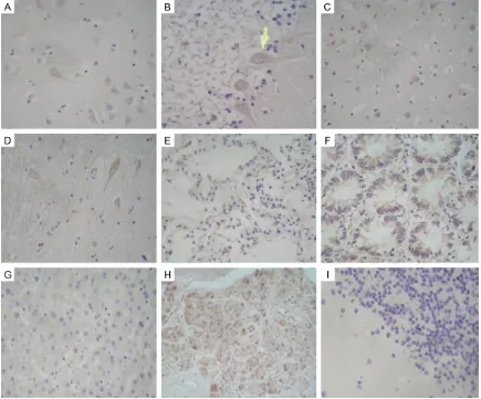
[image:4.612.89.524.72.433.2]To analyze mRNA expression of human Nrn1 in human tissues, an analysis of Nrn1 expression Figure 2.Detection of Nrn1 in human tissues by immunohistochemistry. Areas of positive immunoreactivity are brown and indicated by arrows; nuclei are stained violet with hematoxylin. A. Hippocampus (positive control); B. Cerebellum; C. Cortex; D. Spinal cord; E. Alveolar epithelium; F. Colonic gland; G. Liver; H. Pancreatic epithelium; I.
using the CMT panel comprising 16 normal human adult tissues was performed. The sta-tistics and clustering heat map analysis revealed that Nrn1 mRNA was highly expressed in the brain, and the skeletal muscle, lung, thy-mus, pancreas, planceta, liver and heart tis-sues showed a similar level with it; lower levels were detected in the spleen, testicles, ovary, small intestine, but there was no detectable expression in the kidneys, colon, prostate or leukocytes (Figure 1C, 1D). To analyze the Nrn1 protein expression in these human tissues, some of the tissues with Nrn1 mRNA expres-sion were picked out for immunohistochemical staining. The results indicated that Nrn1 was expressed in multiple tissues and cell types, including hippocampal neurons (Figure 2A), Purkinje cells (Figure 2B), cortical neurons (Figure 2C), spinal cord neurons (Figure 2D), alveolar epithelial cells (Figure 2E), colonic
gland cells (Figure 2F), liver tissue (Figure 2G), and intestinal gland cells (Figure 2H).
Nrn1 is localized in plasma membrane lipid rafts of mature neuronal cells
In order to investigate the subcellular localiza-tion of Nrn1, we performed a bioinformatics analysis and prediction including SignalP pre-diction and TMHMM v.2.0. The results of the SignalP prediction revealed that Nrn1 was a signal peptide (Figure 3A), while the TMHMM v.2.0 tool predicted that Nrn1 sequences were distributed outside the cell membrane without a transmembrane helix (Figure 3B). This evi-dence indicates that Nrn1 is a membrane-linked protein. Since the previous experiments indicated high levels of Nrn1 in brain tissue, we used SH-SY5Y human neuroblastoma cells to confirm the association of Nrn1 with cell mem
branes and lipid rafts by immunofluorescence analysis. Samples were probed with antibodies against Nrn1 and the lipid raft marker caveolin 1. Nrn1 was mostly concentrated at the plasma membrane of SH-SY5Y cells and colocalized with caveolin 1 (Figure 3C). To further confirm these observations, membrane proteins were extracted from the cells and analyzed by west-ern blotting. Caveolin 1 and Nrn1 were both present in the membrane fraction (Figure 3C). Nrn1 expression level was disordered in 13 kinds of human cancers
Since in our previously study, the expression of Nrn1 was not only present in the nervous sys-tem but also in various normal human tissues. The function of Nrn1 in these tissues outside the nervous system remains unclear. Here, by exploring The Cancer Genome Atlas (TCGA) a database for human caner transcriptome, we investigated the expression level in 31 kinds of human cancers. Compared to the normal tis-sues, the results revealed that Nrn1 was sig- nificantly downregulated in BLCA; BRCA; CESC; COAD; GBM; KIHC; KIRP; LGG; READ; UCEC; LUA and OV (*P<0.05), but only upregulated in DLBC (*P<0.05) (Figure 4). Survival analysis is widely used in clinical and epidemiological research: in randomized clinical trials for com-paring the efficacy of treatments and in obser -vational (non-randomized) research to deter-mine and test the existence of epidemiological association [29]. To explore the potential
func-phatidylinositol (GPI)-anchored axonal protein that is mainly expressed in the brain and induced by neuronal activity. In this study, we demonstrated that Nrn1 mRNA is expressed in various normal human tissues. Our results indi-cate that Nrn1 is expressed in the placenta, heart, lungs, skeletal muscle, and spleen at a level similar to that in brain. Nrn1 mRNA was detected in the prostate, heart, small intestine, ovary, thymus, pancreas, liver, and testes with a lower level, but there was no detectable expression in the kidneys, colon, or leukocytes. These results suggest that Nrn1 may have var-ied roles in multiple tissues, not just in the brain. Nrn1 is acknowledged as a neurotrophic factor and its function in other tissues is unclear. Nrn1 may participate in various func-tions and biological processes depending on its tissue distribution. For example, the high level of Nrn1 mRNA in placental tissue suggests that it may play an important role in early embryonic development [30].
Signal peptide is important for protein trans-portation and localization (26). Our bioinformat-ics analysis showed that Nrn1 is distributed outside the cell membrane, which depends on a signal peptide or GPI anchor but not on a transmembrane helix. We speculate that Nrn1 is a secreted protein or a membrane-linked pro-tein with autocrine and paracrine functions. Nrn1 is a GPI-anchored axonal protein [1] that is associated with lipid rafts [31]. In our study,
Figure 5. A. Integrity of prognostic value of Nrn1 in BLCA; BRCA; CESC; COAD; GBM; KIHC; KIRP; LGG; READ; UCEC; LUA and OV patients. B. Prognostic
value of Nrn1 in DLBC patients. HR: hazard ratio, CI: confidence interval,
logRank P<0.01.
tions of Nrn1 in these human tumors, a survival analysis was performed. A combina-tion of the overall survival analysis of the 12 kinds of human tumors with Nrn1 downregulation revealed that patients with high levels of Nrn1 present a long term sur-vival (*P<0.01) (Figure 5A). But there is no significant effect on DLBC patients (Figure 5B).
Discussion
Nrn1/CPG15 is a neurotroph-ic factor and
glycosylphos-Figure 4. Expression level of Nrn1 in cancers including 31 kinds of cancers as indicated in the figure and paired
[image:7.612.90.377.112.254.2]Wide distribution of neuritin and human cancers
we detected Nrn1 at the plasma membrane by immunocytochemistry and Western blotting and found that it colocalized with the lipid raft marker caveolin 1.
Nrn1 distribution in various normal human tis-sues indicated a novel biological process explo-ration of Nrn1. But these potential functions of Nrn1 are still unknown. Data have shown that the neurotrophin family is always involved in the regulation of tumorigenesis like nerve growth factor (NGF), which functions as either supporting or suppressing tumor growth de- pending on the tumor type [32]. Thus, we spec-ulate that the effect of the wide distribution of Nrn1 in human tissues may be associated with tumor development. In this study, 31 catego-ries of human cancer data were included to investigate the correlation of Nrn1 expression and tumorigenesis. The results showing that Nrn1 was significantly downregulated in a total of 12 kinds of human cancers and the survival analysis of these 12 kinds of human cancers indicated that Nrn1 may play an important role in suppressing tumor growth or development. One of the interesting findings was that Nrn1 was significantly upregulated only in DLBC, a kind of Lymphatic hematopoietic malignancy tumor, but there is no significant effect of Nrn1 on DLBC patients’ survival.
In conclusion, this study demonstrated that Nrn1 was expressed in a variety of human tis-sues (brain, skeletal muscle, lung, thymus, pan-creas, planceta, liver and heart tissues) and especially concentrated in the lipid raft of cell membranes. We also provide the first evidence that Nrn1 is associated with a variety human cancers including BLCA; BRCA; CESC; COAD; GBM; KIHC; KIRP; LGG; READ; UCEC; LUA; OV; DLBC, and can act as a tumor suppressor gene. This study provides some powerful evidence to make Nrn1 a new biomarker and a target of these cancers.
Acknowledgements
We extend our sincere gratitude to Dr. Jin Zhao, Dr. Xiaolin Pan, and Dr. Jiangmei Qin for their immense assistance during the study. We are grateful to the staff at the Department of Pathology, The First Affiliated Hospital of Shi-hezi University School of Medicine, for technical support. This work was supported by NSFC Grant 31060136 (Huang Jin), 307600633 (Huang Jin) and 30260029 (Huang Jin).
Disclosure of conflict of interest
None.
Address correspondence to: Dr. Jin Huang, Depart- ment of Biochemistry, The Key Laboratory of Xinjiang Endemic & Ethnic Diseases, School of Medicine, Shihezi University, Shihezi 832002, Xinjiang, China. Tel: +86-0993 205-7882; E-mail: bio-huangjin@ shzu.edu.cn
References
[1] Naeve GS, Ramakrishnan M, Kramer R, Hev-roni D, Citri Y and Theill LE. Neuritin: a gene induced by neural activity and neurotrophins that promotes neuritogenesis. Proc Natl Acad Sci U S A 1997; 94: 2648-2653.
[2] Nedivi E, Wu GY and Cline HT. Promotion of dendritic growth by CPG15, an activity-induced signaling molecule. Science 1998; 281: 1863-1866.
[3] Cantallops I, Haas K and Cline HT. Postsynap-tic CPG15 promotes synapPostsynap-tic maturation and presynaptic axon arbor elaboration in vivo. Nat Neurosci 2000; 3: 1004-1011.
[4] Choi Y, Lee K, Ryu J, Kim HG, Jeong AY, Woo RS, Lee JH, Hyun JW, Hahn S, Kim JH and Kim HS. Neuritin attenuates cognitive function im-pairments in tg2576 mouse model of Alzheim-er’s disease. PLoS One 2014; 9: e104121. [5] Javaherian A and Cline HT. Coordinated motor
neuron axon growth and neuromuscular syn-aptogenesis are promoted by CPG15 in vivo. Neuron 2005; 45: 505-512.
[6] Sharma TP, Liu Y, Wordinger RJ, Pang IH and Clark AF. Neuritin 1 promotes retinal ganglion cell survival and axonal regeneration following optic nerve crush. Cell Death Dis 2015; 6: e1661.
[7] Wang H, Li X, Shan L, Zhu J, Chen R, Li Y, Yuan W, Yang L and Huang J. Corrigendum: recombi-nant hNeuritin promotes structural and func-tional recovery of sciatic nerve injury in rats. Front Neurosci 2017; 11: 65.
[8] Gao R, Li X, Xi S, Wang H, Zhang H, Zhu J, Shan L, Song X, Luo X, Yang L and Huang J. Exoge-nous neuritin promotes nerve regeneration af-ter acute spinal cord injury in rats. Hum Gene Ther 2016; 27: 544-554.
[9] Kalinski AL, Sachdeva R, Gomes C, Lee SJ, Shah Z, Houle JD, Twiss JL. mRNAs and protein synthetic machinery localize into regenerating spinal cord axons when they are provided a substrate that supports growth. J Neurosci 2015; 35: 10357-10370.
[11] Sharma N, Marzo SJ, Jones KJ and Foecking EM. Electrical stimulation and testosterone dif-ferentially enhance expression of regenera-tion-associated genes. Exp Neurol 2010; 223: 183-191.
[12] Seale P, Ishibashi J, Holterman C and Rudnicki MA. Muscle satellite cell-specific genes identi -fied by genetic profiling of MyoD-deficient myo -genic cell. Dev Biol 2004; 275: 287-300. [13] Kojima N, Shiojiri N, Sakai Y and Miyajima A.
Expression of neuritin during liver maturation and regeneration. FEBS Lett 2005; 579: 4562-4566.
[14] Nedivi E, Javaherian A, Cantallops I and Cline HT. Developmental regulation of CPG15 ex-pression in Xenopus. J Comp Neurol 2001; 435: 464-473.
[15] Fujino T, Lee WC and Nedivi E. Regulation of cpg15 by signaling pathways that mediate syn-aptic plasticity. Mol Cell Neurosci 2003; 24: 538-554.
[16] Putz U, Harwell C and Nedivi E. Soluble CPG15 expressed during early development rescues cortical progenitors from apoptosis. Nat Neu-rosci 2005; 8: 322-331.
[17] Zhang L, Zhao Y, Wang CG, Fei Z, Wang Y, Li L, Li L and Zhen HN. Neuritin expression and its relation with proliferation, apoptosis, and an-giogenesis in human astrocytoma. Med Oncol 2011; 28: 907-912.
[18] Yang Z, Zhao T, Liu Y, Gong Z, Cheng S and Yang Q. Identification of new HLA-A*0201-re -stricted cytotoxic T lymphocyte epitopes from neuritin. J Neurooncol 2013; 114: 51-58. [19] Yuan M, Li Y, Zhong C, Li Y, Niu J and Gong J.
Overexpression of neuritin in gastric cancer. Oncol Lett 2015; 10: 3832-3836.
[20] Raggo C, Ruhl R, McAllister S, Koon H, Dezube BJ, Fruh K and Moses AV. Novel cellular genes essential for transformation of endothelial cells by Kaposi’s sarcoma-associated herpes -virus. Cancer Res 2005; 65: 5084-5095. [21] Han D, Qin B, Liu G, Liu T, Ji G, Wu Y and Yu L.
Characterization of neuritin as a novel angio-genic factor. Biochem Biophys Res Commun 2011; 415: 608-612.
[22] Yuan B, Shen H, Su T, Lin L, Chen T and Yang Z. A novel nanoparticle containing neuritin pep-tide with grp170 induces a CTL response to inhibit tumor growth. J Neurooncol 2015; 125: 23-32.
[23] Feng YA, Liu TE and Wu Y. microRNA-182 inhib-its the proliferation and migration of glioma cells through the induction of neuritin expres-sion. Oncol Lett 2015; 10: 1197-1203. [24] Huang J, Yuan H, Lu C, Liu X, Cao X and Wan M.
Jab1 mediates protein degradation of the Rad9-Rad1-Hus1 checkpoint complex. J Mol Biol 2007; 371: 514-527.
[25] Tang Z, Li C, Kang B, Gao G, Li C and Zhang Z. GEPIA: a web server for cancer and normal gene expression profiling and interactive anal -yses. Nucleic Acids Res 2017; 45: W98-W102. [26] Sonnhammer EL, von Heijne G and Krogh A. A
hidden Markov model for predicting trans-membrane helices in protein sequences. Proc Int Conf Intell Syst Mol Biol 1998; 6: 175-182. [27] Krogh A, Larsson B, von Heijne G and Sonn-hammer EL. Predicting transmembrane pro-tein topology with a hidden Markov model: ap-plication to complete genomes. J Mol Biol 2001; 305: 567-580.
[28] Petersen TN, Brunak S, von Heijne G and Nielsen H. SignalP 4.0: discriminating signal peptides from transmembrane regions. Nat Methods 2011; 8: 785-786.
[29] Flynn R. Survival analysis. J Clin Nurs 2012; 21: 2789-2797.
[30] Wang X, Liu C, Xu F, Cui L, Tan S, Chen R, Yang L and Huang J. Effects of neuritin on the migra-tion, senescence and proliferation of human bone marrow mesenchymal stem cells. Cell Mol Biol Lett 2015; 20: 466-474.
[31] Ikezawa H. Glycosylphosphatidylinositol (GPI)-anchored proteins. Biol Pharm Bull 2002; 25: 409-417.