Original Article
Cardiac hypertrophy is positively regulated by long
non-coding RNA PVT1
Yi-Hui Yu*, Zuo-Ying Hu*, Ming-Hui Li*, Bing Li, Zhi-Mei Wang, Shao-Liang Chen
Department of Medicine, Division of Cardiovascular Medicine, Nanjing First Hospital, Nanjing Medical University, Nanjing 210000, China. *Equal contributors.
Received January 9, 2015; Accepted February 27, 2015; Epub March 1, 2015; Published March 15, 2015
Abstract: The aim of this study was to determine whether long non-coding RNA PVT1 can participate in the regula-tion of cardiac hypertrophy. A C57BL/6 mouse cardiac hypertrophic model was established using transverse aortic constriction (TAC). The animals subjected to sham operation were used as controls. Transcripts of PVT1 were ana-lyzed in hearts of model and sham control groups after TAC for 4 weeks using quantitative real-time PCR (qRT-PCR).
Additionally, to investigate whether PVT1 was involved in cardiac hypertrophy, 1 μM angiotensin II (Ang II) was used
to induce hypertrophy and PVT1 siRNA was performed in the cultured neonatal mouse cardiac cardiomyocytes. Cell size was measured by cell surface area and total protein content analyses in response to Ang II treatment. Moreover, some hypertrophic markers including atrial natriuretic peptide (ANP), B-type natriuretic peptide (BNP), and
beta-myosin heavy chain (β-MHC) were also quantified using qRT-PCR. As a result, PVT1 was up-regulated by 2.5-fold (P
< 0.05) in hypertrophic hearts after TAC for 4 weeks as compared to sham group. In addition, siRNA of endogenous
PVT1 in cardiomyocytes significantly reduced (P < 0.05) Ang II-induced increase of cell size in terms of cell surface area (by 5.6-fold) and total protein content (by 23.0%). PVT1 siRNA also obviously attenuated Ang II-induced ANP
and β-MHC expression by 40.9% and 41.5%, respectively (P < 0.05), but had no effect on BNP mRNA expression. Our results demonstrated that PVT1 was essential for the maintenance of cell size of cardiomyocytes and might play a role in the regulation of cardiac hypertrophy.
Keywords: Cardiac hypertrophy, PVT1, long non-coding RNA, cardiomyocytes, transverse aortic constriction
Introduction
Heart failure is still one of the leading causes of mortality and morbidity in the world, even if there has been great improvement in heart dis-ease treatment [1]. Cardiac hypertrophy, a remodeling of myocardium marked by the enlargement of cardiomyocytes, often leads to progression to heart failure and is an indepen-dent and major risk factor for cardiovascular disease [2, 3]. Although great advances in the
identification of genes and signaling pathways
involved in cardiac hypertrophy have been shown [4, 5], additional regulatory mechanisms
remain to be identified.
Previous studies have demonstrated that microRNAs (miRNAs), which are some ~ 22 nucleotide long, evolutionary conserved and noncoding RNA molecules, are implicated in various cardiovascular diseases, including
car-diac hypertrophy and heart failure [6-8].
Furthermore, miRNA expression profiling stud -ies have shown that many miRNAs are aber-rantly expressed and play either pro- or anti-hypertrophic activity roles in the process of cardiac hypertrophy [3, 9-11]. Therefore, miR-NAs have the potential to become one of useful clinically diagnostic and therapeutic targets for heart disease.
noncoding RNAs, protects the heart from path-ological hypertrophy [17]. In addition, some other lncRNAs, such as 2900055J20Rik [18]
and CHRF [19], have also been identified to be
involved in regulation of cardiac hypertrophy. These observations motivate further investiga-tion of the value of lncRNAs in the cardiac hypertrophy.
The plasmacytoma variant translocation 1
(PVT1) gene, originally identified as a transcrip -tional unit from a human homologous sequence to Pvt1 [20], is a lncRNA (1.9 kb) that encodes a number of alternative transcripts and a host
gene for several miRNAs [21]. When amplified
and overexpressed, PVT1 can increase cell pro-liferation and inhibit apoptosis, indicating that it is an anti-apoptotic molecule [22]. Therefore, PVT1 is frequently a target of genetic gains and
amplifications in various cancers [22-25].
Although several previous reports have
identi-fied the functional roles of PVT1 implicated in
the pathogenesis of the human diseases men-tioned above, the role that this gene may play in the development of cardiac hypertrophy is not yet known.
To shed light on a potential effect of PVT1 on hypertrophy, we analyzed its expression level in hearts of hypertrophy model mice subjected to transverse aortic constriction (TAC). With small interference RNA (siRNA) approach, we further demonstrated an essential role of PVT1 in the regulation of angiotensin II (Ang II)-induced increase of cardiomyocytes size.
Materials and methods
Cardiac hypertrophy animal model
To determine the expression changes of PVT1 in hypertrophic hearts we applied a well-estab-lished mouse cardiac hypertrophy model by
TAC as described [3, 26]. Briefly, C57BL/6 mice
of 8-10 weeks old (22-24 g) were anesthetized by intraperitoneal injection of 3.6% chloral hydrate. Under sterile conditions, a longitudinal incision of 2-3 mm was made in the proximal sternum to allow visualization of the aortic arch and then transverse aorta binding constriction was performed with an overlaying blunted 27-gauge needle and a 6-0 silk suture. After that, the needle was quickly removed to create
a defined constriction. Moreover, a sham surgi -cal operation was performed in which the
trans-verse aorta was exposed but not banded. All animal experiments in this study were per-formed with the approval of the Animal Care Committee of Laboratory Animal Centre &
Nanjing Hospital Affiliated to Nanjing Medical
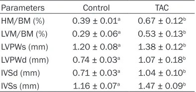
University (SYXK2009-0015). Cardiac hyper-trophy was evaluated by echocardiography analysis of heart size, including left ventricular posterior wall thickness at end-systole (LVPWs), left ventricular posterior wall thickness at end-diastole (LVPWd), interventricular septum thick-ness at end-diastole (IVSd) and interventricular septum thickness at end-systole (IVSs) as out-come indicators. The body mass (BM), heart mass (HM), left ventricular mass were also measured to calculate the ratios of HM/BM and LVM/BM.
Neonatal mouse cardiomyocyte culture
Neonatal mouse cardiomyocytes were pre-pared as described previously [1, 27]. In brief, hearts were obtained from 1 to 3 days old C57BL/6 mice and placed in ice-cold phos-phate-buffered saline (PBS) solution. After repeated rinsing, the ventricular tissues were minced with scissors and enzymatically disso-ciated with 0.25% trypsin (Beyotime, China) at 37°C under 100 rpm rotation. After dissocia-tion, cardiomyocytes were enriched using Percoll (Amersham) step gradients, and then cultured in DMEM (Hyclone) with 10% fetal bovine serum (FBS, Hyclone) and seeded into six-well plates.
Hypertrophic stimulation and small interfer-ence RNA (siRNA)
To induce hypertrophy, cardiomyocytes were
stimulated with 1 μM Ang II as previously
described [28]. The Stealth™ siRNAs targeting PVT1 (CCUGCAUAACUAUCUGCUUTT) and a scrambled control siRNA were synthesized and cells were transfected with the siRNAs using lipofectamine 2000, according to the manufac-turer’s instructions (Invitrogen). The obtained cardiomyocytes were plated at a density of 5 × 10⁵ cells and cultured for 24 h in serum-con-taining media, and then divided into 4 groups: control group, sequentially cultured in complete medium; starvation group, subjected to 6 h serum starvation; (starvation + Ang II) group,
Table 1. The sequences of the primers used for qRT-PCR
Primer Sequences (5’-3’) PVT1 Forward CCTCTTGGTCCCTGATGCA
Reverse GATTCCCATGCCTCTCATCCT ANP Forward TGAGTGAGCAGACTGAGGAA Reverse TGGATCTTCGTAGGCTCCGA BNP Forward ACAGAAGCTGCTGGAGCTGA
Reverse CCGATCCGGTCTATCTTGTG
β-MHC Forward TATCGATGACCTGGAGCTGA
Reverse AGTATTGACCTTGTCTTCCTC GAPDH Forward ACAGCAACAGGGTGGTGGAC
Reverse TTTGAGGGTGCAGCGAACTT
ANP: atrial natriuretic peptide; BNP: B-type natriuretic peptide; β-MHC: beta-myosin heavy chain; GAPDH: glyceraldehydes-3-phosphate dehydrogenase.
PVT1 siRNA transfection for 24 h prior to
stimu-lation of 1 μM Ang II. The serum-free medium
was replaced 6 hours after starvation by the regular culture medium.
Immunostaining and measurement of cell surface area
After experimental treatment for 72 h, cardio-myocytes were subsequently washed three
times with cold PBS and then fixed with 4% paraformaldehyde for 10 min. After fixation,
cells were washed three times with cold PBS and permeabilized with 0.5% Triton X-100 in
PBS for 10 min. Non-specific binding of the fixed cells was blocked by incubation in 5% BSA
solution for 1 h at room temperature, followed
by incubation with anti-α-actinin antibody
(1:200; Beyotime, China) at 4°C overnight. After washing, the cells were incubated with Alexa Fluor™ 568-conjugated goat mouse anti-body (1:1000; Molecular Probes, Eugene, OR) at 37°C for 1 h. Hoechst 33342 (Beyotime, China) staining was used to visualize the nucle-us. The cells were examined and photographed
using a fluorescence microscope (Olympus,
Japan) and cell surface areas were measured using NIH Image J software. Approximately 100 cells were counted from randomized captured images and regarded as an independent exper-iment, and three independent experiments were performed.
Total protein concentration measurement
To assess cardiomyocytes hypertrophy in all groups, total protein content was also mea-sured [29]. After 72 h of experimental treat-ment, cardiomyocytes were lysed in RIPA sam-ple buffer (Boster, China), scraped, and collected in cold lysis buffer, and protein con-centration was measured by Bradford assay (Bio-Rad Laboratories, Hercules, CA).
Quantitative real-time PCR (qRT-PCR)
[image:3.612.91.289.94.242.2]Total RNA was isolated from both hearts of car-diac hypertrophy mouse model and cultured neonatal mouse cardiac cardiomyocytes of all groups using RNeasy mini kit (Qiagen, Germany). First-strand cDNA was synthesized from 1 µg of total RNA using SuperScript II Figure 1. The expression level of PVT1 in hearts of transverse aortic constriction (TAC) mouse model and sham control after 4 weeks (n = 4 for each group). The different letters (a and b) indicated
sig-nificant differences (P < 0.05). Table 2. Outcome indicators for cardiac
hypertrophy
Parameters Control TAC
HM/BM (%) 0.39 ± 0.01a 0.67 ± 0.12b
LVM/BM (%) 0.29 ± 0.06a 0.53 ± 0.13b
LVPWs (mm) 1.20 ± 0.08a 1.38 ± 0.12b
LVPWd (mm) 0.74 ± 0.03a 1.07 ± 0.18b
IVSd (mm) 0.71 ± 0.03a 1.04 ± 0.10b
[image:3.612.92.289.322.415.2]RNase H-Reverse Transcriptase (Invitrogen) according to the manu-facturer’s protocol. The sequences of primers (Shenggong Bioengi- neering Co., Shanghai, China) used
for quantification measurement of
hypertrophic markers atrial uretic peptide (ANP), B-type natri-uretic peptide (BNP), beta-myosin
heavy chain (β-MHC) and PVT1
mRNA were shown in Table 1, and glyceraldehydes-3-phosphate dehydrogenase (GAPDH) was used
as reference gene. The specificity of the PCR amplification was con
-firmed by agarose gel electrophore -sis. Real-time reactions were run and analyzed by using a Real-Time PCR system (Applied Biosystems
ABI Prism 7500). The amplification
was used to calculate the CT value
(ΔCT) of the target genes and the difference between the ΔCT of
those genes and GAPDH gene. In addition, the equation 2-ΔΔCT was
used to determine the relative
amount of mRNA in specific target
genes.
Statistical analysis
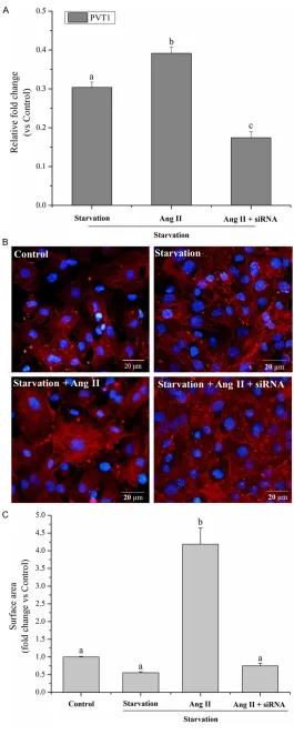
The results were expressed as mean ± SD of at least three inde-pendent experiments. Comparisons among multiple groups were made by analysis of variance (ANOVA) fol-lowed by Tukey post hoc test using SPSS 16.0. Two groups were ana-Figure 2. PVT1 siRNA rescued hyper-trophic responses in cardiomyocytes. A. After 72 h of culture, relative fold change of PVT1 expression in the cul-tured neonatal mouse cardiac cardio-myocyte of starvation group, (starvation + Ang II) group and (starvation + Ang II + siRNA) group vs. control group, respec-tively. B. Representative photograph of cardiomyocytes transfected with PVT1 siRNA after Ang II treatment (Hoechst 33342 staining). C. Cell surface areas of the cardiomyocytes were measured using Image J software (n = 100). Scale
bars, 20 μm. The different letters (a, b and c) indicated significant differences
[image:4.612.90.355.68.730.2]measured by total protein content by Bradford
Assay. Ang II stimulation significantly increased
the protein quantity by 2.0- and 3.0-fold vs. control and starvation groups, respectively (P < 0.05). This was reduced by 23.0% with PVT1 siRNA pretreatment (P < 0.05) vs. non-siRNA preteated Ang II-stimulated cells (Figure 3). PVT1 siRNA attenuated Ang II-induced ANP and β-MHC expression but had no effect on BNP
Ang II treatment dramatically increased
expres-sion levels of ANP and β-MHC, molecular mark -ers of cardiomyocyte hypertrophy, by 4.8- and 3.0-fold (P < 0.05 vs. respective controls for both). PVT1 siRNA transfection markedly
dimin-ished this ANP and β-MHC expression by 40.9%
and 41.5%, respectively, vs. Ang II alone (Figure 4A and 4C). However, Ang II stimulation and PVT1 siRNA pretreatment did not affect BNP expression (P > 0.05, Figure 4B).
Discussion
Previous reports have shown that lncRNAs are involved in several aspects cardiac develop-ment and pathophysiology, including cardiac hypertrophy and heart failure [13, 16, 18, 19]. In present study, we revealed that lncRNA PVT1 had an essential role in cell size maintenance of cardiomyocytes during hypertrophy. Initially, we showed that PVT1 was up-regulated about lyzed by Student’s t test. P < 0.05 was
consid-ered statistically significant.
Results
PVT1 was up-regulatede in hypertrophic hearts
The TAC was applied to induce cardiac hyper-trophy, and development of cardiac hypertro-phy and regression of the established cardiac
hypertrophy were confirmed by echocardiogra -phy analysis. After 4 weeks’ constriction, the
TAC hearts showed significantly higher (P <
0.05) HM/BM, LVM/BM, LVPWs, LVPWd, IVSd and IVSs values than the sham group (Table 2), implying the hypertrophic growth. qRT-PCR analysis demonstrated that the relative
expres-sion level of PVT1 was significantly higher by
2.5-fold (P < 0.05) in TAC than in sham (Figure 1).
Down-regulation of PVT1 reduced the cell sur-face area of cardiomyocytes induced by Ang II
To investigate whether endogenous PVT1
played a significant role in cardiomyocytes
hypertrophy, RNA interference was performed to silence PVT1 gene expression through siRNA. Compared with the starvation and Ang II
treat-ment groups, PVT1 siRNA was sufficient to
[image:5.612.95.347.72.274.2]down-regulate endogenous PVT1 expression in cultured cardiomyocytes by 33.0% and 55.5%, respectively, as analyzed with qRT-PCR (Figure
Figure 3. After 72 h of culture, cardiomyocytes hypertrophy was as-sessed by total protein content measurement in all groups by
Brad-ford Assay. The different letters (a, b and c) indicated significant dif -ferences (P < 0.05).
2A, P < 0.05). Moreover, Ang II
stimu-lation induced significantly increased
(P < 0.05) expression of PVT1 com-pared to starvation group (Figure 2A). The size of cells was measured by relative cell surface areas and showed that the size of cardiomyo-cytes transfected with PVT1 siRNA was obviously reduced by 5.6-fold to baseline level as compared with that of Ang II stimulation (Figure 2B and 2C, P < 0.05), suggesting that the endogenous PVT1 might be required to maintain cell size of cardiomyo-cytes and might be involved in the Ang II-induced cell size enlargement of cardiomyocytes.
Down-regulation of PVT1 diminished Ang II-induced protein content
2.5-fold after TAC for 4 weeks. Then using siRNA approaches in cultured cardiomyocytes, we further revealed that down-regulation of PVT1 in cardiomyocytes could attenuate the Ang II-induced increase of cell size (by cell sur-face area measurement), total protein contents
as well as expression levels of ANP and β-MHC
(molecular markers of cardiomyocyte hyper- trophy).
Mammalian genomes encode numerous
lncRNAs which have been defined to have
important functions in RNA processing,
chro-matin modification, structural scaffolds, and
modulation of apoptosis and invasion [30, 31]. Therefore, unsurprisingly, these molecules are emerging as important players in several human pathologies, including cardiovascular diseases [13]. A recent report has demonstrat-ed that lncRNA CHRF performs as endogenous sponge RNA to regulate cardiac hypertrophy by inhibiting miR-489 expression and activity [19]. A cardioprotective lncRNA Myheart has also
been identified to protect the heart from patho
-logical hypertrophy [17]. In addition, it has been shown that the lncRNA 2900055J20Rik may be involved in regulation of cardiac hypertrophy [18]. In our present study, lncRNA PVT1 was also found to be implicated in hypertrophied heart and hypertrophy of cardiomyocytes. The discovery of the novel lncRNA (PVT1) in cardiac hypertrophy further reveals the function of lncRNAs participating in regulating hypertrophy and may shed new lights on understanding the complex molecular mechanism of cardiac hypertrophy.
[image:6.612.100.312.70.415.2]PVT1 is a lncRNA (1.9 kb) that encodes a num-ber of alternative transcripts [32]. Although there are a few reports showing that PVT1 plays an important role in the pathogenesis of many human diseases [21, 22, 25], it is not yet clear whether PVT1 is involved in the regulation of cardiac hypertrophy. In our study, PVT1 was found to be substantially up-regulated in TAC model and in response to hypertrophic stimula-tion (Ang II), suggesting that the endogenous PVT1 might be required to maintain cell size of Figure 4. After 72 h of culture, relative fold change
of (A) ANP, (B) BNP and (C) β-MHC expression in the
cultured neonatal mouse cardiac cardiomyocyte of treatment groups vs. control group, respectively. The
different letters (a, b and c) indicated significant dif -ferences (P < 0.05). ANP: atrial natriuretic peptide;
BNP: B-type natriuretic peptide; β-MHC: beta-myosin
cardiomyocytes. Inhibition of PVT1 could atten-uate enlargement of cardiomyocytes size, which indicating that PVT1 was essential for regulation of cardiac hypertrophy. However, some research limitations still existed in the present work. The downstream targets of PVT1 and exact molecular mechanism are still
unclear. It is an interesting scientific topic and
we will focus on that in our future research.
Our present study identified for the first time
that PVT1 was essential for cell size mainte-nance of cardiomyocytes and for regulation of cardiac hypertrophy, suggesting that PVT1 might be a potential therapeutic target for car-diac hypertrophy.
Disclosure of conflict of interest
None.
Address correspondence to: Dr. Shao-Liang Chen, Department of Medicine, Division of Cardiovascular Medicine, Nanjing First Hospital, Nanjing Medical University, 68 Changle Road, Nanjing 210000, China. Tel: +86-25-52208048; Fax: +86-2552- 208048; E-mail: [email protected]
References
[1] Song XW, Li Q, Lin L, Wang XC, Li DF, Wang GK, Ren AJ, Wang YR, Qin YW, Yuan WJ. MicroRNAs are dynamically regulated in hypertrophic hearts, and miR-199a is essential for the maintenance of cell size in cardiomyocytes. J Cell Physiol 2010; 225: 437-443.
[2] Cheng Y, Ji R, Yue J, Yang J, Liu X, Chen H, Dean DB, Zhang C. MicroRNAs are aberrantly ex-pressed in hypertrophic heart: do they play a role in cardiac hypertrophy? Am J Pathol 2007; 170: 1831-1840.
[3] Jeong MH, Lee JS, Kim DH, Park WJ, Yang DK.
Identification of novel microRNAs negatively
regulating cardiac hypertrophy. Biochem Bio-phys Res Commun 2012; 428: 191-196. [4] Feng H, Ouyang W, Liu J, Sun Y, Hu R, Huang L,
Xian J, Jing C, Zhou M. Global microRNA
pro-files and signaling pathways in the develop -ment of cardiac hypertrophy. Braz J Med Biol Res 2014: 47: 361-8.
[5] Li M, Wang N, Liu W, Zhi X, Zhang TC. Signaling Pathways in Cardiac Hypertrophy. In: Proceed-ings of the 2012 international conference on applied biotechnology (ICAB 2012). 2014. Springer.
[6] Sayed D, Hong C, Chen IY, Lypowy J, Abdellatif M. MicroRNAs play an essential role in the
de-velopment of cardiac hypertrophy. Circ Res 2007; 100: 416-424.
[7] Gladka MM, Da Costa Martins PA, De Windt LJ. Small changes can make a big difference-mi-croRNA regulation of cardiac hypertrophy. J Mol Cell Cardiol 2012; 52: 74-82.
[8] Tatsuguchi M, Seok HY, Callis TE, Thomson JM, Chen JF, Newman M, Rojas M, Hammond SM, Wang DZ. Expression of microRNAs is dynami-cally regulated during cardiomyocyte hypertro-phy. J Mol Cell Cardiol 2007; 42: 1137-1141. [9] Van Rooij E, Sutherland LB, Liu N, Williams AH,
Mcanally J, Gerard RD, Richardson JA, Olson EN. A signature pattern of stress-responsive microRNAs that can evoke cardiac hypertrophy and heart failure. Proc Natl Acad Sci U S A 2006; 103: 18255-18260.
[10] Jentzsch C, Leierseder S, Loyer X, Flohrschütz I, Sassi Y, Hartmann D, Thum T, Laggerbauer B, Engelhardt S. A phenotypic screen to identify hypertrophy-modulating microRNAs in primary cardiomyocytes. J Mol Cell Cardiol 2012; 52: 13-20.
[11] Martins PaDC, De Windt LJ. MicroRNAs in con-trol of cardiac hypertrophy. Cardiovasc Res 2012; 93: 563-572.
[12] Wapinski O, Chang HY. Long noncoding RNAs and human disease. Trends Cell Biol 2011; 21: 354-361.
[13] Papait R, Kunderfranco P, Stirparo GG, Latro-nico MV, Condorelli G. Long noncoding RNA: a new player of heart failure? J Cardiovasc Transl Res 2013; 6: 876-883.
[14] Schonrock N, Harvey RP, Mattick JS. Long non-coding RNAs in cardiac development and pathophysiology. Circ Res 2012; 111: 1349-1362.
[15] Ounzain S, Crippa S, Pedrazzini T. Small and long non-coding RNAs in cardiac homeostasis and regeneration. Biochim Biophys Acta 2013; 1833: 923-933.
[16] Scheuermann JC, Boyer LA. Getting to the heart of the matter: long non-coding RNAs in cardiac development and disease. EMBO J 2013; 32: 1805-1816.
[17] Han P, Li W, Lin CH, Yang J, Shang C, Nuern-berg ST, Jin KK, Xu W, Lin CY, Lin CJ. A long noncoding RNA protects the heart from patho-logical hypertrophy. Nature 2014. 514: 102-6 [18] Zhang L, Hamad E, Vausort M, Funakoshi H,
Feldman A, Wagner D, Devaux Y. Long non-coding RNAs and cardiac hypertrophy. Cardio-vasc Res 2014; 103: S10-S10.
[19] Wang K, Liu F, Zhou LY, Long B, Yuan SM, Wang Y, Liu CY, Sun T, Zhang XJ, Li PF. The long non-coding RNA CHRF regulates cardiac hypertro-phy by targeting miR-489. Cardiovasc Res 2014; 114: 1377-1388.
lymphoma 2; 8 variant translocation is equiva-lent to the murine pvt-1 locus. EMBO J 1986; 5: 2845.
[21] Beck-Engeser GB, Lum AM, Huppi K, Caplen NJ, Wang BB, Wabl M. Pvt1-encoded microR-NAs in oncogenesis. Retrovirology 2008; 5: 4. [22] Guan Y, Kuo WL, Stilwell JL, Takano H, Lapuk
AV, Fridlyand J, Mao JH, Yu M, Miller MA,
San-tos JL. Amplification of PVT1 contributes to the
pathophysiology of ovarian and breast cancer. Clin Cancer Res 2007; 13: 5745-5755. [23] Shtivelman E, Bishop JM. The PVT gene
fre-quently amplifies with MYC in tumor cells. Mol
Cell Biol 1989; 9: 1148-1154.
[24] Huppi K, Pitt JJ, Wahlberg BM, Caplen NJ. The 8q24 gene desert: an oasis of non-coding tran-scriptional activity. Frontiers in genetics 2012; 3: 69.
[25] Sonoki T. PVT1: A Cancer-associated Non-cod-ing Gene Revisited. ClonNon-cod-ing & Transgenesis 2014; [Epub ahead of print].
[26] Chen X, Wang SC, Cao LH, Yang GQ, Li M, Su JC. Comparison between radial head
replace-ment and open reduction and internal fixation
in clinical treatment of unstable, multi-frag-mented radial head fractures. Int Orthop 2011; 35: 1071-1076.
[27] Sadoshima JI, Jahn L, Takahashi T, Kulik T, Izumo S. Molecular characterization of the stretch-induced adaptation of cultured cardiac cells. An in vitro model of load-induced cardiac hypertrophy. J Biol Chem 1992; 267: 10551-10560.
[28] Hu CM, Chen YH, Chiang MT, Chau LY. Heme oxygenase-1 inhibits angiotensin II-induced cardiac hypertrophy in vitro and in vivo. Circula-tion 2004; 110: 309-316.
[29] Essick EE, Ouchi N, Wilson RM, Ohashi K, Gho-brial J, Shibata R, Pimentel DR, Sam F. Adipo-nectin mediates cardioprotection in oxidative stress-induced cardiac myocyte remodeling. Am J Physiol Heart Circ Physiol 2011; 301: H984-H993.
[30] Mercer TR, Dinger ME, Mattick JS. Long non-coding RNAs: insights into functions. Nat Rev Genet 2009; 10: 155-159.
[31] Ponting CP, Oliver PL, Reik W. Evolution and functions of long noncoding RNAs. Cell 2009; 136: 629-641.