Original Article
JAK2-dependent proliferation of Fas positive
colorectal cancer cells may associate with
poor prognosis in colorectal cancers
Linlin Qu1, Liang He2, Shaomin Wang1, Jing Huang1, Wei Xu1
Departments of 1Clinical Laboratory, 2Surgery, The First Hospital of Jilin University, Jilin, China
Received January 19, 2017; Accepted February 22, 2017; Epub May 1, 2017; Published May 15, 2017
Abstract: JAK2 is considered as an important cytokine mediator in some malignant tumors which can promote tumor growth. Fas is well known as pro-apoptotic receptor on cell membrane which can modulate cell immunity and apoptosis. However, recent studies show that Fas is emerging as a positive modulator in colorectal cancer (CRC) progression. The mystery of Fas makes it worth to explore the way to drive Fas to pro-apoptotic property. The purpose of this research is to investigate the role of JAK2 in Fas (+) CRC cells. We studied the correlation between Fas expression, JAK2 expression and survival in separate human colorectal cancer (CRC) cohorts by Kaplan-Meier survival curve and analysis of gene expression profiles. Fas expression in non-metastasis CRC is associated with poor prognosis. Fas is co-expressed with genes involved in chemokine signaling pathway, antigen processing and presentation, cytokine-cytokine receptor interaction and apoptosis, among which the top is JAK2. IHC on paraffin-embedded CRC tissue sections and western blots for CRC patient-originated cells were probed using Fas and JAK2 antibody, revealing that Fas and JAK2 are co-expressed in CRC tumor tissues as well as in cells. Scoring for IHC and quantification for western blots also link JAK2 to Fas (P < 0.05). In vitro clone formation assays reveal that JAK2 inhibition markedly suppress clone formation capacity in majority of CRC cell lines. JAK2 inhibition alone doesn’t seem to induce apoptosis, and doesn’t seem to cooperate with FasL in apoptosis. These results indicate that JAK2 may serve as a biomarker in predicting prognosis for CRCs. This paper also shows JAK2 may positively regulate CRC stem-like cell proliferation, which emerges to be a novel therapeutic target for Fas (+) CRCs.
Keywords: JAK2, Fas, colorectal cancer, prognosis, mechanism
Introduction
Recently, JAK2 is considered as a promising target in treating many malignant tumors [1]. JAK2 is a critical molecule in that phosphory-lated JAK2 (pJAK2) in activated form can trans-mit signals via JAK/STAT pathway from outside the cells to the inside nucleus involving differ-entiation and cell growth. Given that phosphor-ylation of JAK2 depends on the presence and binding of some immune molecules on cell membranes, we presumed that those colorec-tal cancer cells expressing Fas on the mem-brane may recruit more immune cells with the consequence of more immune molecules pro-duced in tumor microenvironment.
Death receptors have been implicated in apop-tosis in multiple cell types. Among these death
receptors is FasL/Apo-1. Stimulation of Fas activates several cellular signaling pathways. The biofunctions of Fas are regulated through
at least five levels, including extracellular sig -naling, membrane signal transduction (the assembly of DISC), DISC, mitochondrial level, and miRNA. As a consequence, the result of cell fate is either pro- or antiapoptosis [2].
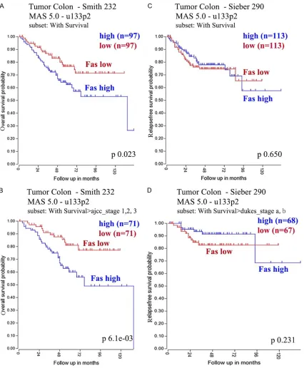
In R2 Single Dataset function, overall survival (OS) data were available in two datasets (Smith et al [16], Sieber et al [17]). Disease-free sur-vival (DFS) data were available in the above dataset. The association of JAK2 or FAS with DFS and OS was determined by using Kaplan Meier curve from R2 Single Dataset function. Median Fas or JAK2 expression levels were used as cutoff values. P values were deter-mined by log-rank test as described in Bewick et al [18].
Fas associated pathways were analyzed first by
choosing the “KEGG Pathway Finder” option and setting the gene correlation (Pearson cor-relation) P-values < 0.01 for each dataset, pro
-viding that Pearson’s correlation coefficient fol -lows t-distribution with n-2 degrees of freedom.
The Fas associated pathways that were signifi -cantly enriched in at least two datasets (chi-square test with continuity correction is per-formed; P-value < 0.01) were identified and
genes were then ordered according to the
sig-nificance, which was determined based on the
combined P values (Stouffer z-trend) that were calculated using the web Meta P application (http://compute1.lsrc.duke.edu/softwares/Me- taP/metap.php). Then Gene Ontology (GO) anal-ysis was done via similar approach to get
sig-nificantly enriched lists of Fas-associated path -ways and genes. The lists of genes were gen- erated then for each dataset.
Using the STRING analysis web-based applica-tion (www.string-db.org), the funcapplica-tional inter-connectivity between the genes on the lists was shown in form of network so that the pro-tein interactions were visualized. The subsets of genes especially the communal subset in
significant association with Fas were identified
via “GeneVenn” web application (http://mcbc. usm.edu/genevenn/) among three data sets.
The heat maps were generated by reimporting those genes into R2 database. Here, the genes were from the largest dataset available (Smith et al, 232 tumors) and the tumors were ordered according to Fas expression levels from low to high.
All patients’ materials were collected after app- roval by patients and in accordance with relat-ed rules and guidelines of Mrelat-edical Ethical Committee of the University Medical Center Utrecht (UMCU).
is required for Fas stimulation and followed by clustering of the membrane receptors and the recruitment of FADD (Fas-associated protein with death domain) [4]. FADD and subsequent caspase-8 activation, together with Fas and FLIP (FLICE-like inhibitory protein), form the death-inducing signaling complex (DISC) [5, 6], which is required for apoptosis [7]. However, Fas receptors not only play an apoptotic role on stimulation but also bring immunological responses to dying cells and their microenvi-ronment [8]. During the process of apoptosis induced by Fas, cells can secret several cyto-kines such as IL-6, IL-8, MCP-1, which can recruit phagocytes and subsequent engulf- ment to dying cells. Nevertheless, Fas is now emerging as an important molecular in promot-ing cell proliferation [9] and/or migration [10-14]. Animal experiments found that loss of Fas reduced cancer incidence as well as tumor bur-den [15], suggesting that Fas may play a posi-tive role in tumorigenesis. Some in vitro studies report that Fas is involved in cell proliferation and survival through activation of some other major tyrosine signaling pathways. Considering that among the pathogenesis of colorectal
tumors, inflammatory bowel diseases (IBD) and
colorectal polyps are major risks where Fas is largely involved [13], and that Fas can act as either a pro-apoptotic or a proliferation media-tor, it is worth to know how colorectal cancer cells respond to Fas stimulation, so as to fur-ther try to make apoptosis instead of prolifera-tion potential through Fas.
In this paper, we show that Fas expression in human colorectal cancers (CRC) is strongly cor-related with JAK2 expression, patients’ surviv-al, especially in non-metastatic colorectal can-cers. We also found that JAK2 plays an impor- tant role in Fas associated gene networks. JAK2 inhibition can suppress tumor colony
for-mation in cell cultures by using JAK2 specific
inhibitors working in medium/Matrigel culturing system.
Materials and methods Bioinformatics study
Wash the slides with PBS-Tween for three times, 5 min for each. Incubate with horserad-ish peroxidase-labeled Bright Vision secondary antibody at room temperature for 1 hour. Detect with DAB and re-stain with HE. Wash, dehydrate and mount the slides. Image acquisi-tion was performed on Nikon Eclipse E800M photomicroscope connected with a computer. A semi-quantitative scoring criterion for IHC was used, in which both staining intensity and positive areas were recorded.
Clone formation assay
Cell cultures medium was refreshed one day before clone formation assay. Immediately before clone formation assay, cell cultures were collected in 50 ml sterilized tubes (BD Falcon). After spheroids sink down, supernatant was aspirated and then 1-2 ml Cell Aggregate Dissociation Medium (eBioscience) added. Spheroids were resuspended in dissociation medium and the tube was incubated in 37°C water bath. Resuspend with a sterilized pipette
every five minutes until spheroids were dissoci -ated completely. Let the dissoci-ated suspen-sion go through 40 µm sterilized cell strainer (Becton Dickinson) and collect single cells from below the strainer. Spinned down and re-sus-pended single-cell suspensions to make a den-sity of 20 cells per µl in culture medium. Gently pipetted a mixture of 50 µl single cell suspen-sion and 50 µl thawed-on-ice liquid Matrigel and seeded onto a well of 6-well or 12-well plates (Costar). Incubate the 6-well or 12-well plates in 37°C for 10 min to make Matrigel hard. The appropriate volume of culture medi-um was applied on Matrigel. Cultures were fed every 3 days and tumor sphere formation was
monitored weekly. Under 10× or 40× magnifi -cation of inverted phase-contrast microscope (EVOS, AMG, USA), images were acquisited to visualize the morphology of sphere, and all spheres were counted, plotted and presented as histogram. Each experiment was performed in triplicate. At least two independent experi-ments were repeated.
Western blot
CRC cells were lysed to get total proteins for each cell line using phosphatase and protease inhibitor diluted in RAS lysis buffer/glycerin phosphate. Protein concentration was analyzed using Bio-Rad protein assay based on Bradford Cell lines and cultures
Human originated colorectal cancer cell lines CRC29, CRC47, CR9, CR16, CRC48, CRC2617, and liver metastatic cell lines L169, L167, L145 were cultured in DMEM/F12 (Gibco) supple-mented with DMEM/F12 supplesupple-mented with 0.5% glucose (Sigma), 1% Ultraglutamine 1 (Lonza), 0.5% Trace element A (Cellgro), 1% Trace element B (Cellgro), 1% Trace element C (Cellgro), 5 mM HEPES Buffer (Lonza), 100 µg/ ml Apo transferrin (Sigma), 20 µg/ml Insulin (Sigma), 5.6 ng/ml Progesterone (Sigma), 4.7 ng/ml Sodium Selenite (Sigma), 1 µg/ml GSH (Sigma), 8.6 µg/ml Putrescine (Sigma), 100 µM
β-mercaptoethanol (Merck), 10 ml of antibiotic-antimycotic (Gibco), 4 µg/ml gentamicin (Invi- trogen), 0.2% lipid mixture (Sigma).
Antibodies, inhibitors and other reagents
The following antibodies were purchased from, Inc, Leiden, The Netherlands: rabbit JAK2 (#3230), rabbit p-STAT3 (#9131), mouse STAT3 (#9139s). Fas antibody rabbit Fas C-20 (sc-715) was obtained from Santa Cruz Biotechno- logy, Inc, Heidelberg, Germany. Secondary per-oxidase-conjugated antibodies were goat anti- mouse-HRP or goat anti-rabbit-HRP obtained from Dako (Glostrup, Denmark). JAK2 inhibitor CEP33779 were provided by Sellechem Incor- poration. Dimethyl sulfoxide (DMSO) was or- dered from Sigma Inc.
Immunohistochemical staining
After excision, samples of primary colorectal
cancer or liver metastasis were fixed in 4% paraformaldehyde and paraffin embedded. Paraffin-embedded tissues were sectioned by
Leica microtome RM 2235 and attached to glass slides. Immunohistochemistry for FAS and JAK2 were stained and scored. Staining was carried out according to standard
immuno-histology protocol from cell signaling. Briefly speaking, deparaffinize and rehydrate the
5% BSA. Blocking procedure requires rotating at room temperature 2 hours. Wash with TBS-0.1% Tween, and then add rabbit anti-human-FAS antibody (1:500), rabbit anti-human-JAK2 antibody (1:1000) and actin on separate trans-ferred PVDF membranes. Incubate at 4°C over-night and wash with TBS-0.1% Tween 3 times, 10 min for each. The PVDF membranes were probed with goat anti-rabbit HRP-labeled sec-method. The protein measurement was done
by a microplate reader at 595 nm wavelength. Comparison to a standard curve provides a rel-ative protein concentration. Load 40 µg of total protein for SDS-PAGE electrophoresis, and then transfer to PVDF membrane. Protein bands can be visualized by ponceau S staining. For FAS and actin, block with TBS-0.1% Tween-5% non-fat milk; for JAK2, block with TBS-0.1%
[image:4.612.88.526.71.481.2]quantify the intensity using Bio-Rad Quantity One analyzing software.
ondary antibody. Detect with ECL detection
[image:5.612.95.521.72.588.2]agent and expose the film. Scan the images and
FAS-centered bioinformatics study was
per-formed first by “KEGG Pathway Finder”
func-tion in R2 database. We searched for FAS-associated cellular processes, pathways, asso-ciated genes in two data sets from two sepa-rate CRC cohorts. The KEGG pathway analysis
reveals several pathways that were significantly
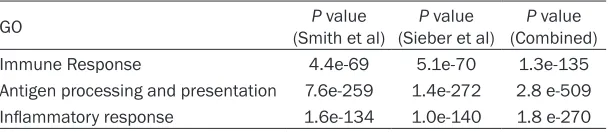
associated with FAS expression level in the two data sets (Table 1). Gene Ontology analysis also reveals that the pathways in association with FAS are functionally involved in immune response, antigen processing and
presenta-tion, inflammatory response (Table 2).
The lists of genes involved in the above pro-cesses were generated by clicking each path-way in R2. Then string analysis was performed by importing each list of genes into string web application tool to get visualized interconnec-tions for proteins. Gene network of the immune response pathway is functionally characterized
by INF-γ and chemokine signaling (data not
shown), which to a large extent depends on JAK2-mediated (data not shown). The proteins in the chemokine signaling network are also
interconnected in inflammatory response (data
not shown). All of the genes that positively cor-related with FAS were shown in heat maps where the tumors were ordered according to FAS expression levels from low to high (Figure 3). They governed chemokine signaling, JAK/ Statistical analysis
Correlation analysis between FAS and JAK2 expression was performed by choosing ‘linear analysis’ function in GraphPad Prime 5.0 soft-ware. Differences between groups were ana-lyzed using ‘unpaired two-sided T-test’ func- tion in GraphPad Prime 5.0 software. Data are expressed as means ± SD. P-value < 0.05 was considered as statistically significant.
Results
High FAS expression is associated with shorter survival in non-metastatic colorectal cancers (CRC)
Our previous results showed that the death receptor FAS stimulates tumor cell invasion in vitro [19]. However, little is known about clinical outcome of high FAS-expressing colorectal
can-cers. Thus, we first analyzed FAS mRNA levels
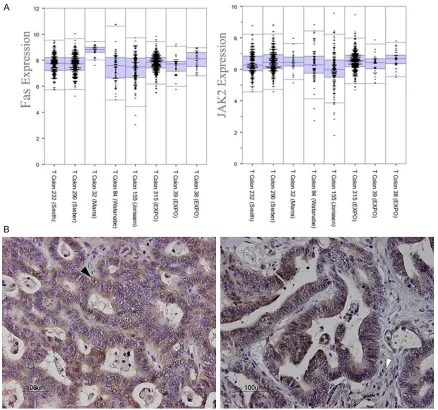
[image:6.612.93.396.85.187.2]in eight separate CRC patient cohorts in order to investigate if FAS expression would corre- late with CRCs prognosis. Although direct com-parison of expression levels between FAS and JAK2 is not available based on mRNA microar-ray data, it is shown that both FAS and JAK2 are expressed at relatively high level in most colorectal tumors (Figure 1A). Immunohistoc- hemical staining for FAS in CRC sections
Table 1. FAS associated pathways by KEGG pathway finder
KEGG pathway (Smith et al)P value (Sieber et al)P value (Combined) P value Chemokine signaling pathway 1.0e-05 3.1e-05 3.5e-09 Antigen processing and presentation 6.9e-05 9.5e-06 5.3e-09
Apoptosis 4.5e-03 0.04 1.4e-03
ECM receptor interaction 9.2e-04 4.0e-04 1.3e-06 T cell receptor signaling pathway 2.1e-04 9.7e-03 1.5e-05 Cytokine cytokine receptor interaction 1.1e-03 9.7e-03 5.0e-05
FAS-associated pathways in each separate tumor cohort were analyzed using KEGG
pathways, which shows pathways significantly associated with FAS expression in two data
sets. Combined P values were calculated using the web tool Meta P software.
Table 2. FAS associated pathways by Gene Ontology (GO)
GO (Smith et al)P value (Sieber et al)P value (Combined)P value
Immune Response 4.4e-69 5.1e-70 1.3e-135
Antigen processing and presentation 7.6e-259 1.4e-272 2.8 e-509 Inflammatory response 1.6e-134 1.0e-140 1.8 e-270
FAS-associated pathways in each separate tumor cohort were analyzed using GO terms function in R2. Combined P values were calculated using the web tool Meta P software.
showed both tumor me- mbrane and cytoplasm staining with moderate to strong staining in 8 of 10 CRC patients (Figure 1B). Then, survival curve was done to assess wh- ether FAS expression wo- uld associate with clini-cal outcome. Using R2 database, FAS associat-ed survival curves were done on several CRC co- horts which demonstrat-ed high FAS expression in primary CRC correlat-ed with poor prognosis (Figure 2).
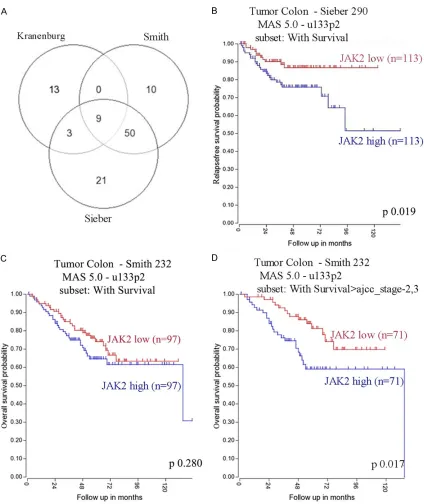
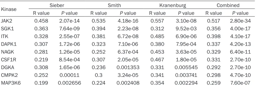
[image:6.612.93.397.252.318.2]0.01), among which JAK2 was on top with com-bined p value 2.8e-34 (Figure 4A; Table 3).
As a member of JAKs, JAK2/STAT3 seems to be an important mediator of chemokine (such as
STAT signaling, inflammation and
epithelial-mesenchymal transition (EMT). Besides, Gene- Venn was performed to three separate CRC cohorts and found 9 genes encoding kinases
[image:8.612.94.518.69.573.2]that were significantly associated with FAS (P <
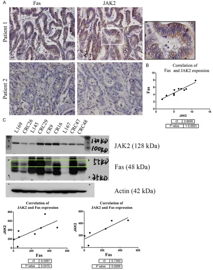
int × mm, and correlation analysis links JAK2 to FAS (P-value < 0.05, r=0.86) (Figure 5C).
JAK2 inhibition suppresses CRC cells clone formation capacity in vitro
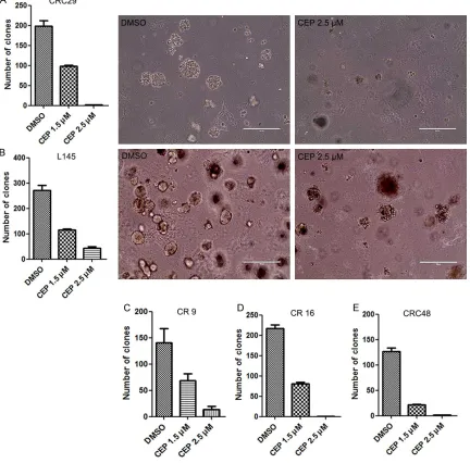
JAK2 was first found as a constitutively activat-ed tyrosine kinase in many myeloproliferative neoplasms (MPNs) [26, 27]. Since high JAK2 expression correlates shorter survival in CRC, we presumed that JAK2 might contribute to CRC cell proliferation. In order to evaluate the role of JAK2 in CRC tumor genesis, single cells were seeded in Matrigel supplemented with culture medium, with or without CEP33779 (JAK2 specific inhibitor) treatment. Here, each condition was done in triplicates. Each experi-ment was performed three times separately. A significant reduction in number of tumor clones were detected in CEP33779 treated group in dose dependent manner, compared with DMSO treated group (Figure 6). Besides, tumor clones in CEP33779 environment was much smaller than those in DMSO environment. These sug-gest that JAK2 inhibition significantly suppress CRC cells clone formation capacity.
Discussion
Finding potential markers for predicting out-comes of colorectal cancer patients would make it of great value for the clinic to seek out other possible target treatment. In this article, we present evidence that colorectal cancer (CRC) tissue express JAK2 and FAS which sig-nificantly associate with disease free survival (DFS) and overall survival (OS) in non-metasta-sis CRC patients.
IL-6) signal in promoting tumor genesis and metastatic progression[22]. Persistent activa-tion of JAK/STAT signaling is implicated in not only tumor genesis but also tumor survival and metastasis[23]. In order to see whether JAK2 expression would correlate with poor survival, we analyzed JAK2 expression in two separate CRC cohorts in a Kaplan Meier curve, and found that high JAK2 expression associate with poor disease free survival (DFS) and overall survival (OS) (Figure 4B-D).
JAK2 has good correlation with FAS both in patients’ slides and cell lines
[image:9.612.91.527.83.229.2]The above gene microarray analysis and bioin-formatics study link JAK2 expression to D95 expression in tumor local immune response, chemokine signaling and CRC progression or metastasis. To test if there was confident cor-relation between JAK2 and FAS, we performed IHC on CRC paraffin-embedded sections and western blots using patient originated cell lines CRC 29, CRC47, CR9, CR16, CRC48, CRC26, and liver metastatic cell lines L169, L167, L145. IHC images suggest that CRC tumor tis-sue with high FAS expression also express high level of JAK2, and vice versa (Figure 5A). Linear analysis for JAK2 and FAS expression reveals JAK2 and FAS expression have good correla- tion (Figure 5B). Next, we lysed CRC cells and performed western blot. We probed western blots (WB) with JAK2 and FAS antibody followed by secondary HRP-conjugated antibody. WB show that both proteins express in majority CRC cell lines at various levels. Using quantity one (Bio rad) software, the protein expression levels on WB were quantified in intensity form
Table 3. Kinases positively associated with FAS
Kinase Sieber Smith Kranenburg Combined
R value P value R value P value R value P value R value P value
JAK2 0.458 2.07e-14 0.535 4.18e-16 0.557 3.10e-08 0.517 2.80e-34
SGK1 0.363 7.64e-09 0.394 2.23e-08 0.312 9.52e-03 0.356 4.00e-17
ITK 0.328 2.55e-07 0.381 6.72e-08 0.485 6.90e-06 0.398 4.10e-17
DAPK1 0.307 1.72e-06 0.323 7.10e-06 0.380 7.95e-04 0.337 4.20e-13
NAGK 0.281 1.26e-05 0.252 6.37e-04 0.453 3.63e-05 0.329 6.40e-11
CSF1R 0.219 8.54e-04 0.307 2.05e-05 0.467 1.80e-05 0.331 2.70e-10
DGKA 0.308 1.65e-06 0.236 0.001353 0.331 0.005545 0.292 2.70e-10
CMPK2 0.252 0.00011 0.3 3.24e-05 0.341 0.003741 0.298 4.70e-10
MAP3K6 0.199 0.002656 0.224 0.002408 0.354 0.002294 0.259 7.60e-07
found that JAK2 may to some degree contrib-ute to colitis-induced colorectal cancer in a mouse model [28]. In the present study, we found that JAK2 expression level is moderate to high in separate cohorts and high JAK2 expres-sion correlates with shorter DFS and OS, sug-gesting that JAK2 may play a positive role in tumor progression and invasion. By using ‘KEGG pathway finder’ software, we analyzed JAK2 is a member of the Janus kinase (JAK)
[image:11.612.93.525.79.505.2]family, which also includes JAK1, JAK3 and TYK2. JAK2 is a non-receptor tyrosine kinase which is widely expressed and residue mostly in cytoplasm, functionally mediating cytokine induced inflammation. Aberrant JAK2 signaling was found in myeloproliferative neoplasms (MPNs). Given that some colorectal tumors are associated with inflammation, researchers
cancers is strongly correlated with FAS expres-sion, patients’ survival, especially in non-meta-static colorectal cancers. We also found that JAK2 plays an important role in FAS associated gene networks. We further show JAK2 inhibi-tion can suppress tumor colony formainhibi-tion in cell cultures by using JAK2 specific inhibitors working in medium/Matrigel culturing system. Importantly, high JAK2 expression levels were found to be correlated with poor overall survival in advanced CRC patients. Thus, these results provide preliminary value to support the further evaluation of JAK2 and FAS as a prognostic bio-marker and novel therapeutic targets in CRCs.
Disclosure of conflict of interest
None.
Address correspondence to: Wei Xu, Department of Clinical Laboratory, The First Hospital of Jilin University, Jilin, China. Tel: +86 431 88782622; Fax: +86 431 85654528; E-mail: [email protected]
References
[1] O’Reilly LA, Tai L, Lee L, Kruse EA, Grabow S, Fairlie WD, Haynes NM, Tarlinton DM, Zhang JG, Belz GT, Smyth MJ, Bouillet P, Robb L, Strasser A. Membrane-bound Fas ligand only is essential for Fas-induced apoptosis. Nature 2009; 461: 659-63.
[2] Yurchenko M, Shlapatska LM, Sidorenko SP. The multilevel regulation of CD95 signaling outcome. Exp Oncol 2012; 34: 153-9.
[3] Cullen SP, Henry CM, Kearney CJ, Logue SE, Feoktistova M, Tynan GA, Lavelle EC, Leverkus M, Martin SJ. Fas/CD95-induced chemokines can serve as “find-me” signals for apoptotic cells. Mol Cell 2013; 49: 1034-48.
[4] Scott FL, Stec B, Pop C, Dobaczewska MK, Lee JJ, Monosov E, Robinson H, Salvesen GS, Schwarzenbacher R, Riedl SJ. The Fas-FADD death domain complex structure unravels sig-nalling by receptor clustering. Nature 2009; 457: 1019-22.
[5] Schleich K, Warnken U, Fricker N, Oztürk S, Richter P, Kammerer K, Schnölzer M, Krammer PH, Lavrik IN. Stoichiometry of the CD95 death-inducing signaling complex: experimen-tal and modeling evidence for a death effector domain chain model. Mol Cell 2012; 47: 306-19.
[6] Dickens LS, Boyd RS, Jukes-Jones R, Hughes MA, Robinson GL, Fairall L, Schwabe JW, Cain K, Macfarlane M. A death effector domain chain DISC model reveals a crucial role for cas-pase-8 chain assembly in mediating apoptotic cell death. Mol Cell 2012; 47: 291-305. the correlation between JAK2 and FAS which
provide evidence of the correlation. This is con-sistent with CRC cell cultures in vitro. Thus, we assume that JAK2 may help tumor growth in FAS expressing CRCs.
Tumor progression and metastasis are two main cause of poor prognosis. In previous study, we found that FAS contributed to CRC cells invasive behavior via cofilin pathway [29]. In some mouse models of CRC liver metastasis, loss of FAS reduces liver metastasis [30, 31]. And except for inducing apoptosis, FAS can also promote tumorigenesis [32], possibly by acti-vating non-apoptotic signaling pathways that stimulate tumor cell proliferation, invasion and metastasis [33, 34]. Interestingly, at least in part, this process depends on FLIP, possibly switching FAS signaling from apoptotic to non-apoptotic JNK or ERK/NFkB signaling [35] instead of FADD caspases activation. Furth- ermore, JAK2 inhibition impairs clone forming capacity of CRC cells, suggesting that JAK2 inhibition blocks surviving pathways including FAS pathway.
In conclusion, this study suggests that JAK2 and FAS are two of the most important mole-cules in CRC tumor progression, and aberrant expression of both proteins are significantly associated with DFS and OS in non-metastatic CRCs. These data contribute to future improve-ment in prognosis for patients with colorectal cancer and provide potential target for antitu-mor therapy. Up to day, there are only limited reports with respect to using JAK2 and FAS as prognostic factor in predicting outcomes for CRC patients after surgery. Therefore, large population-based research is required and more in vitro and in vivo experiments are need-ed to support our findings.
Translational relevance
Bailey CE, Eschrich S, Kis C, Levy S, Washing-ton MK, Heslin MJ, Coffey RJ, Yeatman TJ, Shyr Y, Beauchamp RD. Experimentally derived me-tastasis gene expression profile predicts recur-rence and death in patients with colon cancer. Gastroenterology 2010; 138: 958-968. [17] Jorissen RN, Gibbs P, Christie M, Prakash S,
Lipton L, Desai J, Kerr D, Aaltonen LA, Arango D, Kruhøffer M, Orntoft TF, Andersen CL, Gruidl M, Kamath VP, Eschrich S, Yeatman TJ, Sieber OM. Metastasis-associated gene expression changes predict poor outcomes in patients with dukes stage B and C colorectal cancer. Clin Cancer Res 2009; 15: 7642-7651. [18] Bewick V, Cheek L, Ball J. Statistics review 12:
survival analysis. Crit Care 2004; 8: 389-394. [19] Steller EJ, Borel Rinkes IH, Kranenburg O. How
CD95 stimulates invasion. Cell Cycle 2011; 10: 3857-62.
[20] Bolstad BM, Irizarry RA, Astrand M, Speed TP. A comparison of normalization methods for high density oligonucleotide array data based on variance and bias. Bioinformatics 2003; 19: 185-193.
[21] Smith JJ, Deane NG, Wu F, Merchant NB, Zhang B, Jiang A, Lu P, Johnson JC, Schmidt C, Bailey CE, Eschrich S, Kis C, Levy S, Washing-ton MK, Heslin MJ, Coffey RJ, Yeatman TJ, Shyr Y, Beauchamp RD. Experimentally derived me-tastasis gene expression profile predicts recur-rence and death in patients with colon cancer. Gastroenterology 2010; 138: 958-68.
[22] Kiu H, Nicholson SE. Biology and significance of the JAK/STAT signalling pathways. Growth Factors 2012; 30: 88-106.
[23] Bournazou E, Bromberg J. Targeting the tumor microenvironment: STAT3 signaling. JAK-STAT 2013; 2: e23828.
[24] Snoeren N, Emmink BL, Koerkamp MJ, van Hooff SR, Goos JA, van Houdt WJ, de Wit M, Prins AM, Piersma SR, Pham TV, Belt EJ, Bril H, Stockmann HB, Meijer GA, van Hillegersberg R, Holstege FC, Jimenez CR, Fijneman RJ, Kranenburg OW, Rinkes IH. Maspin is a marker for early recurrence in primary stage III and IV colorectal cancer. Br J Cancer 2013; 109: 1636-47.
[25] Pirooznia M, Nagarajan V, Deng Y. GeneVenn-A web application for comparing gene lists using Venn diagrams. Bioinformation 2007; 1: 420-2.
[26] Levine RL, Pardanani A, Tefferi A, Gilliland DG. Role of JAK2 in the pathogenesis and therapy of myeloproliferative disorders. Nat Rev Cancer 2007; 7: 673-683.
[27] Constantinescu SN, Girardot M, Pecquet C. Mining for JAK-STAT mutations in cancer. Trends Biochem Sci 2008; 33: 122-131. [7] Kischkel FC, Hellbardt S, Behrmann I, Germer
M, Pawlita M, Krammer PH, Peter ME. Cytotox-icity-dependent APO-1 (Fas/CD95)-associated proteins form a death-inducing signaling com-plex (DISC) with the receptor. EMBO J 1995; 14: 5579-88.
[8] Cullen SP, Henry CM, Kearney CJ, Logue SE, Feoktistova M, Tynan GA, Lavelle EC, Leverkus M, Martin SJ. Fas/CD95-induced chemokines can serve as “find-me” signals for apoptotic cells. Mol Cell 2013; 49: 1034-48.
[9] Chen L, Park SM, Tumanov AV, Hau A, Sawada K, Feig C, Turner JR, Fu YX, Romero IL, Lengyel E, Peter ME. CD95 promotes tumour growth. Nature 2010; 465: 492-6.
[10] Barnhart BC, Legembre P, Pietras E, Bubici C, Franzoso G, Peter ME. CD95 ligand induces motilityand invasiveness of apoptosis-resistant tumor cells. EMBO J 2004; 23: 3175-85. [11] Hoogwater FJ, Nijkamp MW, Smakman N,
Stell-er EJ, Emmink BL, Westendorp BF, Raats DA, Sprick MR, Schaefer U, Van Houdt WJ, De Bruijn MT, Schackmann RC, Derksen PW, Medema JP, Walczak H, Borel Rinkes IH, Kranenburg O. Oncogenic K-Rasturns death receptors into metastasis-promoting receptors in human and mouse colorectal cancer cells. Gastroenterology 2010; 138: 2357-67.
[12] Tauzin S, Chaigne-Delalande B, Selva E, Khadra N, Daburon S, Contin-Bordes C, Blanco P, Le Seyec J, Ducret T, Counillon L, Moreau JF, Hofman P, Vacher P, Legembre P. The natural-lyprocessed CD95L elicits a c-yes/calcium/ PI3K-drivencell migration pathway. PLoS Biol 2011; 9: e1001090.
[13] Kleber S, Sancho-Martinez I, Wiestler B, Beisel A, Gieffers C, Hill O, Thiemann M, Mueller W, Sykora J, Kuhn A, Schreglmann N, Letellier E, Zuliani C, Klussmann S, Teodorczyk M, Gröne HJ, Ganten TM, Sültmann H, Tüttenberg J, von Deimling A, Regnier-Vigouroux A, Herold- Mende C, Martin-Villalba A. Yes and PI3K bind CD95 to signal invasion of glioblastoma. Can- cer Cell 2008; 13: 235-48.
[14] O’Reilly LA, Tai L, Lee L, Kruse EA, Grabow S, Fairlie WD, Haynes NM, Tarlinton DM, Zhang JG, Belz GT, Smyth MJ, Bouillet P, Robb L, Strasser A. Membrane-bound Fas ligand onlyis essential for Fas-induced apoptosis. Nature 2009; 461: 659-63.
[15] Tauzin S, Chaigne-Delalande B, Selva E, Khadra N, Daburon S, Contin-Bordes C, Blanco P, Le Seyec J, Ducret T, Counillon L, Moreau JF, Hofman P, Vacher P, Legembre P. The natural-lyprocessed CD95L elicits a c-yes/calcium/ PI3K-drivencell migration pathway. PLoS Biol 2011; 9: e1001090.
[32] Hoogwater FJ, Steller EJ, Westendorp BF, Borel Rinkes IH, Kranenburg O. CD95 signaling in colorectal cancer. Biochim Biophys Acta 2012; 1826: 189-98.
[33] Peter ME, Budd RC, Desbarats J, Hedrick SM, Hueber AO, Newell MK, Owen LB, Pope RM, Tschopp J, Wajant H, Wallach D, Wiltrout RH, Zörnig M, Lynch DH. The CD95 receptor: apop-tosis revisited. Cell 2007; 129: 447-50. [34] Peter ME, Legembre P, Barnhart BC. Does
CD95 have tumor promoting activities? Bio-chim Biophys Acta 2007; 1775: 233-4. [35] Martin-Villalba A, Llorens-Bobadilla E, Wollny
D. CD95 in cancer: tool or target? Trends Mol Med 2013; 19: 329-35.
[28] Seavey MM, Lu LD, Stump KL, Wallace NH, Hockeimer W, O’Kane TM, Ruggeri BA, Dobr-zanski P. Therapeutic efficacy of CEP-33779, a novel selective JAK2 inhibitor, in a mouse mod-el of colitis-induced colorectal cancer. Mol Cancer Ther 2012; 11: 984-93.
[29] Steller EJ, Ritsma L, Raats DA, Hoogwater FJ, Emmink BL, Govaert KM, Laoukili J, Rinkes IH, van Rheenen J, Kranenburg O. The death re-ceptor CD95 activates the cofilin pathway to stimulate tumour cell invasion. EMBO Rep 2011; 12: 931-7.
[30] Li H, Fan X, Stoicov C, Liu JH, Zubair S, Tsai E, Ste Marie R, Wang TC, Lyle S, Kurt-Jones E, Houghton J. Human and mouse colon cancer utilizes CD95 signaling for local growth and metastatic spread to liver. Gastroenterology 2009; 137: 934-44.
![Figure 3. Heatmap analysis for FAS-associated genes and kinases. Heat map lists FAS associated genes in three colorectal cancer cohorts (Smith et al [16], Sieber et al [17], Kranenburg et al [24]) according to FAS expression in the categories including JAK](https://thumb-us.123doks.com/thumbv2/123dok_us/810497.589234/7.792.104.699.77.391/associated-associated-colorectal-kranenburg-according-expression-categories-including.webp)