• No results found

Original Article Endolymphatic sac tumor: clinical, radiological and pathological analyses of four cases

N/A
N/A
Protected

Academic year: 2020

Share "Original Article Endolymphatic sac tumor: clinical, radiological and pathological analyses of four cases"

Copied!
6
0
0

Loading.... (view fulltext now)

Full text

Loading

Figure

Table 1. Clinical features in 4 cases with endolymphatic sac tumor (ELST)
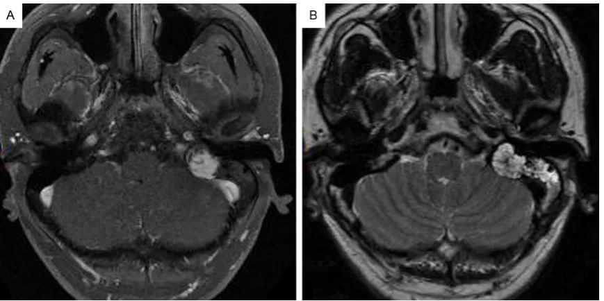
Figure 1. Radiologic characterization of endolymphatic sac tumor (ELST). A: Magnetic resonance images scan re-vealed a 19 mm×18 mm mass in the left petrous segment of temporal bone which was irregular, heterogeneous signal intensity on T1-weighted axial im
Figure 2. Histopathologic characterization of ELST. A: Histopathologic examination of the tumor showed papillary scription factor (TTF)-1and cystic structures (Hematoxylin and eosin, HE, original magnification ×100)
Table 3. Immunohistochemical features in 4 cases with endolym-phatic sac tumor (ELST)

References

Related documents