Original Article
PGE2 regulating Cx43 to affect testosterone synthesis
in rats leydig cell exposed at DBP
Jing Zhang, Yi Zhang, Jin-Chang Zhao, Huan Li
Department of Environmental Hygiene, Institute of Public Health, Beihua University, Jilin, China
Received December 13, 2015; Accepted February 18, 2016; Epub April 1, 2016; Published April 15, 2016
Abstract: To observe the effect of dibutyl phthalate (DBP) on connexin 43 (Cx43) proteins in rat testicular Leydig cells and testosterone secretion induced by Cx43, and to investigate whether Cx43 is involved in the regulation of testosterone by DBP. Purified Leydig cells of the primary culture were divided into 3 groups, 50 µg/ml DBP treatment group, 50 µg/ml DBP+10 µmol/L PGE2 group and DMSO control group, which were processed for 24 hours, respec -tively. Expression of Cx43 on cell membrane was detected by immunofluorescence, and total protein level of Cx43 was evaluated by Western blot. Testosterone production was detected by testosterone kit. Compared with control group, Cx43 expression and testosterone levels were both significantly reduced after DBP processing for 24 hours (P<0.05). Compared with DBP group, the addition of Cx43 enhancer (PGE2) enabled Cx34 expression recovery, but the change of testosterone level was insignificant (P>0.05). Both testosterone and Cx43 can be regulated by DBP. However, the inhibitory effect of DBP on testosterone synthesis in Leydig cells may not be necessarily achieved through the regulation of Cx43 expression.
Keywords: Dibutyl phthalate, Cx43, leydig cells
Introduction
DBP is a plasticizer widely present in every aspect of human life. Because there is no chemical bond between DBP and the polymer matrix, DBP is able to migrate out of the gross product, such as plastics, thus causing environ-mental pollutions [1]. A number of studies have revealed the significant reproductive toxicity to the male induced by DBP. But different from other androgen antagonists, instead of working on androgen receptors (AR) directly, the repro-ductive toxicity of DBP is induced by interfer-ence with the biosynthesis of testosterone in testicular Leydig cells. However, the detailed mechanism of the action still remains unclear [2, 3].
Gap junction intercellular communication (GJIC) is a way of communication between adjacent cells to achieve signal transduction. Extensively present in the testes, GJIC is essential in tes -ticular development, as well as the initiation and maintenance of spermatogenesis [4]. As the predominant GJIC protein expressed in the testes, Cx43 has been reported to play a key
role in spermatogenesis and the regulation of GJIC [5, 6]. Cx43 is also the only protein identi -fied so far expressed in Leydig cells [7], yet its function in testicular Leydig cells has not been extensively studied.
Thought its reproductive toxicity has been broadly reported, investigations of the effect of DBP on testosterone synthesis in testicle Leydig cells remain blank. By using Western blot and immunofluorescence, this study observed the influence of DBP on Cx43 and testosterone, and investigated the involvement of Cx43 change in DBP’s effect on testosterone.
Materials and methods
Materials
-tosterone kit were purchased from Sigma Co. (US). Percoll cell separation media was bought from Pharmacia. 4’,6-diamidino-2-phenylindole (DAPI), mouse anti-rat Cx43 polyclonal antibody and rabbit anti-rat β-actin polyclonal antibody were purchased from Solarbio Co. The rest materials were all domestic analytical reagents. Isolation, identification and treatment expo -sure of leydig cells
The methods reported by Genissel C. et al. [8] were used and improved. One adult male rat, weighted around 300 g, was anesthetized with ether, euthanized by cervical dislocation and disinfected using alcohol. Under sterile condi -tions, the testes were removed and decapsu-lated. After flushed with D-Hanks buffer, the testes were added with 0.5% collagen I and digested in a shaking water bath at 34°C for 5-6 min. Digestion was terminated by adding 10% fetal bovine serum. Supernatant was col-lected after standing 2-3 min and filtered using 200 mesh cell filters. The cells were washed with D-Hanks buffer, centrifuged at 1000 r/min for 5 min and rinsed twice. Cells were added with DMEM/F12 culture media containing 10% fetal bovine serum and pipetted repeatedly to make a 2 ml suspension. 2 ml of each Percoll gradient medium was pipetted and slowly added into a 10 ml centrifuge tube from high to low density (gradients from bottom to top were 70%, 58%, 30% and 5%, respectively), and cell suspension was added to the top. The tube was centrifuged and allowed to stand for 5 min at room temperature. The band of target cells, which was located between 30% and 58% Percoll gradients, was sucked up, centrifuged and washed twice. Cells were counted and diluted into a concentration of 10×105/ml. The
purity of cells was examined by 3β-HSD stain -ing in 6-well plates. The cells were divided into 3 groups, 2 experiment group treated with 50 µg/ml DBP or 50 µg/ml DBP+10 µmol/LPGE, and control group treated with DMSO. Cells were incubated for 24 h at 37°C in a 5% CO2 incubator.
Detection of testosterone levels in leydig cells
Cell culture in each group was collected 24 h after treatment. Testosterone levels were de- tected using radioimmunoassay with testoster-one kits purchased from Sigma Co. (US). Assay was performed following the manufacture pro-tocol of the kit.
Radioimmunoassay of Cx43
After culture for 24 h, old media were discard-ed, and Leydig cells in each group were incu-bated in serum free media for another 6 h. The following steps were performed: cells were fixed in 4% paraformaldehyde at room tempera -ture for 30 min. Rabbit anti-rat Cx43 polyclonal antibody was added to cells (1:100) and incu-bated overnight at 4°C. Cells were added with TRITC labeled IgG and incubated at room tem -perature for 1 h. The nucleus was stained using DAPI (100 ng/ml), incubated at room tempera-ture for 5 min and mounted in 50% buffered glycerol. The cellular expression of Cx43 was observed using fluorescence microscope.
Cx43 expression detected by Western blotting
Cells were collected and lysed completely by sonication in lysis buffer. Cellular protein sam-ples were collected and protein concentration was detected using BCA assay. 50 μg protein sample was loaded in each lane. After SDS-PAGE, samples were transferred to NC mem -brane and incubated with Cx43 primary anti-body overnight (1:100) at 4°C. The membrane was then washed three times using TBST, for 10 min each time. After added with goat anti-rabbit secondary antibody (1:1000), the mem-brane was incubated in dark at room tempera-ture for 1 h. The substrates were illuminated and imaged by infrared scanning system. Quantitative analysis of the intensity of the bands of interest was conducted using Quantity One software.
Statistical analysis
Experimental data were analyzed by SPSS13.0 software. Data were expressed by mean ± standard deviation (_x±s). P<0.05 indicated that difference was statistically significant.
Results
Cultivation and Identification of rat testicular
interstitial cells
above cells reached 3×10⁶ to 4.5×10⁶ (Figure
1).
Results of testosterone levels
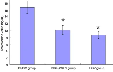
Cells exhibited significant decreased testoster -one levels 24 h after exposed to DBP, com-pared with the control group. However, the tes-tosterone level in cells treated with DBP+PGE2 was not recovered, shown by statistically insig-nificant difference (P>0.05) (Table 1 and Figure 2).
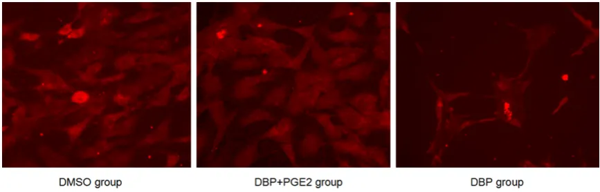
Immunofluorescence assay of Cx43 expres -sion in testicular interstitial cell membrane
Cx43 was extensively expressed among cells, shown by immunofluorescence assay. 24 h after DBP exposure, Cx43 expression on cell membrane was significantly inhibited. In con -trol group, Cx43 was mainly expressed on cell membrane, while the expression was signifi -cantly reduced and mainly present in cytoplasm after DBP exposure. The addition of Cx43 en- hancer PGE recovered Cx43 level close to that of control group (Figure 3).
Cx43 total protein expression in testicular in -terstitial cells
As shown by Western blot, Cx43 total protein express was significantly inhibited 24 h after Leydig cells exposed to DBP, compared with control group. Cx43 total protein level was gradually recovered in the treatment group of DBP+PGE2 compared with DBP alone (Figure 4).
Discussion
[image:3.629.99.531.77.250.2]DPB, widely recognized as an endocrine disrup-tor in the environment, is extensively present in polyvinyl chloride, construction materials, food packaging fixative, detergents, lubricants and medical devices. The US NTP has confirmed that DBP has significant male reproductive tox -icity which can cause rat testicular atrophy, decreased weight, loss of spermatocytes and sperms, degeneration and atrophy of seminifer-ous tubules, genital tract anomalies, etc. The
[image:3.629.98.317.314.386.2]Figure 1. Identification and cultivation of testicular leydig cells.
Table 1. Analysis of testosterone level in different groups (n=6)
Groups Testosterone level (ng/ml)
DMSO group 16.92±1.85
DBP+PGE2 group (50 μg/ml+10 μmol/l) 10.16±1.31*
DBP group (50 μg/ml) 8.73±1.08*
Note: *compared with DMSO group (P<0.05).
[image:3.629.99.294.414.538.2]cause of these changes is mainly associated with the anti-androgen effect of DBP [9, 10]. In this study, we also demonstrated that DBP could reduce testosterone levels.
GJIC is a way of signal communication achieved by gap junction structure formed by connexin proteins. Present between adjacent cells, GJIC plays a key role in the maintenance of cell signal transduction, regulation of cell prolifera-tion and differentiaprolifera-tion, as well as maintenance of tissue homeostasis [11]. Connexin 43 is the predominant gap junction (GJ) protein expre-ssed in the testes. Ralph Brehm et al. using gene knock-out technology, specifically knoc-ked out Cx43 in rat Sertoli cells. The testes of these rats after they reached sexual maturity were only weighed 50% of that in the control group. Shown by Immunohistochemistry test-ing, the Sertoli cells continuously proliferated and exhibited abnormal differentiation; germ cells were absent in adlunimal compartment, while cells in basal compartment were normal;
seroli cells-only syndrome was observed [12, 13]. According to the immunofluorescence results reported by Chen et al., the location of Cx43 expression was similar to that of ZO-1. At the stage 8 of seminiferous epithelium cycle, Cx43 expression changed significantly during the process of renewal of tight junctions [14]. Cx43 is the only connexin protein expressed in Leydig cells. It was shown that the increase of number and volume of Leydig cells was accom-panied by increased level of Cx43 [15].
The authors speculated that DBP might exert an influence toward Cx43 in Leydig cells, and that the regulation of testosterone level by DBP might be mediated by targeting Cx43. In this study, immunofluorescence and Western blot results showed that DBP inhibited Cx43 expres-sion in Leydig cells and the concurrently de- creased testosterone secretion. However, in the group with PGE, immunofluorescence and Western blot demonstrated that Cx43 level was recovered but not accompanied by enhanced
[image:4.629.97.531.79.218.2]Figure 3. Cx43 expression in leydig cells detected by immunofluorescence.
testosterone level, indicating that Cx43 was not involved in the testosterone regulation by DBP. In summary, DBP is able to down-regulate Cx43 expression and testosterone level in testicular Leydig cells, but the influence of DBP on testes are is not through Cx43 expression inhibition. Since Cx43 is extensively expressed in testicu-lar Leydig cells but not regulating testosterone secretion, further investigations are still need-ed to explore the function of Cx43 in Leydig cells.
Acknowledgements
This work was supported by the Science and Technological Project of Jilin Province in china (No.20130101141JC); the nature and science fund from jilin province ministry of education (No.2014211; No.2016071); 2015 National University Student Innovation Program of Bei-hua University.
Disclosure of conflict of interest
None.
Address correspondence to: Dr. Huan Li, Depart- ment of Environmental Hygiene, Institute of Public Health, Beihua University, 3999 Binjiang Road, Jilin 132013, China. Tel: 18604498417; Fax: +86-18604498417; E-mail: [email protected]
References
[1] Adibi JJ, Whyatt RM, Williams PL, Calafat AM, Camann D, Herrick R, Nelson H, Bhat HK, Perera FP, Silva MJ and Hauser R. Charac-terization of phthalate exposure among preg-nant women assessed by repeat air and urine samples. Environ Health Perspect 2008; 116: 467-473.
[2] Thompson CJ, Ross SM and Gaido KW. Di (n-butyl) phthalate impairs cholesterol transport and steroidogenesis in the fetal rat testis through a rapid and reversible mechanism. Endocrinology 2003; 145: 1227-1237. [3] Gray LE Jr, Wilson VS, Stoker T, Lambright C,
Furr J, Noriega N, Howdeshell K, Ankley GT, Guillette L. Adverse effects of environmental antiandrogens and androgens on reproductive development in mammals. Int J Androl 2006; 29: 96-104.
[4] Pointis G, Gilleron J, Carette D and Segretain D. Physiological and physiopathological as-pects of connexins and communicating gap
junctions in spermatogenesis. Philosophical transactions of the Royal Society of London. Philos Trans R Soc Lond B Biol Sci 2010; 365: 1607-1620.
[5] Cyr DG. Connexins and pannexins Coordinating cellular communication in the testis and epi-didymis. Spermatogenesis 2011; 1: 325-338. [6] Cheng CY and Mruk DD. The blood-testis bar
-rier and its implications for male contracep-tion. Pharmacol Rev 2012; 64: 16-64. [7] Risley MS, Tan IP and Farrell J. Gap junctions
with varied permeability properties establish cell-type specific communication pathways in the rat seminiferous epithelium. Biol Reprod 2002; 67: 945-952.
[8] Sadar MD and Andersson TB. Regulation of cy -tochrome P450 in a primary culture of rainbow trout hepatocytes. In Vitro Cell Dev Biol Anim 2001; 37: 180-184.
[9] Mahood IK, Scott HM, Brown R, Hallmark N, Walker M and Sharpe RM. In utero exposure to di(n-butyl) phthalate and testicular dysgenesis: comparison of fetal and adult end points and their dose sensitivity. Environ Health Perspect 2007; 1: 55-61.
[10] Matsumoto M, Hirata-Koizumi M and Ema M. Potential adverse effects of phthalic acid es-ters on human health: A review of recent stud-ies on reproduction. Regul Toxicol Pharmacol 2008; 50: 37-49.
[11] Saez JC, Berthoud VM, Branes MC, Martinez AD and Beyer EC. Plasma membrane channels formed by connexins: their regulation and functions. Physiol Rev 2003; 83: 1359-1400. [12] Sridharan S, Simon L, Meling DD, Cyr DG,
Gutstein DE, Fishman GI, Guillou F and Cooke PS. Proliferation of adult sertoli cells following conditional knockout of the Gap junctional pro -tein GJA1 (connexin 43) in mice. Biol Reprod 2007; 76: 804-812.
[13] Brehm R, Zeiler M, Rüttinger C, Herde K, Kibschull M, Winterhager E, Willecke K, Guillou F, Lécureuil C, Steger K, Konrad L, Biermann K, Failing K and Bergmann M. A sertoli cell-specif -ic knockout of connexin43 prevents initiation of spermatogenesis. Am J Pathol 2007; 171: 19-31.
[14] Li MW, Mruk DD, Lee WM and Cheng CY. Connexin 43 is critical to maintain the homeo-stasis of the blood-testis barrier via its effects on tight junction reassembly. Proc Natl Acad Sci U S A 2010; 107: 17998-18003.