Original Article
MiR-296 promotes colorectal cancer
cells growth through regulating NF-κB
Huiyuan Zhai1,2*, Minghua Sui3*, Lixin Jiang2, Jinchen Hu2, Xiaorui Jiang4, Yuan Yuan5, Mingchuan Li6, Zhenyu Yu6, Sanyuan Hu1
1Department of General Surgery, Qilu Hospital, Shandong University, Jinan, China; Departments of 2 Gastrointesti-nal Surgery, 3Oncology, 4Orthopedics, 5Neurosurgery, 6Anesthesiology, Yantai Yuhuangding Hospital, Yantai, China. *Equal contributors.
Received December 1, 2015; Accepted January 31, 2016; Epub April 1, 2016; Published April 15, 2016
Abstract: Tumor reduced people’s quality of life. Colon cancer is hard to cure. MicroRNA abnormal expression in-duces oncogenesis. We intended to explore miR-296 role in colorectal cancer development. MiR-296 expression in colorectal cancer was tested in clinical samples. MiR-296 overexpression and low expression colorectal cancer cell lines were established to detect NF-κB activation. Western blot was applied to determine P65 and pP65 proteins ex-pression. MTT and Colony formation assay were used to evaluate colorectal cancer cell proliferation. Q-PCR showed that miR-296 overexpressed in colorectal cancer patients, and it also confirmed that miR-296 overexpression and low expression colorectal cancer cell lines were successfully constructed. Western blot revealed that miR-296 over-expression may lead to pP65 level significantly increased, while P65 showed the opposite results. Proliferation assay including MTT and colony formation demonstrated that miR-296 overexpression promoted colorectal cancer cell proliferation, whereas miR-296 low expression weakened cancer cell proliferative ability. MiR-296 promotes colorectal cancer cells growth through regulating NF-κB signaling pathway.
Keywords: miR-296, NF-κB, colorectal cancer
Introduction
Colorectal cancer is considered to have high mortality among common malignant tumors, especially is in East Asia and South Africa. Furthermore, its incidence and mortality is also significantly higher in developing countries compared with developed countries. Colorectal cancer oncome is a multi-factor, multi-step, and complex process that related to genes abnormal expression [1, 2]. However, its poten-tial mechanism has not been fully classified. Many colorectal cancer patients quickly death is due to cancer cell rapid growth. Surgical resection is the potential treatment option with the best prognosis for long term colorectal can-cer patients. Since about 80% of colorectal cancer patients combined with intestinal inflammation, only 10-15% patients can be sur-gically resected [3, 4]. The left patients cannot receive surgical resection because of tumor size and potential of colorectal disease. More importantly, numerous patients may appear
that high AFP is a risk factor of colorectal can-cer recurrence [5, 6]. It is urgently needed to find the potential pathogenesis of colorectal cancer.
nucleus through ectogenic factors, resulting in nucleus phosphorylation to activate the signal-ing pathway, promote inflammation, and impact tumor occurrence and development.
Materials and methods
Clinical information
51 cases of colorectal cancer tissue samples and 50 cases of normal colon tissue speci-mens adjacent to carcinoma in the oncology department of Qilu Hospital, Shandong Uni- versity were collected between July 2014 and December 2014. All the enrolled subjects had signed informed consent and the study was approved by the ethical committee in Qilu Hospital, Shandong University. Clinical staging was based on TNM staging standard published by UICC in 2009. Patient’s medical records and the pathological data were complete.
Q-PCR
Reverse transcription kit was from TaKaRa. The specimen was treated with Trizol and centri-fuged at 12,000 r/min and 4°C for 10 min. Then the supernatant was moved to a new EP tube and let stand for 5 min. Next, the fluid was added with 200 μl chloroform and centrifuged at 12,000 r/min and 4°C for 15 min. After mov-ing the supernatant to a new tube and added with equal amount of isopropanol, the tube was centrifuged at 12,000 r/min and 4°C for 10 min. At last, after removing the supernatant, the tube was added with 75% ethanol (solved in DEPC water) and centrifuged at 7,500 r/min and 4°C for 5 min. Total RNA was solved in 20 μl DEPC water. Real time PCR was applied to detect miR-340 expression in gastric cancer. The primers sequences were as follows:
miR-296, F-GCGAGCTACATTGTCTGCTGGGTT, R-GT- CGAGGGTCCGAGGTATTCCG; U6, F-CGGCGGTA- GCTTATCAGACTGATG, R-CCAGTCGAGGGTCCGA- GGTATT. PCR reaction contained 95°C for 10 min, followed by 40 cycles of 95°C for 30 s, 55°C for 30 s, and 72°C for 30 s. The melting curve was analyzed.
MiR-296 stable transfected cancer cell line
HCT116 cells (purchased from ATCC) in loga-rithm phase were seeded in six-well plate at 3×10⁵. 5 μl lipo2000 was solved in 100 μl FBS free medium and stand for 5 min, while 12 μl miR-296 overexpressed or low expressed plas-mid (purchased from RiboBio) was solved in 100 μl FBS free medium for 5 min. Then the two fluids were mixed together for 20 min and added to cells in 1800 μl FBS free medium. RNA was extracted after 48 h for detection. MiR-296 overexpressed and low expressed plasmids were synthetized by RiboBio.
MTT assay
Single cell suspension in medium with 10% FBS was seeded in 96-well plate at 6,000 cells/200 μl. After HCT116 adherence, 20 μl MTT solution at 5 mg/ml was added to the well for 4 h. After removing the supernatant, 150 μl DMSO was added and the plate was vibrated for 10 min. At last, the plate was read at 490 nm to draw the cell growth curve.
Colony formation assay
100 cells were seeded in the petri dish after counting. The medium was removed after mac-roscopic colony formation. The cells were fixed with 5 ml 4% paraformaldehyde for 15 min. Then the fixing solution was removed and changed to crystal violet staining solution for 10~30 min. At last, the dish was dried in air after washing.
Western blot
Tissue protein was extracted using protein kit purchased from Beyotime and quantified by BCA. Reagent A was mixed with reagent B at 50:1 to form BCA solution. 2 μl cellular super-natant was added to 18 μl PBS and 200 μl AB mixture. The protein was separated by electro-phoresis and transferred to PVDF membrane. After blocked by 5% skim milk for 1 h, the
[image:2.629.99.296.78.199.2]brane was washed by TBST and incubated in primary antibody overnight. On the second day, the membrane was incubated in secondary antibody at room temperature after rewarming and washing. PVDF membrane was developed by ECL luminous fluid.
Results
MiR-296 expression
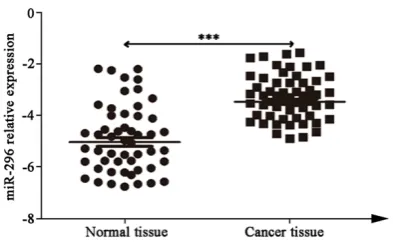
Q-PCR was applied to test miR-296 expression in 51 cases of colorectal cancer tissues and 50 cases of normal colon tissues adjacent to carci-noma. The results showed that miR-296 signifi-cantly overexpressed in colorectal cancer (P < 0.001) (Figure 1). Our results suggested that miR-296 may play an important role in colorec-tal cancer occurrence and development. MiR-296 activated NF-κB signaling pathway At first, we successfully constructed miR-296 overexpressed and low expressed cancer cell lines (Figure 2A and 2B). Western blot show- ed that P65 protein declined, while pP65 level elevated and nuclear import enhanced in miR-296 overexpressed HCT116 cells. They were opposite in miR-296 low expressed HCT116
sion leaded to tumor. MicroRNA received more and more study in colorectal cancer. A variety of miRNAs were discovered in early stage colorectal cancer screening, and some had been recognized as tumor suppressor genes or oncogenes. Colorectal cancer development was a typical multiple factors and multi-step process involving abnormal cell proliferation, apoptosis, invasion, and metastasis. MircoRNA connected to the 3’ UTRs of the base sequence that mainly regulated posttranscriptional level [11, 12]. Previous studies considered miRNAs as tumor suppressor genes or oncogenes main-ly through regulating cell proliferation, apopto-sis, metabolic pathway, and signaling pathways [13]. Many studies reported mircoRNA involved in colorectal cancer occurrence and develop-ment. There were more and more mircoRNAs had been used as tumor markers applied to clinical research, as a study reported that miR-21 can be treated as the marker of colorectal cancer. It was also considered that AFP can be used for colorectal cancer early screening and diabetes diagnosis. Infinite proliferation made tumor continuous division and proliferation, resulting in protein synthesis metabolism fast-er than catabolism and even captured normal cells protein metabolism products. It
exacer-Figure 2. MiR-296 activated NF-κB signaling pathway. A. miR-296 overex-pressed HCT116; B. miR-296 low exoverex-pressed HCT116; C. P65 and pP65 pro-tein expression.
(Figure 2C), suggesting that NF-κB signaling pathway was activated.
MiR-296 overexpression pro-moted cell proliferation
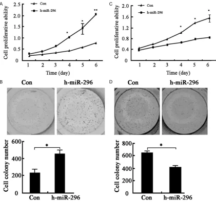
We further tested miR-296 impact on cell proliferation. Proliferation assay including MTT and colony formation demonstrated that miR-296 overexpression promoted co- lorectal cancer cell prolifera-tion (Figure 3A and 3B), whereas miR-296 low expres-sion weakened cancer cell proliferative ability (Figure 3C and 3D).
Discussion
[image:3.629.104.380.79.330.2]expres-bated disease state and leaded to cachexia. Therefore, inducing tumor cells apoptosis became an effective method to restrain tumor unlimited growth. Our results also showed that miR-296 overexpression promoted colorectal cancer cell proliferation. Thus, we aimed to focus on whether miR-296 was related to the speed of apoptosis induction in the future work. Recent studies demonstrated that miR-296 expression was suppressed in normal tissue compared with malignancy, while miR-296 overexpression may contribute to tumor forma-tion and development [14, 15]. For example, someone found miR-296 overexpressed in osteosarcoma cell line significantly increased
[image:4.629.103.525.81.476.2]cell proliferation, migration, in vitro invasion, and metastasis in xenograft mice model [16]. Studies have reported that high miR-296 expression was related to lymph node metasta-sis, high pathological grade, clinical stage, and breast cancer patients’ overall survival [17, 18]. It was also revealed that miR-296 expression aggravated colorectal cancer cell growth, and associated with colorectal cancer prognosis [19, 20]. As widely investigated as a typical inflammatory signaling pathway, NF-κB was found activated in colorectal cancer. In colorec-tal cancer, tumor suppressor gene APC inacti-vation can activate β-catenin importing to nuclear, further activate NF-κB signaling path-way to exacerbating inflammation. Our results
showed that miR-296 overexpressed in colorec-tal cancer compared with paratumor tissue (P < 0.001). Our study further confirmed miR-296 promoted colorectal cancer cell proliferation mainly through activating the NF-κB signaling pathway.
Acknowledgements
Yantai science and technology development project (2013WS216).
Disclosure of conflict of interest
None.
Address correspondence to: Dr. Sanyuan Hu, De- partment of General Surgery, Qilu Hospital, Shan- dong University, 107 Wenhua Xi Road, Jinan 250- 012, China. Tel: +86-15553598263; E-mail: husan- yuanan@163.com
References
[1] Schoppmann A, Tamandl D, Herberger B, Lan-gle F, Birner P, Geleff S, Grunberger T, Schoppmann SF. Comparison of lymphangio-genesis between primary colorectal cancer and corresponding liver metastases. Antican-cer Res 2011; 31: 4605-4611.
[2] Babae N, Bourajjaj M, Liu Y, Van Beijnum JR, Cerisoli F, Scaria PV, Verheul M, Van Berkel MP, Pieters EH, Van Haastert RJ, Yousefi A, Mastro-battista E, Storm G, Berezikov E, Cuppen E, Woodle M, Schaapveld RQ, Prevost GP, Griffio-en AW, Van Noort PI, Schiffelers RM. Systemic miRNA-7 delivery inhibits tumor angiogenesis and growth in murine xenograft glioblastoma. Oncotarget 2014; 5: 6687-6700.
[3] Garouniatis A, Zizi-Sermpetzoglou A, Rizos S, Kostakis A, Nikiteas N, Papavassiliou AG. Vas-cular endothelial growth factor receptors 1,3 and caveolin-1 are implicated in colorectal cancer aggressiveness and prognosis--correla-tions with epidermal growth factor receptor, CD44v6, focal adhesion kinase, and c-Met. Tu-mour Biol 2013; 34: 2109-2117.
[4] Royston D, Jackson DG. Mechanisms of lym-phatic metastasis in human colorectal adeno-carcinoma. J Pathol 2009; 217: 608-619. [5] Dong H, Luo L, Hong S, Siu H, Xiao Y, Jin L,
Chen R, Xiong M. Integrated analysis of muta-tions, miRNA and mRNA expression in glioblas-toma. BMC Syst Biol 2010; 4: 163.
[6] Li X, Liu B, Xiao J, Yuan Y, Ma J, Zhang Y. Roles of VEGF-C and Smad4 in the lymphangiogene-sis, lymphatic metastalymphangiogene-sis, and prognosis in co-lon cancer. J Gastrointest Surg 2011; 15: 2001-2010.
[7] Theocharisa S, Margeli A, Kouraklis G. Peroxi-some proliferator activated receptor-gamma li-gands as potent antineoplastic agents. Curr Med Chem Anticancer Agents 2003; 3: 239-251.
[8] Wei JJ, Wu X, Peng Y, Shi G, Basturk O, Yang X, Daniels G, Osman I, Ouyang J, Hernando E, Pel-licer A, Rhim JS, Melamed J, Lee P. Regulation of HMGA1 expression by microRNA-296 af-fects prostate cancer growth and invasion. Clin Cancer Res 2011; 17: 1297-1305.
[9] Liu ZY, Qiu HO, Yuan XJ, Ni YY, Sun JJ, Jing W, Fan YZ. Suppression of lymphangiogenesis in human lymphatic endothelial cells by simulta-neously blocking VEGF-C and VEGF-D/VEG-FR-3 with norcantharidin. Int J Oncol 2012; 41: 1762-1772.
[10] Lequerica-Fernandez P, Astudillo A, de Vicente JC. Expression of vascular endothelial growth factor in salivary gland carcinomas correlates with lymph node metastasis. Anticancer Res 2007; 27: 3661-3666.
[11] Amiel J, de Pontual L, Henrion-Caude A. miR-NA, development and disease. Adv Genet 2012; 80: 1-36.
[12] Stacker SA, Williams SP, Karnezis T, Shayan R, Fox SB, Achen MG. Lymphangiogenesis and lymphatic vessel remodelling in cancer. Nat Rev Cancer 2014; 14: 159-172.
[13] Weis S, Cui J, Barnes L, Cheresh D. Endothelial barrier disruption by VEGF-mediated Src activ-ity potentiates tumor cell extravasation and metastasis. J Cell Biol 2004; 167: 223-229. [14] Halle C, Lando M, Svendsrud DH, Clancy T,
Holden M, Sundfor K, Kristensen GB, Holm R, Lyng H. Membranous expression of ectodo-main isoforms of the epidermal growth factor receptor predicts outcome after chemoradio-therapy of lymph node-negative cervical can-cer. Clin Cancer Res 2011; 17: 5501-5512. [15] Weis SM, Cheresh DA. Tumor angiogenesis:
molecular pathways and therapeutic targets. Nat Med 2011; 17: 1359-1370.
[16] Nagasaka K, Pim D, Massimi P, Thomas M, To-maic V, Subbaiah VK, Kranjec C, Nakagawa S, Yano T, Taketani Y, Myers M, Banks L. The cell polarity regulator hScrib controls ERK activa-tion through a KIM site-dependent interacactiva-tion. Oncogene 2010; 29: 5311-5321.
[17] Bilder D, Li M, Perrimon N. Cooperative regula-tion of cell polarity and growth by Drosophila tumor suppressors. Science 2000; 289: 113-116.
[19] Folini M, Gandellini P, Longoni N, Profumo V, Callari M, Pennati M, Colecchia M, Supino R, Veneroni S, Salvioni R, Valdagni R, Daidone MG, Zaffaroni N. miR-21: an oncomir on strike in prostate cancer. Mol Cancer 2010; 9: 12.