Original Article
Identification of risk factors for thyroid cancer in
Urumqi, China
Li Zhang1, Xin Ma1, Wenwen Yang1, Wei Wei1, Chao Bai2, Runxiao Zhang1, Ting Huang1,Yao Lu1, Guyaim
Ablez1, Jingjing Yang1
1Department of Endocrinology, Institute of Endocrine, The Second Affiliated Hospital of Xinjiang Medical
University, Urumqi 830063, China; 2Department of Vascular and Thyroid Surgery, The First Affiliated Hospital of
Xinjiang Medical University, Urumqi 830054, China
Received October 28, 2016; Accepted December 6, 2016; Epub January 1, 2017; Published January 15, 2017
Abstract: Aims: Risk factors for thyroid cancer in Urumqi were investigated. Methods: This study enrolled 153 cases of thyroid cancer, 154 cases of benign thyroid nodule and 327 controls between March 2014 and December 2014. Patient demographic information was obtained. Serum total triiodothyronine (TT3), total thyroxine (TT4), thyroid-stimulating hormone (TSH), anti-thyroglobulin antibody (TgAb), thyroid peroxidase antibody (TPOAb), serum
sele-nium concentration (Se), and urinary iodine concentration (UIC) were recorded. Results: Significant difference was
observed in gender, age, and serum TT3, TT4, TSH, TgAb, TPOAb and Se levels as well as UIC in the thyroid cancer,
benign thyroid nodule and control groups (P<0.05). Serum TT3 and TT4 levels were significantly lower in the thyroid
cancer group than that in the benign thyroid nodule and control groups (P<0.001). In addition, the thyroid cancer
group showed significant difference in serum TSH and TgAb levels from the group of benign thyroid nodule (P<0.05). Significantly a higher serum TPOAb level was noted in the groups of thyroid cancer and benign thyroid nodule than
in the control group (P<0.05). Between different serum Se level subgroups, difference was noted in the serum
TT3, TSH and TPOAb levels with significance (P<0.001) instead of TT4 and TgAb. With the rise of serum Se level,
the TT3 level increased while TSH and TPOAb levels decreased. Serum TgAb and TPOAb levels were showed to be
significantly different between groups of different UIC (P<0.05), while serum TT3, TT4 and TSH levels did not differ significantly. Serum TgAb and TPOAb levels increased with the increasing UIC. Multivariate logistic regression analy -sis revealed that female, low Se level and high UIC were risk factors for thyroid cancer. Conclusions: Female, low Se level and high iodine intake were dependent risk factors for thyroid cancer.
Keywords: Thyroid cancer, risk factor, multivariate analysis, thyroid nodule, thyrotropin
Introduction
In recent years, the incidence of thyroid cancer has been rising and thyroid cancer is the most frequent endocrine malignancy [1].The cause of thyroid cancer is not yet clear. In most cases, early diagnosis results in a better prognosis [2]. At the current time, ionizing radiation, exces-sive intake of iodine, female hormone levels and a family history of cancer have been identi-fied to be related to thyroid cancer, however, the mechanism is still not clear. The past years witnessed an increase in the incidence of thy-roid cancer in Urumqi, China. Moreover, thythy-roid cancer attacks more women than men, and the pathological type and age of onset show signifi-cant differences in different ethnic groups. In the current study, patients with thyroid
can-trols in Urumqi were enrolled. Demographic data including age and gender as well as serum total triiodothyronine (TT3), total thyroxine (TT4), thyroid-stimulating hormone (TSH), anti-thyroglobulin antibody (TgAb), thyroid peroxi-dase antibody (TPOAb), serum selenium con-centration (Se), and urinary iodine concentra-tion (UIC) were collected and analyzed to study their associations with thyroid cancer. The risk factors for thyroid cancer in Urumqi were fur-ther identified.
Materials and methods
Study population
roid nodules was confirmed by histopathologi-cal analysis. Between March 2014 and December 2014, patients that underwent thy-roidectomy for thyroid cancer or benign thyroid nodule in the Department of Thyroid and Vascular Surgery, the First Affiliated Hospital of Xinjiang Medical University, were consecutively recruited. Histological type of the thyroid can-cer included in 147 cases of papillary thyroid cancer, 2 cases of follicular thyroid cancer, 2 cases of medullary thyroid cancer, and 2 cases were undifferentiated. In the group of patients with benign thyroid nodule, there were 142 cases of nodular goiter, 6 cases of nodular goi-ter with adenoma, 5 cases of adenoma and 1 case of nodular goiter with Hashimoto’s thyroiditis.
The controls were selected from healthy volun-teers who visited the Second Affiliated Hospital of Xinjiang Medical University for medical exam-ination during the same period, and ultraso- nography was performed for screening. The patients and controls were local residents of Urumqi and had been living in Urumqi for at least 10 years at enrollment.
Exclusion criteria were history of thyroid dis-ease, intake of Se- or iodine-containing drugs and iodinated contrast media in half a year prior to the enrollment, liver and kidney dys-function, and pregnancy.
The study was approved by the institutional review board of the Xinjiang Medical University, and informed consent was obtained from each participant.
Data collection and laboratory test
Patient demographic data including age and gender as well as pathological results were obtained. Serum TT3, TT4, TSH, TgAb, and
nace atomic absorption spectrometry (GFAAS). UIC was measured by As-Ce catalytic spectro- photometry.
Statistical analysis
The statistical analyses were performed using SPSS 17.0 (SPSS Inc, Chicago, IL, USA). The normality test was carried out on quantitative data and normally distributed data were pre-sented as mean ± standard deviation. The stu-dent’s t-test was performed for pairwise com-parison while ANOVA was used for comcom-parison between different groups. The non-normally distributed data were showed as median and interquartile range (IQR) and the rank sum test was performed for comparisons. The quantita-tive data were expressed as absolute number of cases and percentage. The χ2test was
per-formed for pairwise comparison and compari-son between groups. Multivariate logistic re- gression to estimate odds ratios (OR) with 95% confidence interval (CI) was conducted for the risk of thyroid cancer. A P level of <0.05 was regarded as statistically significant.
Results
Demographic characteristics of the study sub-jects
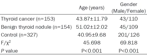
[image:2.612.91.332.97.188.2]Totally, 634 subjects including 153 thyroid can-cers, 154 patients with benign thyroid nodule and 327 controls were enrolled in this study. There were 289 males and 345 females. As shown in Table 1, the age was significantly dif-ferent in the thyroid cancer group compared to the benign thyroid nodule and control groups. Moreover, significant difference was also noted in the age between the benign thyroid nodule and thyroid cancer groups (P<0.001). With regard to the gender, significant differences were observed between the thyroid cancer and
Table 1. Demographic characteristics of the study sub-jects
Age (years) (Male/Female)Gender Thyroid cancer (n=153) 43.87±11.79 43/110 Benign thyroid nodule (n=154) 51.02±12.02 45/109
Control (n=327) 40.95±9.68 201/126
F/χ2 45.698 69.818
P value P<0.001 P<0.001
TPOAb levels as well as Se and UIC were tested and recorded. The normal rang- es of TT3, TT4, TSH, TgAb and TPOAb were 0.80~2 ng/mL, 51~141 ng/mL, 0.27~0.42 μIU/ml, 0~115 IU/mL and 0~34 IU/mL, respectively.
fur-control groups, and between the benign thyroid nodule and control groups.
High TSH, TgAb and TPOAb, and, low TT3 and TT4 levels in thyroid cancer
[image:3.612.88.529.86.156.2]The thyroid hormone and autoantibody levels in different groups were analyzed. As shown in
Table 2, there was significant difference in the levels of TT3, TT4, TSH, TgAb and TPOAb among different groups (P<0.05). The levels of TT3 and TT4 in the groups of thyroid cancer and benign thyroid nodule were significantly lower than that in the control group (P<0.001). Compared with the group of benign thyroid nodule, the levels of serum TSH and TgAb were higher in thyroid can-cer with significant difference (P<0.05). The groups of thyroid cancer and benign thyroid nodule presented with significantly higher level of TPOAb than the control group (P<0.05). Low Se in thyroid cancer
The mean Se of 634 subjects was 118.55 ug/L (range: 76.26~150.93 ug/L). It was 71.32 ug/L (range: 26.79~103.56 ug/L) in the group of thy-roid cancer, 88.34 ug/L (range: 49.95~129.87 ug/L) in the group of benign thyroid nodule and 133.00 ug/L (range: 94.07~169.00 ug/L) in the control group. The Se was significantly lower in thyroid cancer than that in the groups of benign thyroid nodule and controls (P<0.05). According to the different Se levels, subjects were assigned to three subgroups of <100 ug/L, 100~149 ug/L and >150 ug/L (Table 3).
of different Se (P<0.001), while there was no significant difference in the levels of TT4 and TgAb. TT3 increased while TSH and TPOAb decreased as Se increased (Table 4).
High UIC in thyroid cancer
The median UIC was 9.53 μg/L in all 634 sub-jects with a range of 180.40~394.97 μg/L. It was 328.10 μg/L (range: 209.20~571.20 μg/L), 257.20 μg/L (range: 168.85~390.15 μg/L) and 245.55 μg/L (range: 163.83~367.13 μg/L) in the groups of thyroid cancer, benign thyroid nodule and controls, respectively. The UIC was higher in the group of thyroid can- cer than that in the groups of benign thyroid nodule and controls with significant difference (P<0.05).
All subjects were assigned into subgroups according to the criteria on iodine nutrition sta-tus released by the World Health Organization (WHO), the United Nations Children’s Fund (UNICEF), and the International Council for the Control of Iodine Deficiency Disorders (ICCIDD) in 2007. A median UIC<100 μg/L is indicative of insufficient iodine intake (iodine deficiency), 100~199 μg/L of adequate intake, 200~299 μg/L of intakes above requirements, and ≥300 μg/L of excessive intake [3]. Subjects were assigned into subgroups according to the above-mentioned assessment standard crit- eria.
The proportions of patients with thyroid cancer or benign thyroid nodule in subgroups of diff-
Table 2. Thyroid hormone and autoantibody levels in different groups
TT3 (ng/ml) TT4 (ng/ml) TSH (μIU/mL) TgAb (IU/mL) TPOAb (IU/mL) Thyroid cancer (n=153) 1.10 (1.00~1.26)* 88.67 (76.59~96.48)*,# 2.81 (1.82~3.73)*,# 19.66 (13.75~67.99)# 14.63 (8.09~27.18)* Benign thyroid nodule (n=154) 1.11 (1.01~1.25)* 89.90 (86.74~98.52)* 1.85 (1.12~3.43)* 16.98 (12.65~26.26) 13.18 (6.23~19.63)* Control (n=327) 1.78 (1.560~1.98) 96.77 (87.27~106.85) 2.14 (1.56~3.14) 19.47 (14.03~56.06) 7.81 (5.00~14.99)
H value 328.133 48.012 22.404 7.593 54.355
P value P<0.001 P<0.001 P<0.001 P<0.05 P<0.001
[image:3.612.90.379.212.279.2]Note: *P<0.05 versus the control group; #P<0.05 versus the benign thyroid nodule group.
Table 3. Distribution of thyroid cancers, benign thyroid nodule and controls in different serum selenium concentration (Se)
Se ug/L
Total <100 100~149 >150 Thyroid cancer (n, %) 153 102 (66.66) 35 (22.88) 16 (10.46) Benign thyroid nodule (n, %) 154 85 (55.19) 45 (29.22) 24 (15.59) Control (n, %) 327 63 (19.26) 140 (42.81) 124 (37.93)
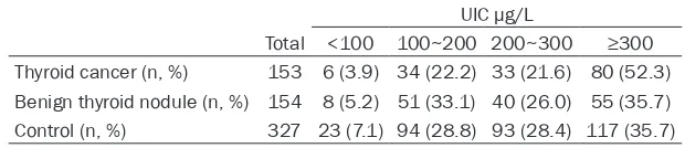
erent UIC were significantly different (P<0.05). A significantly higher proportion of patients with thyroid cancer was noted in the subgroup of excessive iodine intake than other groups (P<0.05,Table 5). Further, the levels of serum TgAb and TPOAb were significantly different in groups of different UIC (P<0.05), while no sig-nificant difference was observed in TT3, TT4 or TSH. The TgAb and TPOAb levels increased as the UIC became higher (Table 6).
Multivariate results
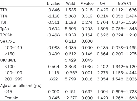
As shown in Table 7, multivariate logistic regres-sion analysis revealed that female (OR=1.874; 95% CI=1.012~3.472), low Se level (OR=0.08; 95% CI=0.078~0.435) and high UIC (OR=0.004; 95% CI=1.548~6.026) were risk factors for thy-roid cancer.
Discussion
Thyroid cancer can occur at any age and the papillary type occurs more often in young women than in men at a ratio of three to one [4]. In the present study, the proportion of papil-lary thyroid cancer was 97.08% in the thyroid cancers and the male/female ratio was 1; 2.56 in the group of patients with thyroid cancer. In the thyroid cancer group, patients had a young-er age than that in the group of benign thyroid nodule with significant difference. Female was demonstrated to be a dependent risk factor for thyroid cancer based on multivariate results. It has been confirmed by animal and cell studies
mote the division of thyroid cancer cells by stimulating mitogen-activated protein kinase (MAPK).
The present study revealed lower TT3, TT4 level and Se, and a higher TSH level in the group of thyroid cancer than control. With the rise of Se, TT3 increased, while TSH and TPOAb decreas- ed. Multivariate results suggested that low Se status is an independent risk factor for thyroid cancer. As a modulator of oncogene expres-sion, Se-containing compound can suppress the division, promote the differentiation and induce programmed death of the cancer cells [7].
[image:4.612.88.526.97.174.2]The conversion of T4 to T3, catalyzed by seleno-proteins, accounts for the major source of T3 in vivo and plays a key role in maintaining the nor-mal thyroid functioning. Se glutathione peroxi-dase can function as oxide reductase and pro-tect thyroid cells from oxidative stress [8]. When Se-deficient status is present, the decrease of selenoproteins can cause abnor-mal thyroid hormone metabolism and reduced serum T3 [9, 10]. Meanwhile, the feedback from Se deficiency can stimulate the increased secretion of TSH which promotes the synthesis of cAMP, activates the cAMP-dependent protein kinase signal transduction system, enhances epidermal growth factor-mediated cell prolifer-ation, stimulates the thyroid follicular cell gro- wth, and functions as a mediator and amplifies the signal in tumor invasion [11, 12].
Table 4. Thyroid hormone and autoantibody levels in different serum selenium concentration (Se) groups (median, IQR)
Se ug/L TT3 (ng/ml) TT4 (ng/ml) TSH (μIU/mL) TgAb (IU/mL) TPOAb (IU/mL)
<100 1.34 (1.28~1.40) 109.99 (90.51~129.48) 3.79 (2.07~5.51) 75.10 (31.16~119.05) 38.14 (26.52~49.78) 100~149 1.58 (1.51~1.66) 118.48 (87.54~149.41) 3.49 (2.04~4.94) 96.26 (40.97~151.55) 31.34 (18.73~43.94) >150 1.64 (1.56~1.72) 115.75 (75.35~156.15) 2.79 (2.48~3.10) 78.33 (39.68~116.99) 27.43 (16.20~38.56)
H value -7.013 -1.922 -2.343 -0.458 -3.590
P value P<0.001 0.065 P<0.001 0.255 P<0.001
Table 5. Distribution of thyroid cancers, benign thyroid nodules and controls in different urinary iodine concentration (UIC)
UIC μg/L
Total <100 100~200 200~300 ≥300
Thyroid cancer (n, %) 153 6 (3.9) 34 (22.2) 33 (21.6) 80 (52.3) Benign thyroid nodule (n, %) 154 8 (5.2) 51 (33.1) 40 (26.0) 55 (35.7) Control (n, %) 327 23 (7.1) 94 (28.8) 93 (28.4) 117 (35.7)
[image:4.612.83.393.221.288.2]pro-The TPOAb level in the groups of thyroid cancer and benign thyroid nodule were significantly higher than that in the control group, and sig-nificant difference was noted in the TPOAb level in groups with different Se. With the increase of Se, TPOAb decreased. It is indicative that Se deficiency can impair the antioxidant capacity of thyroid selenoproteins, leading to the dam-age and even apoptosis of thyroid follicular cell structure. As one kind of autoimmune antigen, thyroid peroxidase (TPO) induces the develop-ment of TPOAb and immune inflammatory dam-age. Nitric Monoxide (NO), released from inflam-matory cells, is found to be increased in papil-lary thyroid cancer and induces the production of high-mobility group box 1 (HMGB1), which stimulates the expression of nuclear factor kappa-light-chain-enhancer of activated B cells (NF-κB) in thyroid cells and maintains the
[image:5.612.89.525.82.178.2]peri-thyroid cancer increased in areas of high iodine intake [14]. The policy of universal salt iodiza-tion has improved the iodine nutriiodiza-tional status in the Urumqi city, which belongs to iodine defi-ciency area, and the iodine nutritional status has been excessive according to the assess-ment criteria established by WHO [15]. With long-term and excessive iodine intake levels, the transcription and expression of sodium iodide symporter (NIS) were decreased, and the iodine uptake capacity of thyroid tissue is impaired [16, 17]. When thyroid homeostasis cannot be maintained by self-regulation, thy-roid function is decreased [18]. The hypotha-lamic-pituitary-thyroid axis is initiated to incr- ease TSH through feedback, and then the thy-roid follicular hyperplasia and nodules are induced [19-21]. Furthermore, the mutations of v-raf murine sarcoma viral oncogene homolog
Table 6. Thyroid hormone and autoantibody levels in different UIC groups (median, IQR)
UIC ug/L TT3 (ng/ml) TT4 (ng/ml) TSH (μIU/mL) TgAb (IU/mL) TPOAb (IU/mL)
<100 1.42 (1.28~1.57) 122.13 (41.23~203.04) 2.52 (1.90~3.15) 44.03 (28.06~60.00) 28.14 (13.85~38.33) 100~200 1.60 (1.52~1.69) 107.23 (75.66~138.81) 4.02 (1.68~6.36) 83.10 (24.85~141.36) 32.22 (23.19~40.73) 200~300 1.58 (1.48~1.66) 121.32 (89.65~153.00) 2.98 (1.96~4.00) 94.49 (47.76~141.22) 36.31 (20.24~52.11) ≥300 1.38 (1.32~1.43) 156.67 (63.01~250.32) 3.40 (2.50~4.30) 127.29 (22.69~231.88) 50.04 (11.82~94.07)
H value 8.555 5.322 3.383 11.627 24.772
P value 0.887 0.647 0.139 P<0.001 P<0.001
Table 7. Odds ratios (OR) and 95% confidence interval (CI) of thyroid cancer associated with anthropometric factors, thyroid hormone, autoantibody, serum selenium concentration (Se) and urinary iodine concentration (UIC)
B value Wald P value OR 95% CI
TT3 -0.846 1.535 0.215 0.429 0.112~1.636
TT4 -1.160 5.880 0.319 0.314 0.058~0.494
TSH -0.351 1.198 0.274 0.704 0.375~1.320
TgAb -0.604 5.693 0.203 1.396 0.785~1.848
TPOAb -0.468 1.939 0.164 0.626 0.324~1.210
Se ug/L 0.218 0.008
100~149 -0.983 4.035 0.000 0.185 0.078~0.435
≥150 -0.409 0.612 0.148 0.664 0.200~1.275
UIC μg/L 5.429 0.045
<100 0.564 3.363 0.036 2.102 1.342~5.120 100~199 1.116 10.363 0.001 2.276 1.165~4.444 200~299 .822 5.799 0.016 3.054 1.548~6.026 Age at enrollment (yrs)
≤45 0.090 0.151 0.697 1.094 0.695~1.723
Female -0.845 12.370 0.000 1.429 1.268~1.688
CI confidence interval.
tumoral inflammatory respo- nse. Low Se status is consid-ered to increase the thyroid cancer risk through mediat-ing immune inflammation.
[image:5.612.92.377.248.459.2]B1 (BRAF) gene are caused and the transcrip-tion and expression of NIS are suppressed, resulting in further impairment of thyroid iodine intake capacity and thyroid cancer [22]. It has been confirmed by animal studies [23] that high iodine intake level can lead to lymphocyte infiltration and follicular cell expansion in thy-roid tissues and inflammatory infiltration will be increased with increasing iodine intake. In the present study, high iodine status has been demonstrated to be an independent risk factor for thyroid cancer. Serum TgAb and TPOAb lev-els increased with the increase of UIC. High iodine status may induce the development of thyroid cancer through immune response. In conclusion, the present study verified Se deficiency as a risk factor for thyroid cancer. However, further investigations are in need to confirm that whether Se supplement can func-tion in the prevenfunc-tion of thyroid cancer. Urinary iodine test can be used as a routine examina-tion. Population-based studies on the risk fac-tors for thyroid cancer are warranted for a more comprehensive and integrated analysis.
Acknowledgements
This work was supported by the Natural Science Foundation of the Xinjiang Uygur Autonomous Region, China (NO.2013211A057).
Disclosure of conflict of interest
None.
Address correspondence to: Chao Bai, Department
of Vascular and Thyroid Surgery, The First Affiliated
Hospital of Xinjiang Medical University, No.137, Liyushan South Road, Urumqi 830054, China. Tel: +86-991-4362719; E-mail: 1481538986@qq.com
References
[1] Chen AY, Jemal A and Ward EM. Increasing in-cidence of differentiated thyroid cancer in the United States, 1988-2005. Cancer 2009; 115: 3801-3807.
[2] Rosario PW, Mineiro Filho AF, Prates BS, Silva LC, Lacerda RX and Calsolari MR. Ultraso- nographic screening for thyroid cancer in sib-lings of patients with apparently sporadic pap-illary carcinoma. Thyroid 2012; 22: 805-808. [3] Bischoff LA, Curry J, Ahmed I, Pribitkin E and
Miller JL. Is above age 45 appropriate for up-staging well differentiated papillary thyroid cancer? Endocr Pract 2013; 19: 995-997.
[4] Kong N, Chen XY, Zhang CY and Han X. Comparative analysis of clinical data among 385 postoperative patients with thyroid benign and malignant nodules. Chin J Endocrinol Metab 2015; 31: 1-3.
[5] Rajoria S, Suriano R, George AL, Kamat A, Schantz SP, Geliebter J and Tiwari RK. Mole- cular target based combinational therapeutic approaches in thyroid cancer. J Transl Med 2012; 10: 81.
[6] Wang X and Wang ZJ. Estrogen, anti-estrogen drugs, and thyroid cancer. Chin J Endocrinol Metab 2014; 30: 1128-1131.
[7] Cui Q, Shang DJ and Zou X. Anticancer activi-ties of selenium compounds. Chinese Journal of Biochemical Pharmaceutics 2004; 25: 247-249.
[8] Liu XY, Feng Y and Liu C. Selenium and thyroid diseases. International Journal of Internal Medicine 2007; 34: 728-731.
[9] Köhrle J, Gärtner R. Selenium and thyroid. Best Pract Res Clin Endocrinol Metab 2009; 23: 815-827.
[10] Duntas LH. Selenium and the thyroid: a close-knit connection. J Clin Endocrinol Metab 2010; 95: 5180-5188.
[11] Roger PP, van Staveren WC, Coulonval K, Dumont JE and Maenhaut C. Signal transd-uc-tion in the human thyrocyte and its perversion in thyroid tumors. Mol Cell Endocrinol 2010; 321: 3-19.
[12] Liu CL, Wu Y and Bi LF. Advances in the thyroid cancer epidemiology and risk factors. Chin J Endemio 2012; 31: 234-236.
[13] Guan HX, Teng WP and Yang SM. Comparative epidemiological study on thyroid cancer in ar-eas with different iodine intake. Natl Med J China 2001; 81: 457-458.
[14] Chen J. Progress of the relationship between thyroid tumor and breast cancer. Chin J Oncol 2011; 21: 148-152.
[15] Zhang MC, Zhang L, Fan Y, Zeng XY, Zhu Y and Abulikemu Y. Relationship between iodine in-take and the prevalence of thyroid disease in Urumqi, Xinjiang. Chin J Endocrinol Metab 2011; 27: 972-974.
[16] Wang K. Effects of iodine deficiency and iodine
excess on thyroid function and the modulating mechanism. Tianjin Medical University 2007. [17] Spitzweg C, Joba W, Morris JC and Heufelder
AE. Regulation of sodium iodide symporter gene expression in FRTL-5 rat thyroid cells. Thyroid 1999; 9: 821-830.
[18] Wang K, Lin LX and Sun YN. Sodium iodine symporter gene expression in thyroid of mice with different iodine intake. Chin J Endocrinol Metab 2009; 25: 264-268.
recep-tor and thyroid carcinoma . Int J Radiat Med Nucl Med 2012; 36: 69-72.
[20] He RZ, Jiang Y, Yu JC and Chen WZ. New prog-ress of BRAF gene and thyroid cancer. Chin J Surg 2016; 54: 237-240.
[21] Franco AT, Malaguarnera R, Refetoff S, Liao XH, Lundsmith E, Kimura S, Pritchard C, Marais R, Davies TF, Weinstein LS, Chen M, Rosen N, Ghossein R, Knauf JA and Fagin JA. Thyrotrophin receptor signaling dependence of Braf-induced thyroid tumor initiation in mice. Proc Natl Acad Sci U S A 2011; 108: 1615-1620.
[22] Guan HX. Spectrum of thyroid disorders in an iodine-excess area and an epidemiological and immunohistochemical study on thyroid cancer. China Medical University 2003. [23] Yuan JH, Zhou XL and Shi XL. Expression of
thy-roid stimulating hormone β splice variant in