Original Article
Multicenter evaluation of membrane-based smear
microscopy for detecting acid-fast bacilli in China
Hui Xia1*, Yanli Yuan2*, Tiejuan Zhang2, Yunlong Bai2, Lifu Zhang3, Yanlin Zhao1, Xiujun Yang3
1National Tuberculosis Reference Laboratory, National Center for Tuberculosis Prevention and Control, Chinese
Center for Disease Control and Prevention, No. 155, Changbai Road, Changping District, Beijing, China; 2Jilin
Research Institute of Tuberculosis Control, 3Ji Lin Provincial Center for Disease Control and Prevention, No. 3145,
Jingyang Road, Changchun District, Jilin Province, China. *Equal contributors.
Received July 5, 2018; Accepted August 23, 2018; Epub November 1, 2018; Published November 15, 2018
Abstract: Objective: To assess the feasibility and reliability of using membrane-based smear microscopy at
periph-eral laboratories in China. Methods: The clinical case control study was conducted in five tuberculosis (TB) dispen -saries from September 2014 to May 2016. The membrane-based microscopy and direct smear microscopy were
performed to compare the sensitivity, specificity, and the examination time for both methods was also analyzed.
Results: A total of 5359 TB suspects were consecutively enrolled from 5 TB dispensaries, and 9915 specimens were
finally analyzed. The sensitivity for membrane-based microscopy and direct smear microscopy was 76.9% (95% CI, 75.4-78.4) and 53.8% (95% CI, 52.1-55.6) respectively, and the specificity was 96.8% (95% CI, 96.4-97.2) and 99.2% (95% CI, 99.0-99.4) respectively. The sensitivity and specificity were both significantly different (P<0.001) between the two methods. The examination time for membrane-based smear microscopy (209.1±112.0 seconds)
was significantly shorter than that for direct smear microscopy (253.1±79.4 seconds) (P<0.05). Conclusions: Mem-brane-based smear microscopy showed higher sensitivity and a shorter examination time in comparison with direct smear microscopy and it could be used at peripheral laboratories in China.
Keywords: Tuberculosis, membrane-based smear microscopy, acid-fast bacilli
Introduction
Tuberculosis continues to be a major health problem in China. Direct smear microscopy is a rapid, simple diagnostic tool used to identify the most infectious cases of TB especially in resource limited countries. It is highly specific but has low sensitivity. The Global Tuberculosis Report (2016) reported that the rate of bacte-riologically confirmed pulmonary cases was only 31% among total pulmonary tuberculosis cases in China [1]. New diagnosis technologies are therefore needed to increase the propor-tion of bacteriologically confirmed pulmonary tuberculosis. Indirect smear can improve the detection of bacilli compared to direct smear. A systematic review reported that processing by a several chemical procedures, followed by centrifugation or overnight sedimentation, was more sensitive than direct microscopy, and th- at the specificity was similar [2]. However, th-ere are few laboratories performing indirect smear microscopy due to its complex
process-es in China. The new membrane-based sme- ar microscopy described in this study simplifi-es the procsimplifi-ess by the liquefaction of sputum and by concentrating the bacteria to a specific high polymer membrane at the bottom of the vessel. The bacilli adsorbed onto the mem-brane can be stained directly in the vessel. The membrane with bacilli is then fixed onto a glass slide and examined under a lighted microscope. This study investigated whether this mem-brane-based smear microscopy method could enhance the diagnosis of TB in China.
Methods
Study population
accord-ing to the guidelines [3]. This study was ap- proved by the Ethical Committee of the Chinese Center for Disease Control and Prevention.
Direct smear microscopy
Direct smears were prepared and stained us- ing the Ziehl-Neelsen method [3]. The results were graded as follows: negative, scanty, 1+, 2+, 3+, 4+.
Membrane-based smear microscopy
The membrane-based smears were prepared from the same specimens used for the direct smear microscopy. The sputum specimen was first digested with the digestion reagent in the specific sealed vessel and vortexed for 3 min -utes. The vessel was then centrifuged at 4500 RPM for 5 minutes in the centrifuge to concen-trate the bacteria. The bacteria were adsorbed to the high-polymer membrane located at the bottom of the vessel after being centrifuged. The supernatant was then discarded, and the membrane was dried by placing the vessel in an oven. The bacteria were fixed onto the mem -brane by adding ethanol to the mem-brane and then staining it with 0.8% carbol fuchsin. The vessel was then placed in the oven for 5 min-utes at 60°C. Methylene blue was added to counterstain the background for 1-3 minutes after decolorization. The membranes adsorbed with the stained bacilli were ejected with a nee-dle from the bottom of vessel and dried com-pletely in the air. The membrane was then stuck to a glass slide using neutral glue with the side adsorbing the bacilli facing the slide to prevent the objective lens from cross-contamination.
Culture
Two specimens were selected for culture with the simple Petroff’s method. In brief, the spu-tum was digested using 4% sodium hydrate for 15 minutes and inoculated. The slants were incubated at 37°C and observed at 3 days and 7 days after inoculation to examine the con -tamination and growth of non-mycobacterium tuberculosis, and then the growth of the myco-bacteria was observed weekly for 8 weeks.
Examination time
To measure the examination time, the exami- nation times were recorded for both methods under routine working conditions. The time
from placing the stained slide under the objec-tive lens to reporting the examination result was measured.
Sterilization effect
Different smear graded sputum samples were used to evaluate the sterilization effect of the digestion process. The sputum was digested with a specific digestion reagent and vortex-ed for 3 minutes, and the suspension was then inoculated onto the slants of modified acid Lowenstein-Jensen (L-J) medium and modified L-J medium. The slants were incubated at 37°C and observed weekly to examine the growth of mycobacteria for 8 weeks.
End-user appraisal
The appraisal survey, a questionnaire, was con-ducted among laboratory technicians.
Definition
Smear positive cases were defined as TB pa-tients with at least one positive smear mic- roscopy result (graded scanty, 1+, 2+, 3+, 4+) from the three specimens. Culture positive ca- ses were defined as TB patients with at least one positive L-J culture result from the two selected specimens. Clinical TB patients were defined as patients with clinical symptoms and chest X-rays indicating TB at intake and who- se clinical symptoms and chest X-rays were improved after two months of anti-tuberculosis treatment.
Statistical analysis
Data were entered into the database using Excel and analyzed using SPSS version 17.0. The sensitivity and specificity of the two smear microscopy methods were calculated in com-parison with culture. McNemar’s test or Pear- son’s chi-square test was used for comparison of the proportions; a t-test was used to com-pare the differences in examination time bet- ween the groups.
Results
the remaining 5311 cases, a total of 10031 specimens were examined using the three laboratory methods simultaneously. Out of the 10031 specimens, 116 (1.2%) specimens had contaminated culture results, and 9915 speci-mens were therefore included in the final analysis.
Detection rates of two smear microscopy methods and culture
Among 9915 specimens, 26.4% (2622/9915), 17.5% (1737/9915) and 31.5% (3125/9915) were positive using a membrane-based sme- ar, direct smear microscopy, and culture, res- pectively.
80.7% (4285/5311) of the cases had three sputum specimens collected. The positive ra- tes with membrane-based smear microscopy were 25.2% (1078/4285) using the spot sp-ecimen, 28.2% (1209/4285) using both the spot and night specimens, and 29.4% (1260/ 4285) using all three samples, which were sig-nificantly higher than the corresponding de-tection rates using direct smear microscopy (15.8% (675/4285), 18.7% (802/4285) and 20.2% (865/4285)) (P<0.001).
The cumulative positive rate of direct smear microscopy of the three specimens was sig-
The sensitivity for membrane-based microsco-py and direct smear microscomicrosco-py was 76.9% (95% CI, 75.4-78.4) and 53.8% (95% CI, 52.1-55.6) respectively, and the specificity was 96.8% (95% CI, 96.4-97.2) and 99.2% (95% CI, 99.0-99.4), respectively (Table 1). The mem-brane-based method increases the sensitivity up to 23%. The differences of the sensitivity and specificity between the two smear micros -copy methods were both significant (P<0.001).
Discrepant results analysis between the two smear microscopy methods
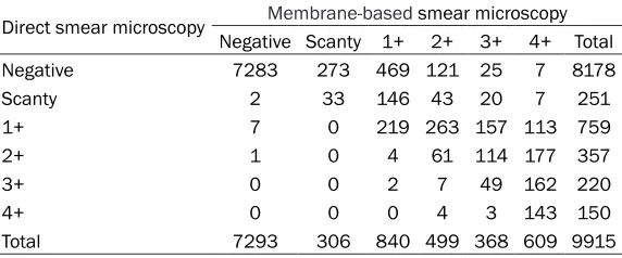
9.1% (905/9915) of specimens had discre-pant qualitative results. 98.9% (895/905) of the specimens were positive using membrane-based smear microscopy and negative using direct smear microscopy. The distributions of the two smear microscopy methods results are shown in Table 2.
[image:3.612.94.524.96.175.2]Among the 895 specimens with membrane-based smear microscopy positive and direct smear microscopy negative results, 30.5% (273/895) were scanty and 52.4% (469/895) were graded 1+. Out of these 895 specime- ns, 81.3% (728/895) were culture positive and thus favor the membrane-based smear results, and only 18.7% (167/895) specimens were cul -ture negative and consistent with direct smear Table 1. Sensitivity and specificity of direct smear microscopy and membrane-based smear micros -copy
Smear microscopy Culture Sensitivity % (95% CI) Specificity % (95% CI)
Positive Negative
Membrane-based smear microscopy Positive 2403 219 76.9 (75.4-78.4) 96.8 (96.4-97.2)
Negative 722 6571
Direct smear microscopy Positive 1682 55 53.8 (52.1-55.6) 99.2 (99.0-99.4) Negative 1443 6735
Table 2. Comparison of grading results between membrane-based smear microscopy and direct smear microscopy
Direct smear microscopy Membrane-based smear microscopy
Negative Scanty 1+ 2+ 3+ 4+ Total
Negative 7283 273 469 121 25 7 8178
Scanty 2 33 146 43 20 7 251
1+ 7 0 219 263 157 113 759
2+ 1 0 4 61 114 177 357
3+ 0 0 2 7 49 162 220
4+ 0 0 0 4 3 143 150
Total 7293 306 840 499 368 609 9915
nificantly lower than the ra-te of the membrane-based smear microscopy from any sputum specimen, 20.2% (865/4285) for direct smear vs. 25.2% (1078/4285) (sp-ot), 26.7% (1145/4285) (ni-ght), 26.7% (1143/4285) (morning) for membrane-ba- sed smear microscopy (P< 0.001).
[image:3.612.92.378.225.344.2]microscopy. For the 167 specimens with a cul -ture negative result, the results of the other specimens from the same patients were fur-ther analyzed. 19.2% (32/167) of the speci -mens were collected from 31 culture positive patients based on the other specimen, and 16.8% (28/167) of specimens were from 26 culture negative but direct smear positive patients based on other specimens. Among the rest of the culture negative and smear negative specimens, 55.1% (92/167) were from 61 patients clinically diagnosed as having pulmo-nary tuberculosis. Only 9.0% (15/167) of speci -mens were collected from 10 patients not diag-nosed with pulmonary tuberculosis based on X-ray results and direct smear microscopy and culture.
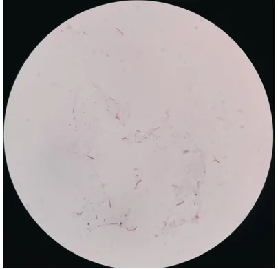
Table 3. The examination time for membra- ne-based smear microscopy was significant-ly shorter than for direct smear microscopy (209.1±112.0 s vs. 253.1±79.4 s, P<0.05). Significantly, the difference in the examina-tion times between the two methods was la- rgely attributable to the shortening of the ex- amination time of the positive slides. The shapes of the acid-fast bacilli and the back-ground of the smear prepared with membra- ne-based smear microscopy are shown in Figure 1.
Sterilization study with clinical sputum sam-ples
90 positive specimens were used for the assessment of the sterilization activity of the digestion reagent in the clinical sputum sam-ples. The results of smear grading of positive specimens were as follows: scanty: 8; 1+: 17; 2+: 23; 3+: 24; 4+: 18. All of the samples treat-ed with the reagent did not show any positives for M. tuberculosis on the L-J medium after 8 weeks incubation.
End-user appraisal
Of the seven laboratory technicians, five gave positive feedback on the ease of the opera- tion process, good observation effects, and short examination time of the new membra- ne-based method. All the technicians sug- gested that this method could be scaled up, but one of them thought that laboratories wi- th heavy daily workloads should not prioriti- ze this method because completing it involv-es more steps compared with direct smear microscopy.
Table 3. Average examination time for direct smear microscopy and membrane-based smear microscopy
Smear result, method Slides, n time (seconds)Examination t P value Negative
Direct smear microscopy 38 291.3±43.0 1.635 0.107
Membrane-based smear microscopy 33 265.5±86.7
Positive
Direct smear microscopy 33 209.1±89.0 2.097 0.04 Membrane-based smear microscopy 38 159.1±109.0
Total
Direct smear microscopy 71 253.1±79.4 2.701 0.008 Membrane-based smear microscopy 71 209.1±112.0
Figure 1. Shapes of AFB and background of smear prepared with membrane-based smear microscopy method.
Among the 10 speci-mens with direct sme- ar microscopy positive and membrane-based smear microscopy nega-tive results, 9 specime- ns were graded scan- ty or 1+. 7/10 specime-ns were culture positi- ve and 3/10 specimens were culture negative.
Examination time
Discussion
A main drawback of direct smear microscopy is its low sensitivity. First, bacilli were not dis-persed evenly in the sputum, so no bacilli were taken out when a small portion of sputum was taken out for direct smear. Secondly, undigest-ed mucoid can superimpose the bacilli, thus masking it during the examination. For the membrane-based smear microscopy method, mucin fibrins were broken down to form a homogenized suspension through liquefaction. A concentration of homogenized suspension could increase the amount of bacilli per millili-ter of sputum. The clear fibrin-free background of the smear improved the examination of the bacilli.
In the present study, membrane-based smear microscopy had a significantly higher sensitivity than direct smear microscopy (76.9% vs. 53.8%). Previous studies reported that the sen -sitivity range of direct smear microscopy was 50%-57% and the sensitivity range of concen -trated smear microscopy was 63% to 89% [4-8]. A study conducted by Peng et al. showed that the sensitivity of the same membrane-based smear microscopy increased (97.3% vs. 55.2%) without a loss of specificity (100% vs. 100%) [9]. That study was conducted in only one high level hospital, and the number of posi-tive specimens was small. In contrast, our study enrolled samples from 5 TB dispensaries and collected more specimens. So our results were more accurate compared with the previ-ous study. The increase in sensitivity with the new membrane-based method is believed to be the result of liquefaction and subsequent concentration with the membrane as an effi -cient adsorption system. The specificity was 99.2% and 96.8% for the direct and the mem -brane-based methods. It was comparable with other studies (96%-99%) [4-6]. A possible ex-planation for specimens with positive smear microscopy results but negative culture results was that these specimens were collected from patients who had taken some anti-tuberculosis drugs before the specimens were collected. So the nonviable bacteria remaining in the sputum were detected by smear microscopy but did not grow in L-J media. Another possible explanation was that this result could have been caused by chance, with only the AFB-containing portion of the sputum used to make a direct smear.
patients and smear positive patients respec-tively based on the results of other specimens, and 55.1% of the specimens collected from 61 clinical diagnosed TB patients. Only a very small proportion (9%) of specimens were collected from patients who were negative for pulmonary tuberculosis based on X-ray result and direct smear microscopy and culture, which indicated that this membrane-based smear microscopy detected more positive results especially for specimens containing few bacilli. 7/10 speci -mens with a positive result by direct smear but negative by the membrane-based smear microscopy method were also found to be posi-tive by culture. It may be possible that the only AFB-containing portion of the sputum was used to make the direct smear by chance. Anyway, the absolute number is very small in compari-son with specimens that are membrane-based smear positive but direct smear negative. The feasibility of using membrane-based sme- ar microscopy was further explored. Membra- ne-based smear microscopy could decrease examination time by 23.9% compared with di-rect smear microscopy, when reporting positi- ve smear results. Some operation procedures, including smear preparation, vortex, and cen-trifugation, can generate potentially infectious aerosols from a sputum sample containing bac-teria. Liquefaction with a chemical reagent effi -ciently sterilizes bacilli in the sputum. The new membrane-based smear microscopy poses a smaller biohazard risk and was accepted by most of the laboratory technicians.
Conclusions
The membrane-based smear microscopy sh- owed higher sensitivity and shorter examina-tion time in comparison with direct smear microscopy, and it could be used at peripheral laboratories in China.
Acknowledgements
The authors acknowledge staff in five study sites in Jilin Province for their hard work in com-pleting this project. This work was supported by the National Science and Technology Ma- jor Project [grant number 2014ZX10003002]; Major State Basic Research Development Program of China [grant number 2014CB- 744403]. The study sponsors had no roles in the study design, in the collection, analysis and
interpretation of data; in the writing of the man-uscript or in the decision to submit the manu-script for publication.
Disclosure of conflict of interest
None.
Address correspondence to: Yanlin Zhao, National Tuberculosis Reference Laboratory, Chinese Center for Disease Control and Prevention, 155 Changbai Road, Changping District, Beijing, China. Tel:
+86-1058900777; Fax: +86-+86-1058900777; E-mail: zhao [email protected]; Xiujun Yang, Ji Lin Provincial Cen- ter for Disease Control and Prevention, No. 3145, Jingyang Road, Changchun District, Jilin Province, China. Tel: +86-18686438518; Fax: +86-43188-
7976967; E-mail: [email protected]
References
[1] Global tuberculosis report 2016. Genova:
World Health Organization; 2016.
[2] Steingart KR, Ng V, Henry M, Hopewell PC,
Ramsay A, Cunningham J, Urbanczik R, Per
-kins MD, Aziz MA, Pai M. Sputum processing
methods to improve the sensitivity of smear microscopy for tuberculosis: a systematic
re-view. Lancet Infect Dis 2006; 6: 664-674.
[3] The manual of standardized operating proce -dure and quality assurance for sputum smear microscopy. In: Zhao YL, editor. Beijing: Chi-nese centre for Disease Control and Preven-tion; 2009.
[4] Bruchfeld J, Aderaye G, Palme IB, Bjorvatn B, Källenius G, Lindquist L. Sputum concentra-tion improves diagnosis of tuberculosis in a setting with a high prevalence of HIV. Trans R
Soc Trop Med Hyg 2000; 94: 677-680.
[5] Angeby KA, Alvarado-Gálvez C, Pineda-García
L, Hoffner SE. Improved sputum microscopy for a more sensitive diagnosis of pulmonary tuber-culosis. Int J Tuberc Lung Dis 2000; 4:
684-687.
[6] Farnia P, Mohammadi F, Zarifi Z, Tabatabee DJ, Ganavi J, Ghazisaeedi K, Farnia PK, Gheydi M,
Bahadori M, Masjedi MR, Velayati AA. Improv-ing sensitivity of direct microscopy for detec-tion of acid-fast bacilli in sputum: use of chitin in mucus digestion. J Clin Microbiol 2002; 40: 508-511.
[7] Uddin MK, Chowdhury MR, Ahmed S, Rahman MT, Khatun R, van Leth F. Comparison of direct versus concentrated smear microscopy in de-tection of pulmonary tuberculosis. BMC Res Notes 2013; 25: 291.
[8] Apers L, Mutsvangwa J, Magwenzi J, Chigara N,
Com-parison of direct microscopy, the concentra-tion method and the mycobacteria growth indi-cator tube for the examination of sputum for
acid-fast bacilli. Int J Tuberc Lung Dis 2003; 7: 376-81.
[9] Peng J, Liu WE, Li HL, Liu QX, Liang XH, Hu K, Jian ZJ, Li YB, Peng WC. Evaluation of our self-designed nanometer silicon membrane sand-wich cup system for diagnosing tuberculosis.
Clin Respir J 2016; 10: 647-652.
[10] Chandra TJ, Selvaraj R, Sharma YV. Same-day sputum smear microscopy for the diagnosis of pulmonary tuberculosis: direct vs.
concentrat-ed smear. Int J Tuberc Lung Dis 2016; 20: