Original Article
microRNA-1471 suppresses glioma cell growth and
invasion by repressing metadherin expression
Liang Meng1,2, Qianxue Chen1, Zhibiao Chen1, Yuefei Wang2, Baowei Ji1, Xiaoxiang Yu2, Jian Ge3
1Department of Neurosurgery, Renmin Hospital of Wuhan University, Wuhan, Hubei, P. R. China; 2Department of Neurosurgery, Tongren Hospital of Wuhan University (Wuhan Third Hospital), Wuhan, Hubei, P. R. China; 3 Depart-ment of Neurosurgery, Hospital of Armed Police Corps in Hubei Province, Wuhan 430060, Hubei, P. R. China
Received August 30, 2018; Accepted October 22, 2018; Epub December 1, 2018; Published December 15, 2018
Abstract: microRNA-1471 (miR-1471) is a newly identified miRNA that is downregulated in breast cancer. However, its biological roles in human tumors are largely unknown. This study aimed to investigate the clinical significance and functions of miR-1471 in glioma. We found miR-1471 expression was significantly reduced in glioma tissues and
cell lines. Forced expression of miR-1471 remarkedly suppressed glioma cell proliferation and invasion. Notably,
metadherin (MTDH) was validated as a direct target of miR-1471 and the restoration of MTDH expression reversed
the inhibitory effects of miR-1471 on glioma cell proliferation and invasion. Also, low miR-1471 expression was a predictor for worse 5-year overall survival of glioma patients. Overall, these results reveal the tumor suppressive role
of miR-1471 in glioma, highlighting the potential to consider miR-1471/MTDH axis as a therapeutic target for the
treatment of glioma in the near future.
Keywords: miR-1471, MTDH, glioma, proliferation, invasion
Introduction
Glioma is the most aggressive and malignant brain tumor type that represents approximately 30% of all brain and central nervous system tumors [1]. The improvements in treatment measures including chemotherapy, radiothera-py and surgery have greatly increase glioma patients’ survival but it is still adverse [2-4]. The main obstacle is the molecular mechanisms behind glioma progression were not fully under-stood [4]. Therefore, investigations on these mechanisms will advance the development of novel therapeutic targets.
Metadherin (MTDH), also knownas the name of
astrocyte elevated gene-1 (AEG-1), has been widely recognized as an important regulator for the malignancy of various human tumors [5]. It has also been reported to be overexpressed in numerous human cancers including oral squa-mous cell carcinoma, glioma, osteosarcoma,
and non-small cell lung cancer since its first identification in 2002 [6-10]. MTDH severs as
critical molecular in a series of human signal
pathways [11-13]. MTDH expression can be
triggered by Ha-Ras to active the
Phos-phoinositide 3-kinase (PI3K)/AKT serine/threo-nine kinase (Akt) pathway [11]. As a result, c-Myc, a transcription factor, is recruited to the
E-box element in the promoter region of MTDH to regulate MTDH transcription [11]. It was found MTDH overexpression reversed the che
-moresistance of trastuzumab in HER2 positive
breast cancer by inhibiting the expression of phosphatase and tensin homolog deleted on chromosome ten (PTEN) through the Nuclear
factor-Kappa B (NF-κB) pathway [12]. Moreover,
the progression of chronic lymphocytic
leuke-mia (CLL) is stimulated by MTDH through the Wnt/β-catenin pathway, indicating MTDH may
be a treatment target for CLL [13]. Furthermore,
expression of MTDH in multiple human cancers
was also found negatively regulated by miRNAs
[7-10]. However, the function of MTDH involved
in the miRNA regulatory network is still poorly understood in glioma [8, 14]. For instance, it was demonstrated that the tumor suppressive role of miR-379 was exerted through inhibiting
MTDH expression by the PTEN/Akt pathway [8].
tar-geting MTDH and revealed the importance of
miR-30b-5p in glioma [14].
miR-1471 is a newly identified miRNA that
reported to function as a crucial role in the
pro-gression of breast cancer [15]. However, wheth
-er or not miR-1471 has a connection with MTDH
in glioma remains unclear. In this study, we
showed miR-1471 expression was significantly
downregulated in glioma tissues and cell lines compared with the noncancerous tissues and normal cell line. Further in vitro functional anal-yses showed that miR-1471 modulates glioma cell proliferation and invasion through targeting
MTDH expression. Our study suggested a novel
therapeutic target for the treatment of glioma.
Materials and methods
Human glioma tissues
Tissues were collected from 30 patients who
underwent treatment at Renmin Hospital of
Wuhan University. These tissues were snap-frozen in liquid nitrogen and then stored at -80°C. Written informed consent was obtained from all the participants. The study protocol was approved by the ethic committee of Renmin
Hospital of Wuhan University. The tumor stage
of these recruited patients was diagnosed
based on the World Health Organization (WHO)
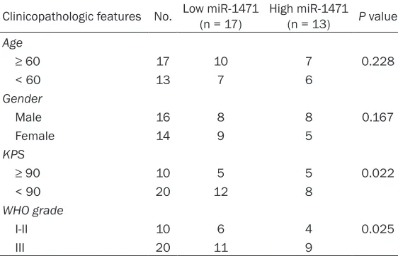
stage and grading system [16]. The clinicopath-ological parameters of these 30 patients were collected and the association with miR-1471
entific, Inc.) at a humidified incubator contain -ing 5% of CO2 at 37°C.
Cell transfection was conducted using Lipo- fectamine 2000 (Invitrogen, Thermo Fisher
Scientific, Inc.) in follow with the
manufactu-rer’s instructions. The synthetic miRNAs includ-ing miR-1471 mimic, miR-1471 inhibitor, and scramble miRNA (miR-NC) purchased from RiboBio Inc. (Guangzhou, China) were used to manipulate the expression levels of miR-1471.
pcDNA3.1-MTDH construct purchased from
GenScript (Nanjing, China) was used to
overex-press MTDH. Cells were incubated to 70% con
-fluence and mixed with miRNAs (100 nM) or MTDH overexpression construct (100 nM) for 48 h. Transfection efficiency was measured by
quantitative real-time PCR (qRT-PCR) or west-ern blot.
RNA isolation and qRT-PCR
RNA samples from the tissue samples and cell lines were extracted using Trizol reagent
(Invitrogen, Thermo Fisher Scientific, Inc.) in
line with the standard protocols. cDNA was syn-thesized using BeyoRTTM cDNA First-strand
syn-thesis kit (Beyotime, Haimen, Jiangsu, China).
[image:2.612.91.372.98.279.2]qRT-PCR was conducted in an ABI 7500 Real-Time PCR system (Applied Biosystems, Foster City, CA, USA) using BeyoFastTM SYBR Green qPCR Mix (Beyotime). U6 small nuclear RNA (U6 snRNA) was used as internal control. Relative
Table 1. Correlations of miR-1471 and clinicopathologic features in glioma patients
Clinicopathologic features No. Low miR-1471 (n = 17) High miR-1471 (n = 13) P value
Age
≥ 60 17 10 7 0.228
< 60 13 7 6
Gender
Male 16 8 8 0.167
Female 14 9 5
KPS
≥ 90 10 5 5 0.022
< 90 20 12 8
WHO grade
I-II 10 6 4 0.025
III 20 11 9
miR-1471: microRNA-1471; KPS: Karnofsky performance scale; WHO: World Health
Organization.
expression was summarized in Table 1.
Cell culture
Glioma cell lines U251, U87, SNB19, A172 and normal hu-
man astrocytes (NHAs) were
purchased from the Cell Bank of Shanghai Life Academy of Science (Shanghai, China). These cells were maintained
in Dulbecco’s Modified Eagle
Medium (DMEM, Thermo Fi-
sher Scientific, Inc., Waltham,
MA, USA) supplemented with
100 U/ml penicillin, 100 μg/
ml streptomycin (Thermo Fi-
sher Scientific, Inc.) and 10%
miR-1471 levels were calculated using the 2-ΔΔCt
method. The sequence of primers used in this study was as follows: miR-1471: F-5’-TGGACC- CTGGTCTACTCCTG-3’, R-5’-CAAGAGCCCCTGTA- CAGCAT-3’; U6 snRNA: F-5’-CTCGCTTCGGCAG- CACA-3’, R-5’-AACGCTTCACGAATTTGCGT-3’. Western blot
Protein samples from the tissue samples and cell lines were extracted using RIPA lysis buffer
supplemented with phenylmethylsulfonyl fluo -ride (Beyotime) according to the manufacturer’s instructions. Protein concentration was ana-lyzed using bicinchoninic acid (BCA) kit (Be- yotime). These samples were separated using 10% SDS-PAGE and transferred onto nitrocel-lulose membranes. Then, the membranes were blocked with 5% fat-free milk and incubated
with primary antibodies of MTDH (ab227981, Abcam, Cambridge, MA, USA) and GAPDH
(ab181602, Abcam). After washing with TBST, the membranes were incubated with second-ary antibody (ab205718, Abcam). Protein bands were visualized using BeyoECL Plus Kit
(Beyotime) and quantified by Quantity One v4.62 software (Bio-Rad, Hercules, CA, USA).
Luciferase assay
The miR-1471 binding site located at
4019-4025 of MTDH 3’-UTR as predicted by
TargetScan. The wild-type (wt) or mutant (mut)
of MTDH 3’-UTR was cloned into a psiCHECK-2
vector (Promega, Madison, WI, USA) to
gener-ate MTDH-wt or MTDH-mut construct. For lucif -erase reporter assay, cells were co-transfected
with MTDH-wt or MTDH-mut and miR-1471
mimic or miR-NC using Lipofectamine 2000. After 48 h of transfection, luciferase activity
transfection and further incubated at 37°C for 2 h. The optical density was measured at 450 nm using a micro-plate analyzer (Bio-Rad). Invasion assay
Cell invasion assay was performed using Transwell invasion assay. The cells (5 × 10⁴ cells) to be investigated were added to the upper chamber in FBS-free DMEM, while the DMEM supplemented with FBS was added to the lower chamber. After incubation for 48 h, the noninvasive cells were scraped and washed with PBS for three times. Then, the membranes
were fixed with 4% paraformaldehyde and
stained with 0.1% crystal violet (Beyotime). The numbers of invasive cells were counted from 5
independent fields.
Statistical analysis
Data were presented as mean ± SD and ana-lyzed using GraphPad Prism 6 (GraphPad Software Inc., San Diego, CA, USA). Student’s t-test or one-way ANOVA and Tukey post-hoc test was used to analyze difference in two or above groups. The association between miR-1471 and clinicopathological parameters was analyzed by chi-square test. Correlations
between miR-1471 and MTDH levels were ana
-lyzed by Pearson’s correlation coefficient. Statistical difference was considered as signifi -cant when P < 0.05.
Results
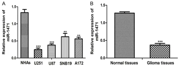
miR-1471 was downregulated in glioma
We first examined the levels of miR-1471 in four
[image:3.612.89.376.72.178.2]glioma cell lines (U251, U87, SNB19, A172) and
Figure 1. Downregulation of miR-1471 in glioma cell lines and tissues. A.
qRT-PCR analysis of miR-1471 expression in glioma cell lines and NHAs cell
line. B. qRT-PCR analysis of miR-1471 expression in glioma tissues and nor-mal tissues. (**P < 0.01, ***P < 0.001) miR-1471: microRNA-1471; qRT-PCR: quantitative real-time PCR.
er assay kit (Promega) acc- ording to the manufacturer’s protocol.
Proliferation assay
Cell proliferation assay was conducted using Cell coun- ting kit-8 (CCK-8, Beyotime) according to manufacturer’s
instruction. Briefly, cells were
incubated in the 96-well plate at the density of 1 × 10⁴ cells/
well. Then, 10 μl CCK-8
dramatically decreased levels of miR-1471
compared with NHAs cell line (Figure 1A). Levels of miR-1471 in four glioma cell lines in descending order were: SNB19, A172, U87, and U251 (Figure 1A). Therefore, the U87 and U251 cell lines were used for the following func-tional studies. Moreover, downregulation of miR-1471 was also found in clinical tumor sam-ples (Figure 1B). We also found low miR-1471
expression was closely associated with WHO
grade (P = 0.025) and Karnofsky performance scale (KPS, P = 0.022) but not associated with age and gender (P > 0.05, Table 1). These results demonstrated that miR-1471 was down-regulated in glioma tissues and cell lines and low miR-1471 expression was correlated with the grade of glioma.
Overexpression of miR-1471 inhibits glioma cell proliferation and invasion
We wanted to investigate the biological roles of miR-1471 in glioma since it was found down-regulated in glioma cell lines. The synthetic miRNAs were transfected into U87 and U251 cell lines and the qRT-PCR analysis results
showed that miR-1471 levels were significantly
increased by miR-1471 mimic but decreased by miR-1471 inhibition compared with the miR-NC (Figure 2A). CCK-8 assay showed that cell pro-liferation in U87 and U251 cell lines was enhanced by miR-1471 inhibitor but decreased by miR-1471 mimic (Figure 2B). Moreover, ecto-pic expression of miR-1471 could inhibit the invasion of U87 and U251 cell lines (Figure 2C). These results showed that miR-1471 overex-pression inhibits glioma cell proliferation and invasion.
MTDH was a direct target of miR-1471
We investigated the potential target of
miR-1471 by TargetScan. MTDH attracted our atten -tion as it contains a putative binding site for miR-1471 (Figure 3A). To investigate whether
miR-1471 could bind to the 3’-UTR of MTDH, a
luciferase reporter assay was conducted. It was found miR-1471 mimic could impair the luciferase activity of cells transfected with
MTDH-wt but not MTDH-mut (Figure 3B). The western blot analysis results revealed that
MTDH protein levels were significantly reduced
by miR-1471 mimic in U87 and U251 cell lines (Figure 3C). Correlation analysis showed that
miR-1471 and MTDH expression levels were
inversely correlated (Figure 3D). These results
showed that MTDH was a direct target of
miR-1471.
MTDH reversed the effects of miR-1471 on glioma cell proliferation and invasion
In light of the above finding between miR-1471 and MTDH, we examined whether MTDH was a
downstream effector for miR-1471 in glioma. Rescue experiments were performed on the
U251 cell line. It was found that MTDH con
-struct significantly enhanced the levels of MTDH in U251 cell line and rescued the inhibi
-tory effect of miR-1471 mimic on MTDH (Figure 4A). Then, we performed CCK-8 and transwell invasion assays to evaluate the effects of
MTDH overexpression on cell proliferation and invasion. Overexpression of MTDH enhanced
glioma cell proliferation and invasion and atten-uated the inhibitory effects of miR-1471 mimic (Figure 4B and 4C). These results showed that
MTDH was a functional target of miR-1471 in
glioma.
Discussion
The progression of human cancers is charac-terized as abnormal status of cell proliferation,
migration, invasion, apoptosis leading to the disruption of normal cell function [17]. Emerging evidence suggested miRNAs had crucial roles in the progression of human can-cers including glioma [7-10, 18-20]. Ding et al. examined miR-122 expression in glioma tis-sues and normal brain tistis-sues and found miR-122 expression was downregulated in glioma tissues compared with the normal brain tis-sues [18]. They also found miR-122 overex-pression could inhibit cell growth and induce apoptosis through regulating the expression of runt-related transcription factors (RUNX2), implicating miR-122/RUNX2 signal might be used as therapeutic targets for glioma [18]. Recently, Gu et al. conducted another set of in vitro experiments showed that miR-384 could inhibit glioma cell malignancy through target-ing cell division cycle 42 [19]. miR-1471 was shown to regulate breast cancer cell
progres-sion [15]. However, the expresprogres-sion pattern and
molecular function of miR-1471 was unclear in glioma.
In this study, we demonstrated the expression
of miR-1471 was significantly downregulated in
glioma tissues and cell lines. Moreover, our results demonstrated that reduced miR-1471
[image:5.612.99.511.68.301.2]expression was correlated with advanced WHO
Figure 3. MTDH is a direct target of miR-1471. A. Schematic representation of MTDH 3’-UTRs showing the putative miR-1471 target site. B. Analysis of the relative luciferase activities of MTDH-wt and MTDH-mut. C. Western blot analysis of MTDH expression in U251 and U87 cells transfected with miR-1471 mimic or miR-NC. D. Pearson’s corre
-lation analysis of miR-1471 and MTDH in glioma. (ns not significant, **P < 0.01, ***P < 0.001) miR-1471: microR
grade and low KPS score. However, the effects
of miR-1471 on the overall survival of glioma patients warrants further study.
The abnormal expression of miRNAs often resulted in malignant cell behaviors [7-10, 18-20]. It was found that miR-1471 levels in
glioma cell lines were significantly lower than in
normal human astrocytes. To study the effects of miR-1471 expression on glioma cell prolifera-tion and invasion, we upregulated and down-regulated miR-1471 expression in glioma cells by miR-1471 mimic and miR-1471 inhibitor. We showed that miR-1471 overexpression sup-pressed glioma cell proliferation and invasion in vitro. It has been widely recognized that the biologic functions of miRNAs are exerted through regulating the expression of
tumor-related genes [7-10, 18-20]. Here, we demon
-strated MTDH contains a putative binding site
for miR-1471 in its 3’-UTR. We further validated
MTDH as a direct target gene of miR-1471 using
luciferase activity report assay and western
blot assay. Additionally, MTDH expression was
found negatively correlated with miR-1471 expression in glioma tissues. Finally,
overex-pression of MTDH stimulates glioma cell prolif -eration and invasion, which is similar to the effects of miR-1471 inhibitor. Surprisingly, we
found MTDH overexpression could partially
reversed the effects of miR-1471 mimic on glio-ma cell behaviors.
In summary, these results demonstrated that miR-1471 functions as a tumor suppressor in
glioma through targeting MTDH. Our results
provided novel insight into the progression of
glioma and proved that the miR-1471/MTDH
axis has the potential to be used as effective therapeutic targets for glioma therapy.
Disclosure of conflict of interest
None.
Address correspondence to: Qianxue Chen, De-
partment of Neurosurgery, Renmin Hospital of
Wuhan University, 99 Zhangzhidong Road, Wuchang
District, Wuhan 430060, Hubei, P. R. China. E-mail:
References
[1] Butowski NA, Sneed PK, Chang SM. Diagnosis and treatment of recurrent high-grade glio-mas. J Clin Oncol 2006; 24: 1273-1280. [2] Stupp R, Mason WP, van den Bent MJ, Weller
[image:6.612.90.527.69.334.2]M, Fisher B, Taphoorn MJ, Belanger K, Brandes AA, Marosi C, Bogdahn U, Curschmann J, Jan-zer RC, Ludwin SK, Gorlia T, Allgeier A, La-combe D, Cairncross JG, Eisenhauer E, Miri-manoff RO; European Organisation for
Figure 4.
Overexpres-sion of MTDH partially
reversed the effects of miR-1471-overexpres-sion on glioma cells. (A) Western blot analysis
of MTDH expression,
(B) Cell proliferation and (C) Cell invasion in U251 cells transfected with miR-1471 mimic
or MTDH construct.
(**P < 0.01, ***P < 0.001) miR-1471:
mi-croRNA-1471; MTDH:
Research and Treatment of Cancer Brain Tu-mor and Radiotherapy Groups; National Can-cer Institute of Canada Clinical Trials Group. Radiotherapy plus concomitant and adjuvant temozolomide for glioblastoma. N Engl J Med 2005; 352: 987-996.
[3] Liang Y, Diehn M, Watson N, Bollen AW, Aldape KD, Nicholas MK, Lamborn KR, Berger MS, Botstein D, Brown PO, Israel MA. Gene
expres-sion profiling reveals molecularly and clinically
distinct subtypes of glioblastoma multiforme. Proc Natl Acad Sci U S A 2005; 102: 5814-5819.
[4] Rao JS. Molecular mechanisms of glioma inva-siveness: the role of proteases. Nat Rev Can-cer 2003; 3: 489-501.
[5] Emdad L, Das SK, Dasgupta S, Hu B, Sarkar D, Fisher PB. AEG-1/MTDH/LYRIC: signaling path -ways, downstream genes, interacting proteins, and regulation of tumor angiogenesis. Adv Cancer Res 2013; 120: 75-111.
[6] Su ZZ, Kang DC, Chen Y, Pekarskaya O, Chao
W, Volsky DJ, Fisher PB. Identification and clon -ing of human astrocyte genes display-ing
ele-vated expression after infection with HIV-1 or exposure to HIV-1 envelope glycoprotein by rapid subtraction hybridization, RaSH. Onco -gene 2002; 21: 3592-3602.
[7] Wang Q, Lv L, Li Y, Ji H. MicroRNA-655 sup -presses cell proliferation and invasion in oral squamous cell carcinoma by directly targeting metadherin and regulating the PTEN/AKT pathway. Mol Med Rep 2018; 18: 3106-3114. [8] Li L, Zhang H. MicroRNA-379 inhibits cell prolif -eration and invasion in glioma via targeting metadherin and regulating PTEN/AKT path-way. Mol Med Rep 2018; 17: 4049-4056. [9] Guo T, Pan G. MicroRNA-136 functions as a
tu-mor suppressor in osteosarcoma via regulat-ing metadherin. Cancer Biomark 2018; 22: 79-87.
[10] Zhang Y, Wang Y, Wang J. MicroRNA-584 inhib-its cell proliferation and invasion in non-small
cell lung cancer by directly targeting MTDH.
Exp Ther Med 2018; 15: 2203-2211.
[11] Lee SG, Su ZZ, Emdad L, Sarkar D, Fisher PB. Astrocyte elevated gene-1 (AEG-1) is a target gene of oncogenic ha-ras requiring phosphati-dylinositol 3-kinase and c-Myc. Proc Natl Acad Sci U S A 2006; 103: 17390-17395.
[12] Du C, Yi X, Liu W, Han T, Liu Z, Ding Z, Zheng Z, Piao Y, Yuan J, Han Y, Xie M, Xie X. MTDH medi
-ates trastuzumab resistance in HER2 positive
breast cancer by decreasing PTEN expression
through an NFκB-dependent pathway. BMC
Cancer 2014; 14: 869.
[13] Li PP, Feng LL, Chen N, Ge XL, Lv X, Lu K, Ding M, Yuan D, Wang X. Metadherin contributes to the pathogenesis of chronic lymphocytic
leuke-mia partially through wnt/β-catenin pathway.
Med Oncol 2015; 32: 479.
[14] Zhang D, Liu Z, Zheng N, Wu H, Zhang Z, Xu J.
MiR-30b-5p modulates glioma cell
prolifera-tion by direct targeting MTDH. Saudi J Biol Sci
2018; 25: 947-952.
[15] Liu X, Zhao T, Bai X, Li M, Ren J, Wang M, Xu R,
Zhang S, Li H, Hu Y, Xie L, Zhang Y, Yang L, Yan
C, Zhang Y. LOC101930370/MiR-1471 axis modulates the hedgehog signaling pathway in breast cancer. Cell Physiol Biochem 2018; 48: 1139-1150.
[16] Komori T. The 2016 WHO classification of tu -mours of the central nervous system: the ma-jor points of revision. Neurol Med Chir (Tokyo) 2017; 57: 301-311.
[17] Hanahan D, Weinberg RA. Hallmarks of can -cer: the next generation. Cell 2011; 144: 646-674.
[18] Ding CQ, Deng WS, Yin XF, Ding XD. MiR-122 inhibits cell proliferation and induces apopto-sis by targeting runt-related transcription fac-tors 2 in human glioma. Eur Rev Med Pharma-col Sci 2018; 22: 4925-4933.
[19] Gu G, Wang L, Zhang J, Wang H, Tan T, Zhang
G. MicroRNA-384 inhibits proliferation migra-tion and invasion of glioma by targeting at CDC42. Onco Targets Ther 2018; 11: 4075-4085.