Gerbode defect in a dog
Carlos F. Agudelo
1, Michal Crha
1, Zeki Yilmaz
2, Branislav Lukac
3*
1Small Animal Clinic, University of Veterinary and Pharmaceutical Sciences Brno, Brno, Czech
Republic
2Department of Internal Medicine, Faculty of Veterinary Medicine, Uludag University, Bursa,
Turkey
3Small Animal Clinic, University of Veterinary Medicine and Pharmacy in Kosice, Kosice, Slovakia
*Corresponding author: [email protected]
Citation: Agudelo CF, Crha M, Yilmaz Z, Lukac B (2019): Gerbode defect in a dog. Veterinarni Medicina 64, 138–143.
Abstract: An intracardiac communication between the left ventricle and the right atrium (Geborde defect) was diagnosed in a 9-year Yorkshire Terrier with a history of chronic exercise intolerance. The history, clinical examina-tion, and diagnostic imaging confirmed the diagnosis and did not reveal evidence of trauma or endocarditis which could lead to this special type of left-to-right shunting. A Gerbode defect is a very rare finding in human beings and animals. In the veterinary literature all reports about this condition were related to thoracic trauma or valvular infection. According to the authors, this would be the first clinical case of congenital Geborde defect in a dog.
Keywords: canine; cushion defect; echocardiography; ventricular septal defect
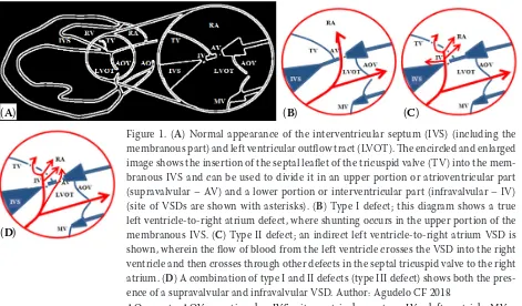
Gerbode defect is a rare type of ventricular septal defect in which there is communication between the left ventricle and right atrium. In human medi-cine, Gerbode defects may account for about 0.08% of all congenital heart diseases (Ramirez et al. 2003; Vijayalakshmi et al. 2013). In general, a ventricular septal defect can be located along the wall of the interventricular septum, but they are more com-monly observed in the upper and posterior part known as the membranous interventricular sep-tum, which separates the aortic vestibule from the lower part of the right atrium and the upper part of the right ventricle. Furthermore, the membra-nous part can be divided by the septal leaflet of the tricuspid valve into the atrioventricular and interventricular parts (Figure 1A). Type I (supra-valvular or atrioventricular) defects occur in the atrioventricular part of the membranous septum resulting in direct communication of the left ven-tricle to the right atrium (Figure 1B). Type II
(infra-valvular or interventricular) defects are indicative of a true ventricular septal defect associated with other congenital changes in the septal leaflet of the tricuspid valve (such as perforations or clefts, fenestration, aneurysmal transformation or inter-growths of valve leaflets to the edges of the defect) (Figures 1B−1D) (Ramirez et al. 2003; Peddle et al. 2008; Karaci et al. 2012). In these cases, shunting of blood is observed not only from the left ven-tricle to the right venven-tricle but also to the right atrium, which is achieved by crossing through the abnormalities in the septal tricuspid leaflet. In some instances, a unique defect involves both supravalvular and infravalvular (type III) abnor-malities (Figure 1D) (Peddle et al. 2008; Sinisalo et al. 2011). Human cases of Gerbode defect can be recognised congenitally or secondary to cardiac surgery (e.g. valvular transplantation), septic endo-carditis, thoracic trauma and myocardial infarction (Ramirez et al. 2003; Peddle et al. 2008; Hezzell et
never suffered from any other condition with the exception of unilateral patellar luxation that was successfully corrected three years earlier. The refer-ring veterinarian found a murmur and prescribed benazepril (0.25 mg/kg p.o., s.i.d.). The dog was up-to-date regarding vaccinations and deworm-ing. On presentation, the dog was mentally alert with pink and moist mucous membranes. There was a mild degree of dental tartar. Auscultation revealed tachycardia (160 bpm) with regular femo-ral pulses. Additionally, a sternal holosystolic heart murmur was detected at the left hemithorax. The respiratory rate was 28 rpm with normal broncho-vesicular sounds. At the time, differential diagno-ses pointed to degenerative mitral valve disease or endocarditis. Diagnostic work-up included cardio-logic examinations such as an electrocardiogram (ECG), thoracic radiographs, blood pressure meas-urement and echocardiography.
A 6-lead ECG (Figure 2) revealed sinus arrhyth-mia with a heart rate of approximately 140 bpm and the presence of Ta waves and occasional tall P waves. Blood pressure obtained from the dorsal (pedal) metatarsal artery using a Doppler ultrasonic system revealed a normal systolic blood pressure of 130 mm Hg. Thoracic radiographs demonstrated a mildly enlarged cardiac silhouette (vertebral heart Figure 1. (A) Normal appearance of the interventricular septum (IVS) (including the membranous part) and left ventricular outflow tract (LVOT). The encircled and enlarged image shows the insertion of the septal leaflet of the tricuspid valve (TV) into the mem-branous IVS and can be used to divide it in an upper portion or atrioventricular part (supravalvular – AV) and a lower portion or interventricular part (infravalvular – IV) (site of VSDs are shown with asterisks). (B) Type I defect; this diagram shows a true left ventricle-to-right atrium defect, where shunting occurs in the upper portion of the membranous IVS. (C) Type II defect; an indirect left ventricle-to-right atrium VSD is shown, wherein the flow of blood from the left ventricle crosses the VSD into the right ventricle and then crosses through other defects in the septal tricuspid valve to the right atrium. (D) A combination of type I and II defects (type III defect) shows both the pres-ence of a supravalvular and infravalvular VSD. Author: Agudelo CF 2018
AO = aorta; AOV = aortic valve; IVS = iterventricular septum; LV = left ventricle; MV = mitral valve; RA = right atrium; RV = right ventricle; VSD = ventricular septal defect (A)
(D)
(B) (C)
al. 2011; Cunningham et al. 2013; Vijayalakshmi et al. 2013). Clinical signs can be seen very early in life due to the presence of a murmur or of clinical signs like difficulty in breathing, exercise intoler-ance, depression, weakness and syncope (Peddle et al. 2008; Vijayalakshmi et al. 2013). Veterinary reports are scarce and limited to canine species. To our knowledge, to this day only four reports of this defect have been published: two of them were secondary to valvular endocarditis (Great Pyrenees Dog and Golden Retriever) (Ramirez et al. 2003; Peddle et al. 2008) and the other two (Newfoundland and Labrador Retriever) were seen after thoracic trauma, interestingly, always associ-ated with an atrial septal defect (Hezzell et al. 2011; Cunningham et al. 2013). We describe a possible congenital Gerbode defect in an older dog unre-lated to endocarditis, surgery or trauma.
Case description
[image:2.595.65.536.96.372.2]left ventricular outflow tract that crossed the pe-rimembranous portion of the septum towards the right ventricle as well as towards the right atrium proximally to the level of the septal tricuspid leaflet (Figure 4). A modified right parasternal 5-chamber view was used to further investigate shunting from left ventricle-to-right atrium, confirming a con-nection between the atrioventricular part of the membranous interventricular septum and the right atrium (Figure 5). Spectral continual Doppler esti-mated a radient through the left-to-right shunting of 3.3 m/s (approximately 44 mm Hg) (Figure 6). Also, mild degenerative changes in the mitral and tricuspid valves were recorded from left apical views. The pulmonary-systemic flow ratio (Qp/Qs) scale = 11) without pulmonary venous congestion.
The trachea presented different degrees of lumen irregularities and there was a dominant bronchial lung pattern (Figure 3). Complete blood count and serum chemistries were unremarkable.
[image:3.595.66.393.96.251.2]Standard echocardiography showed thicken-ing of the caudal leaflet of the mitral valve and a mildly hyperechogenic and elongated valve ap-paratus of the cranial tricuspid leaflet. There was no prolapse of any of the atrioventricular valves. Systolic left atrial size at M-mode was 22.3 mm and the left atrium/aorta ratio was 1.42. The oth-er 2D and M-mode parametoth-ers woth-ere within the normal range (Boon 2011). Colour flow mapping displayed turbulent flow originating within the
Figure 2. ECG tracing revealed sinus arrhythmia with a heart rate of approximately 140 bpm and the presence of Ta waves (black arrows) and occasional tall P waves (asterisks), which may be a change suggesting right atrial overload. Wander-ing sinus pacemaker can also be seen. Paper speed 50 mm/s
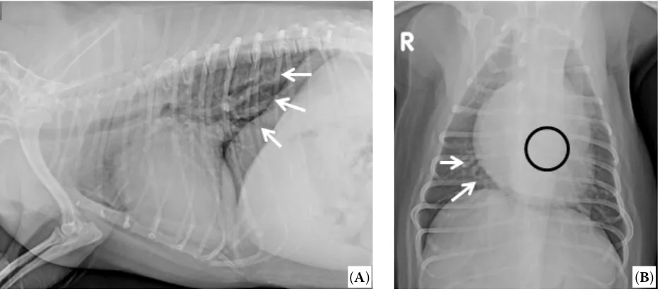
Figure 3. (A) Laterolateral view. Vertebral heart scale measures approximately 11 vertebral bodies. The tracheal lumen can be distinguished as irregular based on its thoracic length. Some spondylosis could also be seen at the 4th, 5th and 6th thoracic vertebral bodies. A lung bronchial pattern also can be seen at caudal lung lobes (arrows). (B) Dorsoventral view. Mildly rounded heart silhouette. There is an apparent mild collapse of the left main bronchus (black circle). Also, the lung bronchial pattern is evident on both sides but is more marked on the right middle lung lobe (white arrows)
[image:3.595.64.531.469.677.2]was determined at 1.2 (< 2 : 1 is haemodynamically insignificant) (Boon 2011). Based on the results of the cardiological examination, a supravalvular (type I) Gerbode defect was diagnosed. The owner declined other more invasive investigations such as contrast studies or catheterisation. The patient was sent home with the same therapy and after six months the follow-up did not show any worsening in clinical or echocardiographic situations.
DISCUSSION AND CONCLUSIONS
The first description of Gerbode defect was reported in humans in 1857 (Meyer 1857). Most Gerbode-type defects in humans are congenital and have been reported in association with other con-genital heart defects, including subaortic stenosis, atrial septal defect and other vascular anomalies (Vijayalakshmi et al. 2013). Interestingly, human patients with congenital Gerbode defects (and oth-er congenital diseases) are at a highoth-er risk of devel-oping infective endocarditis. The risk of infective endocarditis is closely related to abnormal intra-cardiac shunts and high-velocity or turbulent flows (Karaci et al. 2012). In the dog of this report, other congenital or acquired diseases were not observed with the exception of mild degenerative changes in the mitral valve (ACVIM classification B1), a condi-tion that would be more typical of such a patient due to the age and breed.
An acquired Gerbode defect in humans may be secondary to cardiosurgery, endocarditis, thoracic trauma or myocardial infarction. It was first re-ported in the veterinary literature as a necropsy finding by Ramirez et al. (2003). Two of the four reports on Gerbode defects that have been so far described in canine patients were secondary to bac-terial endocarditis (Ramirez et al. 2003; Peddle et al. 2008), and the other two cases were found sec-ondary to thoracic trauma (in one case associated with atrial septal defect, probably also traumatic) Figure 4. (A) Modified right parasternal long axis view at the level of left ventricular outflow tract showing the pres-ence of a ventricular septal defect (arrow) between the left ventricle and right atrium. (B) Simultaneous colour flow mapping (CFM) displaying a left-to-right shunting through the defect. (C) Right parasternal short axis view at the level of the right ventricular outflow tract and aorta. CFM demonstrates abnormal blood shunting through the septal defect from the left ventricle to both the right atrium and right ventricle
[image:4.595.67.534.96.248.2]AO = aorta; LVOT = left ventricular outflow tract; RA = right atrium; RV = right ventricle; RVOT = right ventricular outflow tract; TV = tricuspid valve
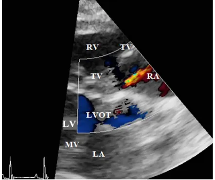
Figure 5. Modified right parasternal long axis view. Colour flow mapping (CFM) reveals a systolic flow from left ventricular outflow tract (LVOT)-to-right atrium (RA) above the tricuspid valve
LA = left atrium; LV = left ventricle; MV = mitral valve; RV = right ventricle; TV = tricuspid valve
[image:4.595.67.288.489.674.2](Hezzell et al. 2011; Cunningham et al. 2013). No traumatic incident or any cardiac infection was described in the history, suspected in the clinical exam nor confirmed with other diagnostic tests in our patient. We assume that the origin in this case was congenital; however, and according to human reports, most supravalvular defects are acquired conditions (Ramirez et al. 2003). This may be due in part to the associated lesions seen on the septal leaflet of the tricuspid valve.
In general, dogs with ventricular septal defect may develop clinical signs normally seen in young dogs (one to two years of age) but can present at an older age with symptoms of generalised weakness, cough, exercise intolerance, syncope, abdominal swelling associated with ascites and pale mucous membranes. Other patients may not present signs at all, and ventricular septal defects are found as incidental findings during other examinations. Dogs with the Gerbode defect may show acute symptomatology due to a destructive perforating endocarditis or blunt trauma leading to rupture of the membranous interventricular septum. Signs of systemic infection (due to endocarditis-like fever, lethargy, anorexia or weight loss), arrhythmia or signs associated with trauma (laboured respira-tion, bleeding or lameness) may precede signs of heart failure (Hezzell et al. 2011; Cunningham et al. 2013). The patient of this report was a geriatric dog with vague symptomatology of exercise intoler-ance, which does not reflect an acute process; how-ever, past valvular infections could have occurred and then subsequently healed. ECG abnormalities
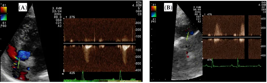
were numerous. We found ECG findings of right atrial overload due to the presence of peaked tall P waves in combination with Ta waves (Tilley 1992). These findings may suggest long-term right-side overload as seen in other reports (Cunningham et al. 2013). Other described ECG findings are VPCs (ventricular premature complexes) (Hezzell et al. 2011) and varying degrees of atrioventricular blocks (Cunningham et al. 2013) or left or right bundle branch blocks (Vijayalakshmi et al. 2013). This may be due to the proximity of ventricular septal defects to the atrioventricular conduction tissue in the membranous interventricular sep-tum or could be secondary to direct damage to the conduction system in the atrioventricular node as seen in canine and human patients (Peddle et al. 2008; Sinisalo et al. 2011). Long-term shunt-ing may lead to biventricular volume overload and ventricle enlargement (Silbiger et al. 2009). To date, the patient is doing well on medical management alone; her estimated Qp/Qs is an indication that pulmonary flow exceeds systemic flow and defines a net left-to-right shunt that remains below the threshold for any other intervention (1.5–2 : 1 is considered indication for intervention). Treatment is usually conservative, and the goals are to pro-long the onset of overt heart disease and manage-ment of the secondary congestive heart failure; however, surgical correction can be performed at specialty referral centres. In humans, the condi-tion is usually repaired surgically, mainly by using either percutaneous Amplatzer ductal or septal occluder devices. The first successful closure of Figure 6. (A) Continuous wave Doppler of the left-to-right ventricular septal defect in a modified right parasternal long axis view at the level of the left ventricular outflow tract. The cursor is located at the supravalvular (type B) septal defect. Maximal instantaneous peak gradient in this patient was approximately 44 mm Hg. (B) Left 5-chamber apical view at the level of the insertion of the septal leaflet of the tricuspid valve. Peak gradient is similar as that obtained from the right side
[image:5.595.65.532.96.241.2]such a defect was reported by Kirby (using hypo-thermia and inflow occlusion) at the Hospital of the University of Pennsylvania in 1956 (Kirby et al. 1957). The first successful series of operations on patients with a left ventricular-to-right atrial shunt and a complete description of this condition was reported by Gerbode et al. (1958). According to the veterinary literature, the proximity to the aortic valve and the ratio device size and patient size makes it difficult to deploy equipment in very small patients. A more invasive approach in this patient was not considered due to the restrictive nature of the defect, lack of clinical signs and the size of the dog.
REFERENCES
Boon JA (2011): Manual of Veterinary Echocardiography. 2nd edn. John Wiley & Sons. 632 p.
Cunningham SM, Lindsey KJ, Rush JE (2013): Acquired Gerbode defect and third-degree atrioventricular block secondary to vehicular trauma in a dog. Journal of Vet-erinary Emergency and Critical Care 23, 637–642. Gerbode F, Hultgren H, Melrose D, Osborn J (1958):
Syn-drome of left ventricular-right atrial shunt. Successful repair of defect in five cases, with observation of brady-cardia on closure. Annals of Surgery 148, 433–446. Hezzell MJ, Dennis S, Lewis DH, Fuentes VL (2011):
Ger-bode defect associated with blunt trauma in a dog. Jour-nal of Veterinary Cardiology 13, 141–146.
Karaci AR, Aydemir NA, Harmandar B, Sasmazel A, Saritas T, Tuncel Z, Yekeler I (2012): Surgical treatment of
infec-tive valve endocarditis in children with congenital heart disease. Journal of Cardiac Surgery 27, 93–98.
Kirby CK, Johnson JJ, Zinsser HF (1957): Successful closure of a left ventricular-right atrial shunt. Annals of Surgery 145, 392–394.
Meyer H (1857): On congenital tightness or occlusion of the pulmonary arterial tract (in German). Virchows Ar-chiv fur pathologische Anatomie 12, 497–532.
Peddle GD, Boger L, Van Winkle TJ, Oyama MA (2008): Gerbode type defect and third degree atrioventricular block in association with bacterial endocarditis in a dog. Journal of Veterinary Cardiology 10, 133–139.
Ramirez GA, De los Monteros E, Rodriguez F, Weisbrode SE, Jaber JR, Herraez P (2003): Left ventricular outflow tract-right atrial communication (Gerbode type defect) associated with bacterial endocarditis in a dog. Veterinary Pathology 40, 579–582.
Silbiger JJ, Kamran M, Handwerker S, Kumar N, Marcali M (2009): The Gerbode defect: Left ventricular to right atrial communication-anatomic, hemodynamic, and echocar-diographic features. Echocardiography 26, 993–998. Sinisalo JP, Sreeram N, Jokinen E, Qureshi SA (2011):
Ac-quired left ventricular-right atrium shunts. European Journal of Cardiothoracic Surgery 39, 500–506.
Tilley LP (1992): Essentials of Canine and Feline Electro-cardiography: Interpretation and Treatment. 3rd edn. Lea
& Febiger, Philadelphia. 348 p.
Vijayalakshmi IB, Narasimban C, Simha P (2013): Ventric-ular septal defects. In: Vijayalakshmi IB, Syamasundar P, Chugh R (eds): A Comprehensive Approach to Congen-ital Heart Diseases. Jaypee Brothers Medical Publishers, New Delhi. 266–291.