ORIGINAL RESEARCH
Benign Ulceration as a Manifestation of Soft
Tissue Radiation Necrosis: Imaging Findings
J.M. Debnam A.S. Garden L.E. Ginsberg
BACKGROUND AND PURPOSE: The purpose of this study was to review CT imaging findings of soft tissue mucosal ulceration in patients following radiation treatment for head and neck malignancies and to correlate these with patient outcomes.
MATERIALS AND METHODS: The CT examinations in 20 patients with soft tissue ulceration after radiation therapy for treatment of head and neck cancer were reviewed. External beam radiation therapy was completed between 3 and 61 months (mean, 11.5 months) before the initial diagnosis of soft tissue ulceration. In all 20 patients, the initial diagnosis was made or confirmed on CT examination.
RESULTS: Of the 20 ulcerations, 12 did not demonstrate enhancement, and the results of biopsy in 9 of these 12 were negative. Of the 12 nonenhancing ulcerations, biopsy was not performed in 3, but they have been followed clinically and radiologically for 15.7 months without evidence of recurrence. Of the 20 ulcerations, 8 demonstrated adjacent enhancement, and the results of a biopsy in 4 were positive for recurrent cancer and negative in 2; these 2 have been followed for 16.3 months without evidence of recurrence. Biopsy was not performed in 2 ulcerations, but they have been followed for 15.0 months without evidence of recurrence.
CONCLUSION:For soft tissue ulceration occurring after radiation treatment, if there is no enhancement or clinical evidence of recurrence, it is likely benign and follow-up without biopsy seems warranted. If the ulceration is associated with adjacent enhancement, then differentiation between radiation necro-sis and recurrent tumor is difficult. In these cases, correlation with clinical examination with close interval follow-up is necessary if a biopsy is not performed.
S
quamous cell carcinoma of the head and neck occurs in approximately 40,000 patients annually in the United States1and is often treated with radiation therapy. The radia-tion treatment causes endothelial damage and fibrosis, leading to impairment of vascular and lymphatic flow.2This impair-ment produces hypoxic, hypocellular, and hypovascular tis-sue, which is unable to maintain normal tissue turnover3,4 resulting in tissue necrosis, infection, and ulceration.4-6 Ulcer-ation is defined as a defect, or excavUlcer-ation, of the surface of a tissue or organ, which is produced by the sloughing of inflam-matory necrotic tissue.7On CT, soft tissues within the treat-ment field may demonstrate inflammatory swelling, fragmen-tation, and ulceration.8However, recurrent tumor may also present clinically as a soft tissue ulceration,9-11leading to dif-ficulty in the diagnosis and management of these patients.To our knowledge, no previous reports on the imaging appearance of soft tissue ulcerations that occur in patients after radiation treatment of head and neck cancer have been published in the radiology literature. The purpose of this study was to review the CT imaging findings of benign and malig-nant soft tissue ulceration in 20 patients who had undergone radiation therapy for head and neck cancer and to correlate with the patients’ outcomes.
Materials and Methods
The institutional review board approved this study and waived the requirement for informed consent. We reviewed the clinical records and imaging findings of 20 patients who underwent radiation therapy as part of the treatment of head and neck cancer and were diagnosed with a mucosal ulceration between September 2001 and August 2005. Of 20 patients, 13 were men and 7 were women with an age range of 46 to 78 years (mean, 61 years). Primary tumors included squa-mous cell carcinoma in 19 patients of the oropharynx (n⫽12), oral tongue (n⫽3), larynx (n⫽3), and hypopharynx (n⫽1). One patient had poorly differentiated carcinoma of the nasopharynx. A total of 17 patients underwent radiation therapy at our institution (60 –72 Gy; mean, 68.5 Gy, in 30 – 42 fractions), and 3 patients underwent radia-tion therapy at an outside instituradia-tion, where the radiaradia-tion dose was not available in the clinical notes. The radiation therapy was com-pleted between 3 and 61 months (mean, 11.5 months; median, 5.5 months) before the initial diagnosis of soft tissue radiation necrosis in all 20 patients.
A total of 20 CT examinations with evidence of a mucosal ulcer-ation, obtained as part of routine posttreatment follow-up between January 2001 and January 2006, were evaluated in the 20 patients for initial characterization of the soft tissue ulcerations. The ulcerations were originally detected on both clinical and radiologic examinations, 13 of them on radiologic examination alone because 3 of these were not clinically evident; 4 were not available on clinical examination and later on radiologic examination between 1 and 3 months (mean, 1.8 months) after the clinical diagnosis. The CT examinations were per-formed on scanners (Light Speed; GE Healthcare, Milwaukee, Wis) after intravenous administration of contrast material. Imaging in the axial plane was performed with the following section thicknesses: 1.25 mm (n⫽11), 2.5 mm (n⫽3), 3.75 mm (n⫽1), and 5.0 mm (n⫽5). Soft tissue and bone windows were evaluated in all patients. In 15 of the 20 patients, a biopsy was performed on the ulceration, results confirming either radiation necrosis or recurrent tumor. The remain-Received April 26, 2007; accepted after revision October 3.
From the Department of Radiology (J.M.D., L.E.G.) and the Department of Radiation Oncology (A.S.G.), University of Texas M.D. Anderson Cancer Center, Houston, Tex.
Paper previously presented at: Annual Meeting of the American Society of Neuroradiology, May 3, 2006; San Diego, Calif.
Please address correspondence to J. Mathew Debnam, MD, University of Texas M.D. Anderson Cancer Center, 1515 Holcombe Blvd., Unit 370, Houston, TX 77030; e-mail: matthew.debnam@mdanderson.org
ing 5 patients were followed up clinically and radiologically between 8 and 30 months (mean, 15.4 months). Five positron-emission tomog-raphy (PET)/CT examinations were obtained in 4 patients, 2 before (1–2 weeks; mean, 1.5 weeks) and 3 after (1–2.5 months; mean, 1.7 months) biopsy.
Results
Sixteen patients complained of pain or odynophagia, or both. Two patients did not complain of pain but did report weight loss; one had a percutaneous endoscopic gastrotomy tube and the other was receiving a liquid diet. The remaining 2 patients were asymptomatic. Eighteen of the ulcerations occurred at the same location as the primary tumor. One patient was treated with a total laryngectomy followed by radiation ther-apy and developed an ulceration in the base of the tongue. The other patient had a squamous cell carcinoma of the left false vocal cord and developed ulceration in the epiglottis.
The types of ulcerations with their enhancement character-istics (enhancing or nonenhancing) and results of the biopsy for recurrent tumor (positive, negative, not performed) have been divided into the following 5 categories. Patient data are provided in the accompanying Table.
Category 1: Nonenhancing Ulcerations, Negative Biopsy Results (nⴝ9)
Nine ulcerations did not demonstrate enhancement, and re-sults of the biopsy were negative (Fig 1). Eight were followed clinically and radiologically between 5 and 28 months (mean, 16.2 months) without evidence of recurrence. The final pa-tient in this group underwent a total laryngectomy for airway compromise related to the radiation therapy. Pathologic ex-amination reported no evidence of recurrent tumor within the ulceration and in the other submitted specimens.
Two of the nonenhancing ulcers with negative biopsy re-sults underwent a PET/CT examination, which demonstrated [18F]fluorodeoxyglucose (18F-FDG) avidity. The first patient
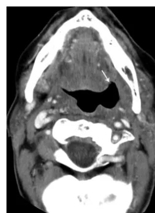
had a negative biopsy result 1 week before an 18F-FDG PET/CT (standardized uptake value [SUV], 5.5 mg/mL) and a second negative biopsy result 1 month after the PET/CT. This patient has subsequently been followed up for 14 months clin-ically and radiologclin-ically, with CT examinations demonstrat-ing no evidence of tumor recurrence. The second patient (Fig 2AandB) underwent biopsy of a nonenhancing ulceration of the right lateral part of the oral tongue because of increase in pain and clinical suspicion of a recurrent tumor. The results of this biopsy were negative, and a PET/CT scan obtained 2.5 months later was suspicious for a recurrent tumor (SUV, 12 mg/mL). The results of a clinical examination were consistent with a healing ulcer, and the patient was observed. Although the ulcer was nonenhancing and seemed to be regressing on Fig 1.Patient 3, a 46-year-old man status post 7 months of radiation ther-apy (72 Gy) for treatment of a squamous cell carci-noma of the left orophar-ynx, with a nonenhancing ulceration with negative biopsy results (category 1) in the left oropharynx ( ar-rows). This patient has been followed for 11.5 months since initial diag-nosis without evidence of tumor recurrence. Summary of clinical data of patients with soft tissue ulceration
Patient
No. Category Age/Sex
Rad Dose (Gy)
Months After
XRT Location of Ulceration Enhancement Biopsy Results
1 1 55/M 66 4.5 Left palatine tonsil None Negative
2 1 53/M 66 8.5 Left palatine tonsil None Negative
3 1 46/M 72 7 Left oropharyngeal wall None Negative
4 1 58/M 70 6.5 Left vocal cords None Negative
5 1 59/F 60 3.5 Right oral tongue None Negative
6 1 50/M OSH 5 Left base of tongue None Negative
7 1 61/F OSH 12 Epiglottis None Negative
8 1 59/M 70 3.5 Epiglottis None Negative
9 1 51/F 70 3 Right base of tongue None Negative
10 2 70/M 60 3.5 Left lateral nasopharynx None N/A
11 2 75/F 70 5 Left oropharyngeal wall None N/A
12 2 64/M 70 5 Right floor of mouth None N/A
13 3 46/M 70 7 Left pyriform sinus Submucosal Positive
14 3 63/F 76 9 Midline oral tongue Submucosal Positive
15 3 70/M 66 6.5 Right oral cavity Nodular Positive
16 3 54/M 66 5 Left palatine tonsil Submucosal Positive
17 4 78/F 72 10.5 Right retromolar trigone Heterogeneous Negative
18 4 70/M OSH 61 Base of tongue Peripheral/Nodular Negative
19 5 64/M 70 5 Left oropharyngeal wall Peripheral N/A
20 5 74/F 70 6 Left oropharynx Peripheral N/A
Note:—Rad indicates radiation; OSH, outside hospital; XRT, external beam radiation therapy; N/A, biopsy not performed. N.B.—Categoric description of ulcerations is author’s terminology.
HEAD
&
NECK
ORIGINAL
[image:2.594.54.538.58.284.2]imaging, because of clinical persistence of the ulceration and associated pain, a second biopsy (13 months after the first biopsy) was performed, the results of which were also negative for a recurrent tumor.
Category 2: Nonenhancing Ulcerations, Biopsy Not Performed (nⴝ3)
There were 3 ulcerations that were without enhancement and in which a biopsy was not performed. These patients were followed up clinically and radiologically for 8 to 30 months (mean, 15.7 months) without evidence of recurrence (Fig 3). One of these patients developed osteoradionecrosis (ORN) of the hyoid bone that was separate from an ulceration of the right base of the tongue.
Category 3: Enhancing Ulcerations, Positive Biopsy Results (nⴝ4)
Of the 20 ulcerations, 4 demonstrated adjacent enhancement (submucosal [n⫽3], nodular [n⫽1]), biopsy results were positive for recurrent disease (Fig 4A), and these patients un-derwent further treatment of recurrent tumor. Two of these patients, both with oral cavity carcinoma, developed ORN of the mandible. One of the areas of ORN involved the anterior midline part of the mandible, separate from the ulceration of the oral tongue, and the other in the right mandibular ramus adjacent to an ulceration of the right oral cavity.
A PET/CT examination was also performed in 2 of the en-hancing (submucosal [n⫽2]) ulcerations with positive bi-opsy results at approximately the same time of bibi-opsy (2 weeks before and 1.5 months after biopsy), which demonstrated 18F-FDG avidity (SUV, 6 and 4.6 mg/mL, respectively) (Fig 4B).
Category 4: Enhancing Ulcerations, Negative Biopsy Results (nⴝ2)
Results of a biopsy on 2 ulcerations with enhancement (pe-ripheral and nodular [n⫽1], heterogeneous [n⫽1]) were negative for recurrent tumor (Fig 5) and were subsequently followed clinically and radiologically for 13 and 20 months (mean, 16.5 months) without evidence of recurrence. One of
these patients developed ORN of the mandibular ramus, ad-jacent to an ulceration, which extended into the right retro-molar trigone.
Category 5: Enhancing Ulcerations, Biopsy Not Performed (nⴝ2)
In 2 of the ulcerations with enhancement (peripheral [n⫽2]), a biopsy was not performed because these were not suspicious on clinical examination. They were followed clinically and ra-diologically for 14 and 20 months (mean, 17 months) without evidence of recurrence (Fig 6).
Discussion
Our findings that all 12 ulcerations without adjacent enhance-ment failed to demonstrate evidence of recurrent tumor, ei-Fig 2.Patient 5, a 59-year-old woman status post 3.5 months of radiation therapy (60 Gy) for treatment of squamous cell carcinoma of the oral tongue, with a nonenhancing ulceration with negative biopsy results (category 1). This patient has been followed for 16 months since initial diagnosis without evidence of tumor recurrence.A, Contrast-enhanced CT with ulceration in the right lateral part of the oral tongue (arrows). B,PET/CT with 18F-FDG activity (SUV, 12 mg/mL) along the posterior border (arrows) on the ulceration, suspicious for a recurrent tumor.
[image:3.594.53.371.45.252.2] [image:3.594.338.495.461.678.2]ther with biopsy results or on follow-up imaging, suggest that an ulceration without adjacent enhancement is benign and is likely radiation induced. When ulcerations demonstrate adja-cent enhancement, careful observation is required if biopsy is not performed because 4 of 8 ulcerations with adjacent en-hancement demonstrated recurrent tumor, whereas the other 4 ulcerations were free of tumor on biopsy results or on fol-low-up clinical and radiologic evaluations.
The risk for injury related to radiation necrosis is greatest during the first 6 to 12 months after radiation therapy.12,13 Our results were similar, with radiation therapy completed between 3 and 61 months (mean, 11.5 months; median, 5.5 months) before the initial radiologic diagnosis of soft tissue ulceration. However, most necroses and many recurrences
oc-cur within 2 years after radiation therapy,14 so the time of onset of the ulceration is usually not helpful in distinguishing between radiation injury and recurrent tumor.
Imaging of the head and neck in patients treated for cancer is routinely performed to evaluate for recurrent tumor and is complementary to physical examination. CT examinations can evaluate the underlying soft tissues, which cannot be visu-alized on physical examination. Hermans et al15found that CT examination detected treatment failure in 12 (41%) of 29 of patients at the primary site, a mean of 5.5 months before re-sults of clinical examination in laryngeal and hypopharyngeal carcinoma.
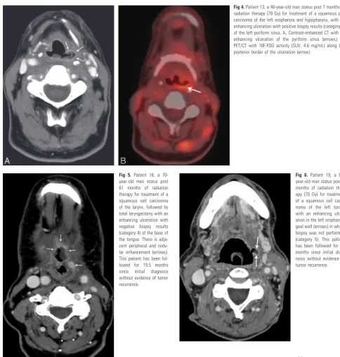
Osteoradionecrosis is a known complication of radiation therapy for malignant tumors of the head and neck.16-18Four of the patients in our series developed osteoradionecrosis, which was adjacent to the soft tissue ulceration in 2 patients (categories 2 and 3) and was remote from the ulceration in the Fig 4.Patient 13, a 46-year-old man status post 7 months of radiation therapy (70 Gy) for treatment of a squamous cell carcinoma of the left oropharynx and hypopharynx, with an enhancing ulceration with positive biopsy results (category 3) of the left pyriform sinus.A,Contrast-enhanced CT with an enhancing ulceration of the pyriform sinus (arrows). B, PET/CT with 18F-FDG activity (SUV, 4.6 mg/mL) along the posterior border of the ulceration (arrow).
Fig 5. Patient 18, a 70-year-old men status post 61 months of radiation therapy for treatment of a squamous cell carcinoma of the larynx, followed by total laryngectomy with an enhancing ulceration with negative biopsy results (category 4) of the base of the tongue. There is adja-cent peripheral and nodu-lar enhancement (arrows). This patient has been fol-lowed for 19.5 months since initial diagnosis without evidence of tumor recurrence.
[image:4.594.50.535.38.545.2]other 2 patients (categories 3 and 4). Overall, we did not find the presence or absence of associated osteoradionecrosis to be beneficial in differentiating soft tissue ulcerations as benign or harboring a recurrent tumor.
PET/CT is now often used to follow patients after treat-ment of malignant tumors of the head and neck, with a re-ported specificity of 18F-FDG-PET ranging between 67% and 93% for recurrent squamous cell carcinoma.19-22An SUV after therapy of more than 323,24or 425mg/mL is regarded as posi-tive for a recurrent tumor. In our series, 4 patients underwent a PET/CT examination, and all 4 studies were suspicious for a recurrent tumor. The results of 2 of these studies were true-positives (SUV, 4.6 and 6 mg/mL) for recurrent tumor, both in cases in which the ulceration had adjacent enhancement. The results of 2 cases were false-positives (SUV, 5.5 and 12 mg/ mL), and in both cases, there was no adjacent enhancement around the ulceration. The false-positive result was likely re-lated to radiation-induced pharyngitis,26 in which therapy causes an inflammatory or leukocytic infiltrate with 18F-FDG uptake in macrophages, fibroblasts, and granulation tis-sue.27-29Therefore, a positive PET/CT examination must cor-relate with information from the physical examination and cross-sectional imaging because nonspecific inflammation may often be 18F-FDG avid, as illustrated by Quon et al.21Our results further suggest that PET/CT may not be necessary if no enhancement around a soft tissue ulceration develops after radiation therapy. It should also be noted that the results of a biopsy obtained before a PET/CT examination may lead to a false-positive study.30
Before biopsy of a soft tissue ulceration is performed, in-formation from the CT, PET/CT, and physical examination should be compared because deep biopsies can aggravate ex-isting necrosis,31further complicating imaging findings. Our results suggest that ulceration without enhancement is benign and can be followed without the need for biopsy.
Conclusion
On CT examination, soft tissue radiation necrosis may have the appearance of an ulceration, possibly with adjacent en-hancement. From a radiologic prospective, if the ulceration is free of adjacent enhancement, then it is likely radiation in-duced and benign, and continued monitoring without biopsy or PET/CT examination seems warranted. If the ulceration is associated with adjacent enhancement, then differentiation between radiation necrosis and recurrent tumor is difficult. In these cases, correlation with clinical examination with close interval follow-up is necessary if biopsy is not performed. The radiologist should be aware of the CT finding of a benign ul-ceration as a side effect of radiation treatment of malignant tumors of the head and neck to avoid misinterpreting this finding as a recurrent tumor or other process. Knowledge of recent radiation treatment, surgical history, and clinical exam-ination will assist the radiologist in making the correct diag-nosis of benign soft tissue radiation necrosis.
References
1. Carvalho AL, Nishimoto IN, Califano JA, et al.Trends in incidence and prog-nosis for head and neck cancer in the United States: a site-specific analysis of the SEER database.Int J Cancer2005;114:806 –16
2. Alexander FW.Micropathology of radiation reaction in the larynx.Ann Otol Rhinol Laryngol1963;72:831– 41
3. Marx RE.Osteoradionecrosis: a new concept for its pathophysiology.J Oral Maxillofac Surg1983;41:283– 88
4. Mathes SJ, Alexander J.Radiation injury.Surg Oncol Clin N Am1996;5:809 –24 5. Ben-Yosef R, Kapp DS.Persistent and/or late complications of combined
ra-diation therapy and hyperthermia.Int J Hyperthermia1992;8:733– 45 6. Gibbs IC, Le QT, Shah RD, et al.Long-term outcomes after external beam
irradiation and brachytherapy boost for base-of-tongue cancers.Int J Radiat Oncol Biol Phys2003;57:489 –94
7.Dorland’s Illustrated Medical Dictionary.Philadelphia: W.B. Saunders; 1988. 8. Becker M, Schroth G, Zba¨ren P, et al.Long-term changes induced by high-dose
irradiation of the head and neck region: imaging findings.Radiographics
1997;17:5–26
9. Agrawal A, deSilva BW, Buckley BM, et al.Role of the physician versus the patient in the detection of recurrent disease following treatment for head and neck cancer.Laryngoscope2004;114:232–35
10. Quillin SP, Balfe DM, Glick SN. Pharyngography after head and neck irradiation: differentiation of postirradiation edema from recurrent tumor.
AJR Am J Roentgenol1993;161:1205– 08
11. Epstein JB, Feldman R, Dolor RJ, et al.The utility of tolonium chloride rinse in the diagnosis of recurrent or second primary cancers in patients with prior upper aerodigestive tract cancer.Head Neck2003;25:911–21
12. Epstein JB, Wong FL, Stevenson-Moore P.Osteoradionecrosis: clinical expe-rience and a proposal for classification.J Oral Maxillofac Surg1987;45:104 –10 13. Beumer J, Harrison R, Sanders B, et al.Osteoradionecrosis: predisposing
fac-tors and outcomes of therapy.Head Neck Surg1984;6:819 –27
14. Mancuso AA, Hanafee WN.Oral cavity and oropharynx including tongue base, floor of mouth and mandible.In: Mancuso AA, Hanafee WN, eds. Com-puted Tomography and Magnetic Resonance Imaging of the Head and Neck.2nd ed. Baltimore: Williams and Wilkins; 1985:371–75
15. Hermans R, Pameijer FA, Mancuso AA, et al.Laryngeal or hypopharyngeal squamous cell carcinoma: can follow-up CT after definitive radiation therapy be used to detect local failure earlier than clinical examination alone? Radiol-ogy2000;214:683– 87
16. Bedwinek JM, Shukovsky LJ, Fletcher GH, et al.Osteonecrosis in patients treated with definitive radiotherapy for squamous cell carcinomas of the oral cavity and naso-and oropharynx.Radiology1976;119:665– 67
17. Beumer J 3rd, Curtis T, Harrison RE.Radiation therapy of the oral cavity: sequelae and management, part 2.Head Neck Surg1979;1:392– 408 18. Morrish RB Jr, Chan E, Silverman S Jr, et al.Osteonecrosis in patients
irradi-ated for head and neck carcinoma.Cancer1981;47:1980 – 83
19. Kunkel M, Fo¨rster GJ, Reichert TE, et al.Detection of recurrent oral squamous cell carcinoma by [18F]-2-fluorodeoxyglucose-positron emission tomogra-phy: implications for prognosis and patient management. Cancer
2003;98:2257– 65
20. Fogarty GB, Peters LJ, Stewart J, et al.The usefulness of fluorine 18-labelled deoxyglucose positron emission tomography in the investigation of patients with cervical lymphadenopathy from an unknown primary tumor.Head Neck
2003;25:138 – 45
21. Quon A, Fischbein NJ, McDougal IR, et al.Clinical role of 18F-FDG PET/CT in the management of squamous cell carcinoma of the head and neck and thy-roid carcinoma.J Nucl Med2007;48:58S– 67S
22. Farber LA, Benard F, Machtay M, et al.Detection of recurrent head and neck squamous cell carcinomas after radiation therapy with 2–18F-fluoro-2-de-oxy-D-glucose positron emission tomography.Laryngoscope1999;109:970 –75 23. Kapoor V, Fukui MB, McCook BM.Role of 18FFDG PET/CT in the treatment of head and neck cancers: principles, technique, normal distribution, and ini-tial staging.AJR Am J Roentgenol2005;184:579 – 87
24. Sakamato H, Nakai Y, Ohashi Y, et al.Monitoring of response to radiotherapy with fluorine-18 deoxyglucose FDG PET of head and neck squamous cell car-cinomas.Acta Otolaryngol1998;538:254 – 60
25. Kitagawa Y, Sadato N, Azuma H, et al.FDG PET to evaluate combined intra-arterial chemotherapy and radiotherapy of head and neck neoplasms.J Nucl Med1999;40:1132–37
26. Goerres GW, von Schulthess GK, Hany TF.Positron emission tomography and PET CT of the head and neck: FDG uptake in normal anatomy, in benign lesions, and in changes resulting from treatment.AJR Am J Roentgenol
2002;179:1337– 43
27. Kostakoglu L, Goldsmith SJ.PET in assessment of therapy response in patients with carcinoma of the head and neck and of the esophagus.J Nucl Med
2004;45:56 –58
28. Kubota R, Yamada S, Kubota K, et al.Intratumoral distribution of fluorine-18-fluorodeoxyglucose in vivo: high accumulation in macrophages and granulation tissues studied by microautoradiography.J Nucl Med1992;33:1972– 80 29. Reinhardt MJ, Kubota K, Yamada S, et al.Assessment of cancer recurrence in
residual tumors after fractionated radiotherapy: a comparison of fluorode-oxyglucose, L-methionine and thymidine.J Nucl Med1997;38:280 – 87 30. Terhaard CH, Bongers V, van Rijk PP, et al.F-18-fluoro-deoxy-glucose
positron-emission tomography scanning in detection of local recurrence af-ter radiotherapy for laryngeal/pharyngeal cancer.Head Neck2001;23:933– 41 31. Hermans R, Pameijer FA, Mancuso AA, et al.CT findings in