Original Article
Ganoderma lucidum
polysaccharides improve renal
aging through upregulating SIRT1 expression
Ying-Ying Han1*, Peng Shen2*, Jie Hao1, Wen-Xiu Chang1
Departments of 1Nephrology, 2Rheumatology, Tianjin First Center Hospital, Tianjin 300192, P.R. China. *Equal contributors.
Received November 22, 2017; Accepted May 3, 2018; Epub July 15, 2018; Published July 30, 2018
Abstract: Ganoderma lucidum polysaccharides (GL-PS) are characterized by antioxidant activity and a protective
role in the immune system. However, it is unclear whether GL-PS can play a beneficial role in preventing aging-related renal diseases. First, the levels of α-smooth muscle actin (α-SMA) and zinc finger E-box binding homeobox 1
(Zeb1), P16, P21, and sirtuin 1 (SIRT1) were explored in renal tissues of senescence-accelerated-resistant (SAMR1)
mice and (senescence-accelerated prone mouse) mice. Enhanced α-SMA, Zeb1, P16, and P21 expression was identified in aging mice compared with younger control mice, indicating increased epithelial-mesenchymal transi
-tion (EMT) and senescence. After GL-PS treatment, the corresponding protein levels were significantly suppressed.
Furthermore, GL-PS treatment could enhance expression of SIRT1 in the kidneys of aging mice. To further determine
whether HG-induced senescence and EMT were achieved via enhanced SIRT1 expression, a specific siRNA target
-ing SIRT1 was selected. Compared with NC, silenc-ing of SIRT1 induced expression of P16, P21, Zeb1, and α-SMA
in TCMK-1 cells occurred even with GL-PS pre-incubation. These data indicate that SIRT1 plays a key role in GL-PS regulated senescence and EMT of the aging kidney. In summary, these results indicate that GL-PS suppressed EMT and senescence in the renal tissues is mainly achieved via upregulating SIRT1.
Keywords: Kidney, senescence, Ganoderma lucidum polysaccharides, SIRT1
Introduction
The number of newly diagnosed cases of chron-ic kidney disease (CKD) is growing each year among the elderly [1]. CKD is characterized by decreased glomerular filtration rate, protein-uria, or structural kidney disease [2]. It is esti-mated that the average prevalence of CKD is three to five times higher than that of young and middle-aged populations [3]. Morphological and functional changes that accompany kidney aging include glomerulosclerosis, interstitial fibrosis, tubular atrophy, vascular sclerosis, and loss of renal function in the elderly [4]. The pri-mary reason for kidney aging is attributed to cellular senescence, which is defined as the reduction of cell proliferation even in the pres-ence of ample space, nutrients, and growth factors in the culture [5, 6]. Therefore, to eluci-date the underlying mechanism by which cellu-lar senescence and fibrosis is regulated during kidney aging is of great importance.
SIRT1, an oxidized form of nicotinamide ade-nine dinucleotide (NAD+)-dependent deacety-lases and mono-ADP-ribosyltransferases, and it belongs to the mammalian sirtuin family [7]. Studies have indicated the important role of SIRT1 in stress response, metabolism, and life- span regulation [7, 8]. It has been suggested that activation of SIRT1 protects mice from diet-induced obesity, metabolic disorders and oxida-tive injury [9]. In contrast, inactivation of SIRT1 results in increased cell injury and diabetic nephropathy [10, 11].
Ganoderma lucidum, a famous herbal medicine in China, is featured in traditional Chinese med-icine for over 1,000 years [12, 13]. G. lucidum
dam-age and cell senescence by activating SIRT1, thereby indicating the potential of GL-PS in the treatment of renal diseases.
Materials and methods
Animals
Sixty old SAMP8 and fifteen 3-month-old senescence-accelerated-resistant (SAMR1) pathogen- and virus-free mice, which are con-sidered to be the control-strain of SAMP8, were purchased from Beijing HFK Bioscience Co., Ltd. (Beijing, China). These SAMP8 mice were randomly divided into two groups: GL-PS groups and the blank control group. SAMR1 mice were considered as the control group. 98% purity GL-PS was purchased from Shaanxi Ciyuan Biotech Co., Ltd. (Xi’an, China). Briefly, GL-PS groups were intra-gastrically given 100 mg/kg of GL-PS every day for 30 days, respectively. The same volume of saline was provided to the model and control groups. The mice were euthanatized while the kidney were excised for immunohistochemistry staining, hematoxylin and eosin staining. Animal care and experimen-tal procedures were implemented in accor-dance with the animal committee of Tianjin First Center Hospital.
Cell culture
Mouse renal tubular epithelial (TCMK-1) cells was obtained from the cell bank of Shanghai Biology Institute, Chinese Academy of Science (Shanghai, China) and were cultured in RPMI-1640 supplemented with 10% fetal bovine serum (FBS), streptomycin (100 mg/ml) and penicillin (100 U/ml) at 37°C in a humidified atmosphere containing 5% CO2.
High glucose or hydrogen peroxide treatment of endothelial cells
At 80% confluence, the cultures were switched to serum-free medium containing 0.1% bovine serum albumin (BSA) and treated with 25 mM glucose (HG) or 5 mM glucose (NG) media for 72 h.
Cell cycle analysis
TCMK-1 cells were plated onto 6-well plates. After treatment, cells were harvested by tryp-sinization without EDTA, washed 3 times with ice-cold PBS, and fixed with 70% ethanol
over-night at 4°C. Cell cycle analysis was performed using RNaseA and PI staining by flow cytometry. Experiments were performed in triplicate. The percentage of cells in each cycle phase was assessed using FlowJo software.
RNA extraction
Total RNA was extracted from renal tissues using TRIzol reagent according to the manufac-turers’ instructions (Thermo Fisher Scientific, Inc., Waltham, MA, USA).
Quantitative reverse transcriptase-polymerase chain reaction (qRT-PCR)
Total RNA was reverse-transcribed using Takara microRNA Reverse Transcription Kit (Takara Bio, Inc., Otsu, Japan) with specific primers for SIRT6. Subsequently, the PCR amplification was performed. One mg of cDNA was used for qPCR using SYBR green Master mix (Roche Diagnostics, Basel, Switzerland) on a Roche Lightcycler 480 (Roche Diagnostics) at 95°C for 10 min followed by 50 cycles of 95°C for 10 sec, specific annealing temperature 55°C for 10 sec, 72°C for 5 sec; 99°C for 1 sec; 59°C for 15 sec; 95°C for 1 sec; then cooling to 40°C. Relative mRNA expression was normal-ized against the endogenous control, GAPDH, using the Δ-Δ Cq method [18].
Transient transfections
Shortly before transfection, 0.4-1.6×10⁵ cells
were seeded in per well of a 24-well plate with 0.5 ml of RPMI-1640 culture medium contain-ing serum and antibiotics. At the same time, specific siRNA targeting SIRT1 or negative con-trol (Genepharma) were pre-incubated with HiperFect transfection reagent (QIAGEN) at room temperature for 10 min. Then, the com-plex was transfected into TCMK-1 cells at a final concentration 5 nM. After transfection, the cells and complexes were incubated under nor-mal growth conditions for 48 h.
Immunofluorescence
wash-ing with PBS for three times (five minutes per time), the slides were incubated with TRITC-conjugated anti-mouse IgG (1:100 diluted in PBS with 1% BSA) for 1 hour at room tempera-ture. Three times after washing the slides in PBS, the slides were incubated with Hoechst 33258 (10 μg/mL) for 5 min. Then, the slides were washed again and examined using a fluo-rescence microscope.
Western blotting
Tissues were lysed in 1 mL of 1× RIPA Buffer containing 1 μL leupeptin (Amresco LLC, Solon, OH, USA), 1 μL aprotinin (Amresco), and 10 μL phenylmethylsufonyl fluoride (Amresco). A total of 60-100 μg of the extracted proteins was sep-arated by 6-12% SDS-polyacrylamide gel elec-trophoresis (Bio-Rad, Hercules, CA, USA) and electrotransferred (DYCP-40C; Liuyi Instrument Factory, Beijing, China) onto nitrocellulose me- mbranes (Merck Millipore, Billerica, MA, USA). The membranes were blocked with casein for 1 hour at room temperature and subsequently incubated with the following primary antibodies at 4°C overnight: anti-SIRT1 (Rabbit; Abcam, Cambridge, MA, USA) at 1:1000. After being washed with Tris-buffered saline containing
sted by trypsinization without EDTA, washed 3 times by ice-cold PBS, and fixed with 70% etha-nol overnight at 4°C. Cell cycle analysis was performed using RNaseA and PI staining by flow cytometry. Experiments were done in tripli-cate. The percentage of cells in each cell cycle phase was assessed using FlowJo software.
Senescence-associated β-galactosidase (SA-β-gal) staining
Cryostat sections (4 μm) were fixed in 0.2% glu-taraldehyde and 2% formaldehyde at room tem-perature for 15 minutes, and then washed in PBS and incubated in freshly prepared SA-β-gal staining solution (1 mg/mL X-gal, 40 mM citric acid/sodium phosphate (pH 6.0), 5 mM potas-sium ferrocyanide, 150 mM NaCl, and 2 mM MgCl2) at 37°C without CO2 overnight. The tis-sue sections were counterstained with eosin and examined under a microscope.
Statistical analysis
[image:3.612.90.385.70.306.2]All experiments were repeated at least three times. The results are expressed as the mean ± SD. Statistical analyses were performed using analysis of variance with SPSS software, ver-sion 17.0 (SPSS Inc., Chicago, IL, USA), and a
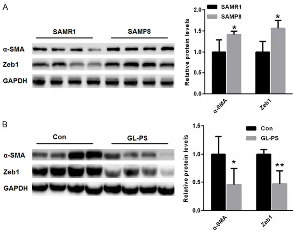
Figure 1. GL-PS treatment improved age-related EMT in the kidney of SAMP8
mice. A. Compared with that of SAMR1 mice, expression of α-SMA and Zeb1
was much higher in the kidneys of SAMP8 mice. B. After GL-PS treatment in
SAMP8 mice, renal expression of both α-SMA and Zeb1 was lower than in the
saline control group. *p<0.05, **p<0.01 vs. control.
0.1% Tween (TBST) 20, the membranes were probed with horseradish peroxida- se-conjugated anti-mouse or anti-rabbit IgG (1:1000 dilution; Beyotime Institute of Biotechnology, Shanghai, China). After washing again with TBST, the bands were visualized using an enha- nced chemiluminescence system (DP2-BSW; Olym- pus, Tokyo, Japan) and den-sitometry was performed using ImageJ (Wayne Ras- band, National Institutes of Health, Bethesda, MD, USA).
Cell cycle analysis
GL-PS treatment amelio-rated renal cell senescence in aging kidneys
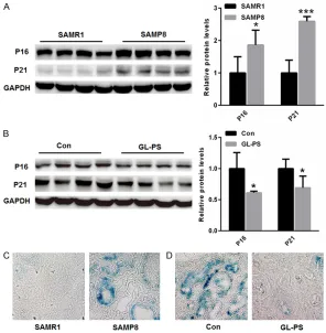
Next, we evaluated whether cell senescence is mitigat-ed by GL-PS treatment in aging kidneys. It is well known that P16 and P21, two markers of cell cycle arrest in the process of cell senescence [17]. Western blot analysis indicated that renal expression of P16 and P21 was increased in SAMP8 mice than that of SAMR1 mice (Figure 2A). However, renal expression of P16 and P21 was signifi-cantly reduced in GL-PS gro- up than in the saline group of SAMP8 mice (Figure 2B). Similar results were identi-fied through SA-β-gal stain-ing. As shown in Figure 2C
[image:4.612.90.386.72.374.2]and 2D, more senescence cells were observed in the kidneys SAMP8 mice than that of SAMR1 mice, but GL-PS treatment decreased senescence cells in SAMP8 mice. Our data suggested that GL-PS remediates cel-lular senescence in aging kidney.
Figure 2. GL-PS treatment ameliorated renal cell senescence in aging kidneys. (A) Western blot analysis indicated that renal expression of P16 and P21 was increased in SAMP8 mice than that of SAMR1 mice. (B) Renal expression of
P16 and P21 was significantly reduced in GL-PS group than in saline group of SAMP8 mice. (C) SA-β-gal staining showed that more senescence cells were
observed in the kidneys SAMP8 mice than that of SAMR1 mice, (D) but GL-PS treatment decreased senescence cells in SAMP8 mice. *p<0.05, **p<0.01, ***p<0.001 vs. control.
GL-PS treatment enhanced SIRT1 expression
Western blot analysis indicated that the expres-sion of SIRT1 was decreased in the renal tis-sues of SAMP8 mice than that of SAMR1 mice (Figure 3A). However, GL-PS treatment incre- ased the expression of SIRT1 in the kidneys than that of control (Figure 3B).
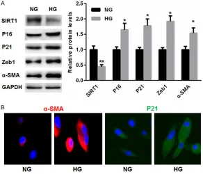
High glucose-induced senescence and EMT was accompanied by reduced SIRT1 expres-sion in TCMK-1 cells
Furthermore, we established in vitro TCMK-1 cellular senescence models by 25 mM high cose treatment. Compared with NG, high glu-cose treatment induced the expression of P16, P21, Zeb1, and α-SMA in TCMK-1 cells. Addi- tionally, HG suppressed expression of SIRT1 in TCMK-1 cells to less than that of NG (Figure 4A). Comparable results with respect to α-SMA level of p<0.05 was considered statistically
significant.
Results
GL-PS treatment improved age-related EMT in the kidney of SAMP8 mice
and P21 in HG group were obtained with immunofluo-rescence staining (Figure 4B).
GL-PS alleviated HG-induced senescence and EMT via upregulating SIRT1 expression
To further determine wheth-er HG-induced senescence and EMT is achieved via enhancing SIRT1 expres-sion, a specific siRNA tar-geting SIRT1 was selected. Compared with NC, silenc-ing of SIRT1 induced the expression of P16, P21, Zeb1, and α-SMA in TCMK-1 cells even with GL-PS pre-incubation (Figure 5A). Fur- thermore, flow cytometry analysis indicated enhan- ced G0-G1 cell cycle arrest in TCMK-1 cells transfected with si-SIRT1 compared with that of NC, even with GL-PS pre-incubation (Fig- ure 5B). These data indi-cate that SIRT1 plays a key role in GL-PS regulated senescence and EMT.
Discussion
[image:5.612.89.385.70.320.2]EMT is the process in which differentiated epithelial ce- lls are converted into matrix-producing fibroblasts [18]. Recent studies have indi-cated that EMT is a key con-tributor in the process of kidney fibrosis and decre- ased renal function [19]. Multiple factors, including transforming growth factor- β (TGF-β), epidermal growth factor (EGF) and vascular endothelial growth factor (VEGF), are considered to result in tissue homeosta-sis, thereby triggering EMT and progressive fibrosis [20]. Based on the above
Figure 3. GL-PS treatment enhanced SIRT1 expression. A. Western blot analy-sis indicates that the expression of SIRT1 was decreased in the renal tissues of SAMP8 mice than that of SAMR1 mice. B. GL-PS treatment increased the expression of SIRT1 in the kidneys than that of control. *p<0.05, **p<0.01 vs. control.
Figure 4. High glucose-induced senescence and EMT was accompanied by re-duced SIRT1 expression in TCMK-1 cells. A. Compared with NG, high glucose
treatment induced expression of P16, P21, Zeb1, and α-SMA, but suppressed
SIRT1 expression in TCMK-1 cells. B. Comparable results with respect to
α-SMA and P21 in HG group were obtained with immunofluorescence staining.
[image:5.612.91.383.402.652.2]studies, early therapeutic interventions may be helpful in the conversation of cellular senes-cence and EMT process. In line with previous studies, our data showed that EMT was enhanced in the kidneys of older rats than that of the younger rats, indicating that EMT is increased along with the senescence as a func-tion of age. Given that EMT is attributed to the decline in renal function with age, we investi-gated the effect of GL-PS on age-related EMT in our rat models. We found that GL-PS reduced the number senescent cells in the kidneys and improved age-related EMT process, indicating a protective role of GL-PS in kidney aging. Multiple research studies have indicated that common hallmarks of aging kidney include cel-lular senescence, upregulation of P16 and P21, and enhanced SA-β-gal activity [21]. To under-stand age-related changes in kidney function, it is important to explore the expression of P16 and P21 in the renal tissues. Compared to young rats, increases in expression of P16 and P21 were observed in the older rats. In con-trast, treatment with GL-PS significantly de- creased the levels of P16 and P21 in the kid-neys of SAMP8 mice.
These above observations have led us to the
EMT and cellular senescence in aging kidneys. Here, we mainly focused on SIRT1, a key lon-gevity gene [22]. Previous studies have sug-gested that SIRT1 protects kidney cell injury from various cellular stresses [23, 24]. Further- more, the podocyte-specific loss of SIRT1 aggravates diabetic kidney injury [7]. However, whether SIRT1 is involved in GL-PS-induced improvement of aging kidney has never been explored. Therefore, in this study, we sought to determine the effects of SIRT1 in age-induced kidney injury after GL-PS treatment. In line with previous studies, renal reduction of SIRT1 was identified in the kidneys of aging mice. Not sur-prisingly, GL-PS treatment increased SIRT1 expression in the kidney of aging mice. We pro-pose that increased levels of SIRT1 may improve EMT and cellular senescence in the renal tissues of aging mice. This is, to our knowledge, for the first study to demonstrate an effect of GL-PS on SIRT1 expression in the aging kidney.
[image:6.612.94.524.75.358.2]In the in vivo experiments, we showed that SIRT1 was upregulated by GL-PS treatment in the aging mice. To verify whether it is the major contributor of GL-PS-improved renal function, EMT and cellular senescence of TCMK-1 cells in vitro was induced with high glucose [25, 26].
Figure 5. GL-PS alleviated HG-induced senescence and EMT via upregulating SIRT1 expression. A. Compared with NC, silencing of SIRT1 induced expression of P16, P21, Zeb1
and α-SMA in TCMK-1 cells
glucose for 48 hours results in significant EMT, cellular senescence, and reduced levels of SIRT1 expression. In contrast, pre-incubation with GL-PS alleviated high glucose-induced EMT and cellular senescence, and enhanced SIRT1 expression. Moreover, silencing of SIRT1 could induce EMT and senescence even in GL-PS treated cells. These results indicate that GL-PS suppresses EMT and senescence in the renal tissues mainly via upregulating SIRT1. In conclusion, this study shows the protective effects of GL-PS on renal senescence and aging-related EMT. Therefore, GL-PS could be used as an early prevention and treatment in the protection of kidney function among the elderly.
Acknowledgements
The present study was supported by the Science and Technology Foundation of Tianjin Municipal Health Bureau (No. 2015047).
Address correspondence to: Dr. Wen-Xiu Chang, Department of Nephrology, Tianjin First Center Hospital, 24 Fukang Road, Nankai, Tianjin 300192, P.R. China. Tel: +23626600; Fax: 86-22-23626199; E-mail: [email protected]
References
[1] Glassock RJ and Rule AD. Aging and the kid-neys: anatomy, physiology and consequences
for defining chronic kidney disease. Nephron
2016; 134: 25-29.
[2] Glassock RJ, Denic A and Rule AD. The conun-drums of chronic kidney disease and aging. J Nephrol 2017; 30: 477-483.
[3] Coresh J, Astor BC, Greene T, Eknoyan G and Levey AS. Prevalence of chronic kidney disease and decreased kidney function in the adult US population: third national health and nutrition examination survey. Am J Kidney Dis 2003; 41: 1-12.
[4] Anderson S, Halter JB, Hazzard WR, Himmel-farb J, Horne FM, Kaysen GA, Kusek JW,
Nay-field SG, Schmader K, Tian Y, Ashworth JR,
Clayton CP, Parker RP, Tarver ED, Woolard NF and High KP; workshop participants. Predic-tion, progression, and outcomes of chronic kid-ney disease in older adults. J Am Soc Nephrol 2009; 20: 1199-1209.
[5] Campisi J and d’Adda di Fagagna F. Cellular se-nescence: when bad things happen to good cells. Nat Rev Mol Cell Biol 2007; 8: 729-740.
[6] Evan GI and d’Adda di Fagagna F. Cellular se-nescence: hot or what? Curr Opin Genet Dev 2009; 19: 25-31.
[7] Chuang PY, Cai W, Li X, Fang L, Xu J, Yacoub R, He JC and Lee K. Reduction in podocyte SIRT1 accelerates kidney injury in aging mice. Am J Physiol Renal Physiol 2017; 313: F621-F628. [8] Kume S, Maegawa H and Koya D.
[Sirt1-medi-ated autophagy in aging kidney]. Nihon Jinzo Gakkai Shi 2012; 54: 73-77.
[9] He W, Wang Y, Zhang MZ, You L, Davis LS, Fan H, Yang HC, Fogo AB, Zent R, Harris RC, Breyer MD and Hao CM. Sirt1 activation protects the mouse renal medulla from oxidative injury. J Clin Invest 2010; 120: 1056-1068.
[10] Hou X, Xu S, Maitland-Toolan KA, Sato K, Jiang B, Ido Y, Lan F, Walsh K, Wierzbicki M, Verbeur-en TJ, CohVerbeur-en RA and Zang M. SIRT1 regulates hepatocyte lipid metabolism through activat-ing AMP-activated protein kinase. J Biol Chem 2008; 283: 20015-20026.
[11] Kitada M, Kume S, Imaizumi N and Koya D. Resveratrol improves oxidative stress and pro-tects against diabetic nephropathy through normalization of Mn-SOD dysfunction in AMPK/SIRT1-independent pathway. Diabetes 2011; 60: 634-643.
[12] Sliva D. Cellular and physiological effects of ganoderma lucidum (reishi). Mini Rev Med Chem 2004; 4: 873-879.
[13] Dudhgaonkar S, Thyagarajan A and Sliva D.
Suppression of the inflammatory response by
triterpenes isolated from the mushroom gano-derma lucidum. Int Immunopharmacol 2009; 9: 1272-1280.
[14] Wang J, Wang Y, Liu X, Yuan Y and Yue T. Free radical scavenging and immunomodulatory ac-tivities of ganoderma lucidum polysaccharides derivatives. Carbohydr Polym 2013; 91: 33-38. [15] Xu Z, Chen X, Zhong Z, Chen L and Wang Y.
Ganoderma lucidum polysaccharides: immu-nomodulation and potential anti-tumor activi-ties. Am J Chin Med 2011; 39: 15-27.
[16] Peinado H, Olmeda D and Cano A. Snail, Zeb and bHLH factors in tumour progression: an al-liance against the epithelial phenotype? Nat Rev Cancer 2007; 7: 415-428.
[17] Leontieva OV and Blagosklonny MV. CDK4/6-inhibiting drug substitutes for p21 and p16 in senescence: duration of cell cycle arrest and MTOR activity determine geroconversion. Cell Cycle 2013; 12: 3063-3069.
[19] Liu Y. New insights into
epithelial-mesenchy-mal transition in kidney fibrosis. J Am Soc
Nephrol 2010; 21: 212-222.
[20] Schmitt R and Cantley LG. The impact of aging on kidney repair. Am J Physiol Renal Physiol 2008; 294: F1265-1272.
[21] Yue Z, Rong J, Ping W, Bing Y, Xin Y, Feng LD and Yaping W. Gene expression of the p16 (INK4a)-Rb and p19(Arf)-p53-p21(Cip/Waf1) signaling pathways in the regulation of hema-topoietic stem cell aging by ginsenoside Rg1. Genet Mol Res 2014; 13: 10086-10096. [22] Kong D, Zhan Y, Liu Z, Ding T, Li M, Yu H, Zhang
L, Li H, Luo A, Zhang D, Wang Y, Wang S, Zhang Z, Zhang H, Huang X, Yao P, Ding Y and Liu Z. SIRT1-mediated ERbeta suppression in the en-dothelium contributes to vascular aging. Aging Cell 2016.
[23] Simoncini S, Chateau AL, Robert S, Todorova D, Yzydorzick C, Lacroix R, Ligi I, Louis L, Bach-elier R, Simeoni U, Magdinier F, Dignat-George F and Sabatier F. Biogenesis of pro-senescent microparticles by endothelial colony forming cells from premature neonates is driven by SIRT1-dependent epigenetic regulation of MKK6. Sci Rep 2017; 7: 8277.
[24] Imperatore F, Maurizio J, Vargas Aguilar S, Busch CJ, Favret J, Kowenz-Leutz E, Cathou W, Gentek R, Perrin P, Leutz A, Berruyer C, Sieweke MH. SIRT1 regulates macrophage self-renew-al. EMBO J 2012; 36: 2353-2372.
[25] Lee YJ and Han HJ. Troglitazone ameliorates high glucose-induced EMT and dysfunction of SGLTs through PI3K/Akt, GSK-3beta, Snail1, and beta-catenin in renal proximal tubule cells. Am J Physiol Renal Physiol 2010; 298: F1263-1275.