ISSN Online: 2163-0585 ISSN Print: 2163-0569
DOI: 10.4236/ojmn.2018.81007 Jan. 19, 2018 84 Open Journal of Modern Neurosurgery
Surgical Management of Lumbar and
Thoracolumbar Spinal Fractures: Indications,
Surgical Technique and Evaluation on a Series
of 64 Patients Treated with Percutaneous
Posterior Osteosynthesis Combined with
Kyphoplasty or Anterior Arthrodesis
Romuald Kouitcheu
1,2*, Drogba Landry
2, N’da Hermann Adonis
2, Diallo Moussa
1,
Melot Anthony
1, Troude Lucas
1, Roche Pierre-Hugues
11Department of Neurosurgery, CHU-HÔPITAL NORD, Marseille, France 2Department of Neurosurgery, CHU-YOPOUGON, Abidjan, Ivory Coast
Abstract
Introduction: Fractures of the lumbar spine and thoracolumbar junction are common in spinal trauma. The aim of this work is to analyze the nature of the indications, the morbidity as well as the results of these treatment regimens. Patients and Methods: A retrospective, single-center study, based on a re-view of 64 patients with lumbar spine and thoracolumbar junction fractures (T10-L2) without neurological disorders, was collected in the neurosurgery department of the North Hospital and University Hospital (CHU), Marseille over a period of 2 years from January 2015 to December 2016. Posterior per-cutaneous osteosynthesis were more or less associated with kyphoplasty pre-ceded anterior arthrodesis. Clinical and radiological endpoints were collected at least 6 months later. Results: The mean follow-up was 9.5 months (6 - 24). The clinical evaluation found a mean VAS at last follow-up at 14/100 (0 - 30) and an average Oswestry score at the last follow-up at 88%. The initial average vertebral kyphosis went from 13˚ to 4˚ at the last follow-up with a correction loss of 1˚, an absolute gain of 8˚. No postoperative neurological complications were noted in our series. Conclusion: The implementation of a two-step the-rapeutic strategy with anterior reconstruction in Magerl’s lumbar spine or A3.3 thoracolumbar junction fractures allows effective and long-lasting cor-rection of lumbar lordosis and thoracic kyphosis, and obtaining a balanced How to cite this paper: Kouitcheu, R.,
Landry, D., Adonis, N.H., Moussa, D., An-thony, M., Lucas, T. and Pierre-Hugues, R. (2018) Surgical Management of Lumbar and Thoracolumbar Spinal Fractures: Indi- cations, Surgical Technique and Evaluation on a Series of 64 Patients Treated with Percu- taneous Posterior Osteosynthesis Combined with Kyphoplasty or Anterior Arthrodesis. Open Journal of Modern Neurosurgery, 8, 84-100.
https://doi.org/10.4236/ojmn.2018.81007
Received: November 30, 2017 Accepted: January 16, 2018 Published: January 19, 2018
Copyright © 2018 by authors and Scientific Research Publishing Inc. This work is licensed under the Creative Commons Attribution International License (CC BY 4.0).
DOI: 10.4236/ojmn.2018.81007 85 Open Journal of Modern Neurosurgery spine in the sagittal plane. Our functional results are close to normal, with low morbidity and a low complication rate.
Keywords
Anterior Arthrodesis, Spine Fracture, Kyphoplasty, Percutaneous Osteosynthesis
1. Introduction
The optimal therapeutic management of lumbar spine fractures and thoraco- lumbar (T10-L2) type A and B2 junction of Magerl [1] without neurological signs remains controversial. Orthopedic treatment gives good immediate results with a low rate of complications [2]. Long-term results are variable with a recur-rence of initial deformity [3] [4]. Surgical treatment allows for better reduction and faster ambulation of patients [5]. Percutaneous vertebroplasty or kyphop-lasty techniques can treat A1 compression fractures and even A3 burst fractures
[6] [7] and can be performed in addition to pedicle screw osteosynthesis [8].
Percutaneous posterior surgery techniques have been developed to reduce mus-cle trauma, blood loss and length of hospital stay. The placement of percutane-ous pedicle screws has been described for some years [9] but the placement of stems has necessitated the development of specific ancillaries. This posterior arthrodesis allows short-term reduction and stabilization of the fracture site. However, pseudarthrosis related to highly comminuted fractures prompts some centers to perform preventive surgical treatment of complementary anterior sta-bilization. Few data are available on the practices and results of this two-step strategy, later and later. The aim of this study is to analyze, on a retrospective monocentric series of lumbar spine fractures and thoracolumbar junction, the nature of the indications, the morbidity as well as the results of these therapeutic regimens.
2. Patients et Methods
A retrospective, single-center study, based on the review of 64 files of patients operated for a fracture of the lumbar spine or thoracolumbar junction (T10-L2), collected in the neurosurgery department of the North Hospital and University Hospital (CHU), Marseille over a period of 2 years from January 2015 to De-cember 2016. Posterior percutaneous osteosynthesis more or less associated with kyphoplasty preceded a corporectomy performed either by retroperitoneal lum-botomy or a left retropleural thoracotomy. The corporeal reconstruction is done by means of an expandable tubular cage containing a spongy graft taken at the expense of the corporectomy product.
2.1. Inclusion Criteria
DOI: 10.4236/ojmn.2018.81007 86 Open Journal of Modern Neurosurgery without neurological disorders (Frankel E) [10], treated with percutaneous post-erior osteosynthesis more or less associated with kyphoplasty or arthrodesis were included earlier. These techniques were used for A1 and A3 fractures with ver-tebral kyphosis between 10˚ and 20˚, and for A2 (diabolo) and B2 (Chance) fractures. A minimum follow-up of 6 months was necessary for the review.
2.2. Exclusion Criteria
Patients with a history of thoracic or lumbar spinal fusion, the presence of neu-rological disorders and posterior wall recoil greater than 40% were excluded.
2.3. Epidemiological Criteria
The most affected age groups are [50 - 60 [with 17 cases, or 26.56%, followed by the [40 - 50] age group 21.87%. Fractures of the lumbar spine and thoracolum-bar junction appear rarer in the older ones. In our series, there are only 5 pa-tients in the age group [70 - 80 [7.81% of cases. The average age of our papa-tients was 49.8 years (18 - 91 years) with a median of 51 years.
Our series consisted of 44 men (68.75%) and 20 women (31.25%). A male predominance was observed with a sex ratio of 0.45 women for one man.
Fractures of the lumbar spine and thoracolumbar junction were caused by various accidents, with a clear predominance of falls in our context. Falling height is the leading cause and represents 39 cases in our series, a percentage of 60.95% of all etiologies. These are accidental falls in 35 cases (54.7%) and 4 cases of attempted autolysis of which 2 patients were followed for a psychiatric illness (6.25% of cases). Road accidents accounted for 20 injuries, accounting for 29.73% of all etiologies. Workplace accidents: We observed 3 cases, a percentage of 4.7%. The attacks were reported by 2 patients in our series, i.e. 3.1%.
2.4. Radiological Criteria
The distribution of vertebral lesions was as follows: 9 cases in T10, 5 cases in T11, 13 cases in T12, 20 in L1, 7 in L2, 4 in L3, 3 in L4 and 3 cases in L5. In 54 cases (84.4%), the lesion was located at the thoracolumbar junction (T10-L2) with a clear predominance of lesions in the L1 and T12 vertebrae
The distribution of fracture types according to the Magerl classification [1] was as follows: There were 4 A1.3 type lesions, 12 A2 type (10 A2.2 and 2 A2.1), 35 type A3 (16 A3.1, 3 A3.2 and 16 A3.3) and 13 of type B2. In 35 cases (54.68%) the fracture was of type A3 according to Magerl classification. The recoil of the posterior wall for A3 was in all cases less than 40%.
2.5. Radiological Evaluation
DOI: 10.4236/ojmn.2018.81007 87 Open Journal of Modern Neurosurgery and at the last follow-up. Thus, vertebral kyphosis (VK) and regional angle (RA) were measured and the corrected regional angle (CRA) was calculated by sub-tracting the standard regional angle from the AR [11]. After the first posterior surgical time, a new scanner assessed the residual stability according to Mac Cormack’s classification [12]. The position and eventual mobilization of the material was also studied.
2.6. Clinical Criteria
In the initial management, the inventory of associated spinal lesions was added to the other potential lesions (skull, thorax, abdomen, pelvis, limbs). Twenty one patients (32.81%) had associated lesions: nine in the limbs, three in the pelvis, six in the thorax and three in the skull. None of the patients presented neurological signs and all were classified Frankel stage E [10].
The pain was assessed pre- and postoperatively immediately and at the last follow-up on a visual analogue scale (VAS). The overall functional assessment was performed using the Oswestry score [13] as well as the ability to return to the previous professional activity.
2.7. Surgical Techniques
The ventral decubitus for posterior time and right lateral decubitus for the pre-vious time was systematic. The percutaneous character of posterior fixation and its extent were noted. The duration of each intervention was quantified. Per and postoperative complications have been described as well as the need for surgical revision. The delay between the two posterior and anterior procedures was ana-lyzed.
Kyphoplasty: This is a non-invasive method. The injection of cement under scopic control allows the stability of the fracture. In case of significant deforma-tion, the associated placement of a balloon or expander (spine Jack®), (Figure 1) allows the reduction of deformity by raising the vertebral plateau. This method has an immediate analgesic effect, and stops the progression of the spinal defor-mity without allowing the control of disco-ligament instability.
DOI: 10.4236/ojmn.2018.81007 88 Open Journal of Modern Neurosurgery
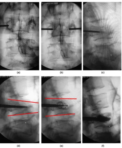
Figure 1. 38 year old man, L3 type A3.1 fracture of magerl, without neurological deficit. Treated by
ky-phoplasty with spine jack (VEXIM) (our series). (a) scopic control of the trocar when it approaches the pe-dicle; ((b), (c)) scopic control of the trocar when it leaves the pedicle (face and profile); (d) L3 type A3.1 fracture of Magerl with spine jack placement; (e) scopic control of the reduction of the fracture; (f) scopic control after cement injection.
DOI: 10.4236/ojmn.2018.81007 89 Open Journal of Modern Neurosurgery
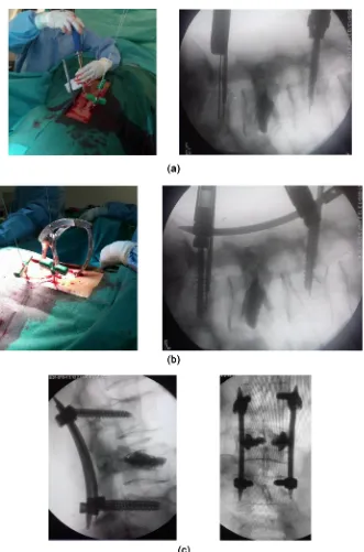
Figure 2. same patient with L3 type A3.1 fracture of Magerl. Posterior percutaneous
os-teosynthesis L2-L4 by SEXTANT (Our series). (a) Pedicular sight percutaneous (insertion of guide pins). Placement along the pins of dilator tubes of progressive diameter. Percu-taneous screwing along the guide pins. Scopic control; (b) percuPercu-taneous placement of the rods with the SEXTANT; (c) final scopic control of profile and face of screws and rods.
re-DOI: 10.4236/ojmn.2018.81007 90 Open Journal of Modern Neurosurgery peated for the ipsilateral screw.
In the sagittal plane, the upper screw was directed up and forward, the lower screw down and forward. In the opposite case, the travel of the screw heads was limited, the assembly of the ancillary is impossible. The acceptable limit is the parallelism of the upper and lower screws. In the horizontal plane, the marker was the pedicle on the frontal scopies which was likened to a ring. There are two essential times when penetrating the trocar. When entering the pedicle, the tro-car should be located at the lateral part of the ring. At the level of the vertebral wall, he must have been at his center or medial part. The two screw holders were then secured with the rod holder shaped compass. A graduated mark is used to measure the length of the rod required. A proximal incision of 15 mm was made and the adapted rod introduced. The path had to be done without forcing. The correct positioning of the rod in the screw heads was verified by scopic controls of face, profile and three quarters. Both screwdrivers were positioned on the heads and allow to complete the reduction by a compression maneuver. The bolts were then tight and broken after a scopic check. The contralateral pedicles were instru-mented in the same way. The closure was carried out without drainage.
Circumferential strategy, complementary anterior arthrodesis: Distraction and lordosis allow the reduction of thoracolumbar fractures. However, previous loss of substance or reduction in the intervertebral disc may lead to pseudarthrosis and thus to disassembly or loss of correction [14] [15]. To prevent this compli-cation, a second surgical time of anterior arthrodesis can reinforce the fusion. The analysis of the CT images after the first procedure allows the indication of the circumferential arthrodesis. The finding of ductal crowding by an earlier fragment also justifies the use of anterior arthrodesis. The arthrodesis can be supported by the placement of an iliac tricortical graft or an intervertebral cage. On a technical level, the patient was placed in the right lateral decubitus posi-tion. The approach is either retroperitoneal lumbar and retrorenal lumbar lum-botomy or thoracic retropleural thoracotomy. For left retroperitoneal and retro renal lombotomy, the incision, about 8 cm long, follows the 12th rib and extends
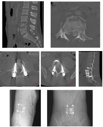
towards the umbilicus. Dissection of the subcutaneous planes allowed access to the muscles of the abdominal wall. After opening, the retroperitoneal space was released to the spine in front of the psoas muscle. Particular care was taken in the hemostasis of the segmental lumbar vessels at a distance from the foramina. The psoas muscle was retracted and the level was detected at the image inten-sifier. After discectomy, corporectomy and flattening of the trays, an expandable tubular cage was put in place (VLIFT©, Stryker® company) (Figure 3). Its height and angulation of its trays are adjustable. She was filled with cortico-cancellous autograft from the corporectomy product. Before placement of the cage, anterior decompression of the spinal canal was performed. The correct positioning of the implant was controlled under scopie, and the approach of closure closed plan by plan on suction drain.
DOI: 10.4236/ojmn.2018.81007 91 Open Journal of Modern Neurosurgery
Figure 3. Posterior percutaneous osteosynthesis of a Burst L4 fracture by Medtronic
Sex-tant (L3-L5) + L4 corporectomy by left lombotomy and placement of a V-LIFT (strycker). (Our series).
of thromboembolic complications. The complications described are vascular (involvement of the primary iliac vessels in L4/L5 or the aorta), plexus (involve-ment of the lower hypogastric plexus responsible for vaginal dryness and retro-grade ejaculation), thromboembolic, digestive (ileus reflex, hernia, peritoneal gap), infectious.
3. Resultats
3.1. Duration of Hospitalization
as-DOI: 10.4236/ojmn.2018.81007 92 Open Journal of Modern Neurosurgery sociated with kyphoplasty. An average of 20 days, with extremes ranging from 7 to 39 days for posterior percutaneous osteosynthesis associated with anterior route. Patients who were hospitalized for more than 10 days all had associated lesions or were waiting for additional anterior pathway programming.
3.2. Duration of Intervention
The median duration of intervention was variable depending on the type of sur-gery. It is for posterior percutaneous osteosynthesis only 95 minutes (50 - 235); 115 minutes (55 - 179) for percutaneous posterior osteosynthesis associated with kyphoplasty and 250 minutes (200 - 332) for percutaneous posterior osteosyn-thesis associated with anterior approach. The median delay between the two operative times was 9.5 days (3 to 28 days).
3.3. Surgical Indications
Our series consists of 33 patients (51.57%) who had percutaneous posterior os-teosynthesis associated with kyphoplasty (PPO + K); of 19 patients (29.68%) with posterior percutaneous osteosynthesis alone (PPOA) and 12 patients (18.75%) with percutaneous posterior osteosynthesis associated with anterior approach (PPO + A). In the first group of 19 patients, the majority of indications were for type B2 fractures; the second group of 33 patients, the fractures were of type A3.1 and in the 3 group of 12 patients, all fractures were of type A3.3
(Table 1).
3.4. Clinical Results
The mean follow-up was 9.5 months (6 - 24) with a median of 6 months. Mean VAS at D2 postoperative was 37/100 (range 0 - 70). The average VAS at last follow-up is 14/100 (0 - 30). All patients with an VAS at 30 have a follow-up of less than 1 year.
[image:9.595.192.541.554.742.2]The Oswestry score at the last follow-up is on average 88% (65 - 100) with a median of 90. The three patients with a score lower than 70 have a follow-up of
Table 1. Indications according to the type of lesion (Magerl).
n Type of fracture (Magerl) Intervention types:
PPOA PPO + K PPO + A
4 A1.3 1 3 0
2 A2.1 2 0 0
10 A2.2 4 6 0
16 A3.1 2 14 0
3 A3.2 3 0 0
16 A3.3 1 3 12
13 B2 9 4 0
DOI: 10.4236/ojmn.2018.81007 93 Open Journal of Modern Neurosurgery less than 1 year.
Resumption of work: Fifty-one patients returned to work (80%), thirty-nine of whom had the same activity as preoperatively and twelve with reclassification. Thirteen patients did not return to work.
Subjective Results: Forty-six (71.88%) patients were very satisfied with the surgery and eighteen (28.13%) were satisfied. None of the patients were dissatis-fied or very dissatisdissatis-fied with the surgery.
Complications and recoveries: A total of 366 screws were placed percuta-neously posteriorly and 12 tubular cages by anterior route. In seven patients, 15 screws cause the pedicular cortical to break into the control CT without clinical translation. There is therefore 4.1% malposition of the screws. No septic com-plications occurred. There was no dismantling or breaking of the equipment. No root lesion or CSF leak was found.
One patient underwent percutaneous posterior osteosynthesis in the presence of worsening of the vertebral kyphosis on the control CT after kyphoplasty alone. This intervention was performed after a period of 6 months by minimally invasive technique.
Equipment removal: There have been three ablations of posterior osteosyn-thesis material for the time being. Two patients are eager to make it happen, de-spite the absence of significant pain. N˚1 comment: morbidity in the investiga-tion
3.5. Radiological Results
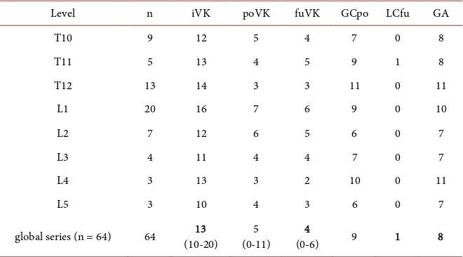
The results of vertebral kyphosis (VK) and corrected regional angle (RCA) are given for the different setbacks (Table 2 and Table 3). The evolution of VK was analyzed according to the type of lesions (Table 4).
[image:10.595.209.539.506.689.2]Last follow-up scan: Three patients had a control scan with a follow-up at 2
Table 2. Vertebral kyphosis evolution in degrees.
Level n iVK poVK fuVK GCpo LCfu GA
T10 9 12 5 4 7 0 8
T11 5 13 4 5 9 1 8
T12 13 14 3 3 11 0 11
L1 20 16 7 6 9 0 10
L2 7 12 6 5 6 0 7
L3 4 11 4 4 7 0 7
L4 3 13 3 2 10 0 11
L5 3 10 4 3 6 0 7
global series (n = 64) 64 (10-20) 13 (0-11) 5 (0-6) 4 9 1 8
DOI: 10.4236/ojmn.2018.81007 94 Open Journal of Modern Neurosurgery
Table 3. Evolution of corrected regional angle in degrees.
Level n iCRA poCRA CRAfu GCpo LCfu GA
T10 9 11 9 10 2 1 1
T11 5 13 8 7 5 0 6
T12 13 12 10 10 2 0 2
L1 20 11 −2 0.5 13 2.5 10.5
L2 7 15 6 8 9 2 7
L3 4 13 3 3 10 0 10
L4 3 12 5 7 7 2 5
L5 3 14 7 8 7 1 6
global serie (n = 64) 64 14 9 7 8 1.5 6
iCRA: initial corrected regional angle; poCRA: postoperative corrected regional angle; CRAfu: corrected re-gional angle at last follow-up; GCpo: gain in postoperative correction; GAfu: Absolute gain in correction at last follow-up; LCfu: loss of correction at last follow-up.
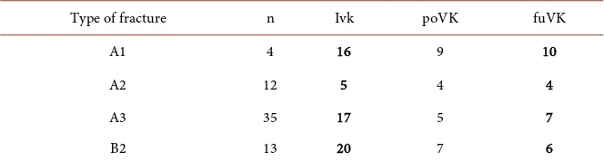
Table 4. Study of vertebral kyphosis in degrees according to the type of fracture.
Type of fracture n Ivk poVK fuVK
A1 4 16 9 10
A2 12 5 4 4
A3 35 17 5 7
B2 13 20 7 6
iVK: initial vertebral kyphosis; poVK: postoperative vertebral kyphosis; fuVK: vertebral kyphosis at last fol-low-up.
years. In all cases, the corporeal consolidation of the fractures is obtained. There is no late mobilization or bankruptcy of the equipment.
4. Discussion
The management of patients with lumbar spine fracture and thoracolumbar junction without neurological disorders is not codified. Indeed, all sorts of treatments are proposed: conservative by corset or supine position, surgery by posterior route, anterior way or combined. [1] [16] [17]
The review of the literature finds no method statistically superior to the other. Thomas [16] concludes that there is no current argument for orthopedic treat-ment or surgical treattreat-ment for non-neurological A3 fractures.
In the only prospective randomized study available, including 47 patients,
Wood [17] found no benefit after surgical treatment of A3 fractures. The latter
also causes a higher complication rate [17]. This conclusion should be mod-erated by the fact that the preoperative VK in this series were only 11.3˚ for or-thopedic treatment and 10.1˚ for surgical treatment, therefore less than the ky-phosis of our patients.
[image:11.595.207.539.325.414.2]DOI: 10.4236/ojmn.2018.81007 95 Open Journal of Modern Neurosurgery allow faster mobilization, shorten hospital stay, decrease pain more quickly and facilitate an early resumption of professional activity. In addition, the sagittal alignment of the spine is of better quality in the surgical series than in the con-servative series. But this state is not correlated with a better functional result.
For many authors [9] [18] [19] [20], posterior percutaneous osteosynthesis is recommended for type A fractures when the VK is greater than 15˚ or 20˚. Some fracture subtypes readily lead to this indication: A2.2 (diabolo) and A3.3 (com-minuted fracture-burst), as well as their location in the lower lumbar spine. In our series, the average VK goes from 13˚ to 4˚ with an average correction loss of 1˚, an absolute gain of 8˚. This result is lower than that of the SOFCOT sympo-sium series of 1996, the average VK goes from 17˚ to 5.9˚, an absolute gain of 11.1˚ for posterior surgery (all lesions combined) [21].
Indeed, despite the technical progress and the aids to surgery, percutaneous pedicle screwing can be complicated by nerve injuries. In our series, we had 4.1% of misplacement on 366 screws placed without neurological repercussions. The incorrect positioning of the screws is encrypted by 2.5% to 15% [16] [20] [22]. The advantage of the percutaneous system is the use of face and profile scopie which secures the implantation gesture of the screws in the pedicles [23].
The use of vertebroplasty allows a consolidation of the fractured vertebral body and thus maintain the reduction of the fracture. This use in spinal trauma on A3 burst fractures is recent. Few series or publications refer to them [7] [8] [24]. None of these studies treated A3 fractures prospectively. One of the classic and far-reaching complications of percutaneous vertebroplasty is leakage of ce-ment into the spinal canal [25]. The frequency of cement leakage also varies with the level of the treated vertebra. In fact, high thoracic lesions have a risk of ab-sconding up to 85.7% [25]. In spinal traumatology, this risk of passage of cement in the canal then compressing the nerve structures is also high given the fracture of the vertebral body that goes to the posterior wall in a burst fracture A3 [6]. Kyphoplasty decreases the risk of cement passage in the canal. It allows a reduc-tion of the fracture thanks to the balloon or an expander. It is of course neces-sary to inject the most precement cement possible to avoid the intraductal pas-sage and to maintain the reduction. The combination of these two techniques makes it possible to propose a solution to Magerl A3 burst fractures. This mini-mally invasive technique allows a quick return home, Muscle trauma is minimal, resulting in simpler operative follow-up and shorter hospital stay. The place-ment of the pedicular screws percutaneously requires an important use of the image intensifier with irradiation of the hands of the operator. Irradiation is however less than during the procedures carried out under scanner control [26]. Fluoro-navigation [27] seems to allow an easy and safe placement of the pedicle screws while decreasing the irradiation. We do not have the experience right now.
DOI: 10.4236/ojmn.2018.81007 96 Open Journal of Modern Neurosurgery more true because we use a short montage that was once the rule in spinal trau-ma. For example, the Sextant * system uses polyaxial screws, which are therefore less rigid, used alone, the loss of reduction could be greater, hence the need to stiffen the reduction of the fracture by complementary techniques of kyphoplas-ty or anterior corporectomy with placement of a tubular cage.
The anterior access to the fractured vertebral body and adjacent disks allows additional bone grafting. When this has already been performed posteriorly, the program corresponds to a circumferential arthrodesis. The increase of the graft surface potentiates the chances of fusion. Discectomies and avascularization of the trabeculae of the vertebrae above and below the lesion level tend to optimize arthrodesis. The use of a tubular implant to contain the graft aimed at providing a stable support to support the anterior reconstruction. Its distractible nature fa-cilitates its installation and promotes its primary hold. The morbidity associated with this additional intervention must be contained. In our series, we did not note a specific complication to lumbar or thoracic surgery by anterior approach. The consequences are rarely severe if we exclude the character
Dysesthetic. The average bleeding of 536 ml is greater than that observed during the posterior time. However, the use of a transfusion was less frequent because surgery was scheduled with anesthetic assessment out of emergency context. Some authors advocate an anterior graft limited to the upper half of the vertebral body and the overlying disc [29]. In our series, both discs were syste-matically included in the arthrodesis. The first realization of a posterior arthro-desis bridging the sub-lesional disc makes, in our opinion, obsolete the conser-vation of this one. This pattern may, however, be of interest when the posterior time corresponds to percutaneous osteosynthesis. Secondary ablation of the posterior material could then restore areas of mobility.
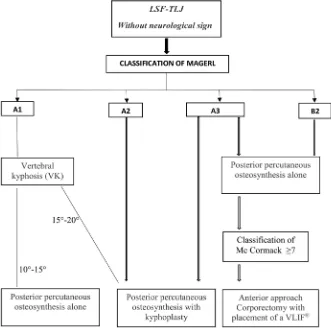
The indication of anterior reconstruction is based on the risk of loss of post-erior correction or nonunion. This reconstruction can be done in one or two surgical times: lateral osteosynthesis and corporeal reconstruction at the same time, or posterior osteosynthesis and remote reconstruction as performed in our series. Several authors show disappointing results after posterior stabilization alone in case of comminuted fracture, with significant rates of loss of correction [18]. A two-step therapeutic strategy was systematically performed in our series for type A3.3 fractures. Mc Cormack’s score [12] was established on a scanner obtained after the first beat, and confirmed the indication of a previous time. Our 2-time choice is opposed to isolated anterior reconstruction strategies, which are considered to be more haemorrhagic in the acute post-traumatic pe-riod, so performing 2 surgical times during the same anesthetic session exposes a greater risk of bleeding due to freshness of the fracture and surgery in acute peri- traumatic context less framed than a programmed surgery. Our clinical and ra-diological findings support a 2-stage circumferential surgical strategy for A3.3 comminuted fractures with a Mc Cormack score ≥ 7.
li-DOI: 10.4236/ojmn.2018.81007 97 Open Journal of Modern Neurosurgery terature [30] (80% to 94%), but our decline is lower. This leads us to the propos-al of a therapeutic propos-algorithm (Figure 4). However, our study has limitations: the number of patients included in this study remains insufficient to obtain a po-werful statistical analysis. We did not perform a randomized, double-blind, con-trolled therapeutic trial that allowed comparative statistical analysis, so we will not be able to say with certainty that one event is linked to another. However, the parameters used allow a descriptive analysis of the results. Radiological anal-ysis is limited by the absence of preoperative data in charge. The comparison can therefore be done only between the different operating times. The same is true for functional scores, which here only reflect a remote situation of spinal trauma. This type of event does not occur in patients initially asymptomatic, it remains difficult to assess the relative value of the scores.
5. Conclusion
[image:14.595.208.539.354.682.2]Posterior percutaneous osteosynthesis more or less associated with kyphoplasty or anterior arthrodesis is an interesting therapeutic option in Magerl type A and B2 thoracolumbar spine and lumbar spine fractures, without neurological dis-orders. The objective and subjective results are satisfactory, with low morbidity
DOI: 10.4236/ojmn.2018.81007 98 Open Journal of Modern Neurosurgery and a low rate of complications. The implementation of a two-step therapeutic strategy with anterior reconstruction in Magerl’s lumbar spine or A3.3 thoraco- lumbar junction fractures allows efficient and long-lasting correction of lumbar lordosis and kyphosis thoracic, and obtaining a balanced spine in the sagittal plane. Mc Cormack’s classification is an applied tool for strategic decision- making.
References
[1] Magerl, F., Aebi, M., Gerstein, S., Harms, J. and Nazarian, S. (1994) A Comprehen-sive Classification of Thoracic and Lumbar Injuries. European Spine Journal, 3, 184-201. https://doi.org/10.1007/BF02221591
[2] Tropiano, P., Huang, R.C., Louis, C.A., Poitout, D.G. and Louis, R.P. (2003) Func-tional and Radiographic Outcome of Thoracolumbar and Lumbar Burst Fractures Managed by Closed Orthopaedic Reduction and Casting. Spine, 28, 2459-2465.
https://doi.org/10.1097/01.BRS.0000090834.36061.DD
[3] Mumford, J., Weinstein, J.N., et al. (1993) Thoracolumbar Burst Fractures. The Clinical Efficacy and Outcome of Nonoperative Management. Spine, 18, 955-970.
https://doi.org/10.1097/00007632-199306150-00003
[4] Denis, F., Armstrong, G.W., Searls, K. and Matta, L. (1984) Acute Thoraco-Lmbar Burst Fractures in the Absence of Neurologic Deficit. Clin Orthop, 189, 142-149. [5] Knop, C., et al. (2000) Surgical Treatment of Injuries of the Thoracolumbar
Transi-tion. 2: Operation and Roentgenologic Findings. Der Unfallchirurg, 103, 1032-1047.
https://doi.org/10.1007/s001130050667
[6] Chen, J.-F. and Lee, S.-T. (2003) Percutaneous Vertebroplasty for the Treatment of Thoraclumbar Spine Bursting Fracture. Surgical Neurology, 62, 494-500.
https://doi.org/10.1016/j.surneu.2003.10.049
[7] Amoretti, N., et al. (2005) Burst Fracture of the Spine Involving Vertebrae Present-ing No Other Lesions. The role of vertebroplasty.Clinical Imaging, 29, 379-382.
https://doi.org/10.1016/j.clinimag.2005.07.006
[8] Verlaan, J.J., Dhert, W.J., Verbout, A.J. and Oner, F.C. (2005) Balloon Vertebrop-lasty in Combination with Pedicle Screw Instrumentation: A Novel Technique to Treat Thoracic and Lumbar Burst Fractures. Spine, 30, E73-E79.
https://doi.org/10.1097/01.brs.0000152162.64015.fb
[9] Lowery, G.L. and Kulkarni, S.S. (2000) Posterior Percutaneous Spine Instrumenta-tion. European Spine Journal, 9, S126-S130. https://doi.org/10.1007/PL00008318
[10] Frankel, H., Hancock, D.O. and Hislop, G. (1969) The Value of Postural Reduction in the Initial Management of Closed Injuries of the Spine with Paraplegia and Te-traplegia. Part I, Paraplegia, 7, 179-192.
[11] Stagnara, P., DeMauroy, J.C. and Dran, G. (1982) Reciprocal Angulation of Verte-bral Bodies in a Sagittal Plane: Approach to the References for the Evaluation of Kyphosis and Lordosis. Spine, 7, 335.
https://doi.org/10.1097/00007632-198207000-00003
[12] McCormack, T., Karaikovic, E. and Gaines, R.W. (1994) The Load Sharing Classifi-cation of Spine Fractures. Spine, 15, 1741-1744.
[13] Fairbank, J.C. and Pynsent, P.B. (2000) The Oswestry Disability Index. Spine, 25, 2940-2952. https://doi.org/10.1097/00007632-200011150-00017
DOI: 10.4236/ojmn.2018.81007 99 Open Journal of Modern Neurosurgery (2007) Anterior Vertebral Body Replacement with a Titanium Implant of Adjusta-ble Height: A Prospective Clinical Study. European Spine Journal, 16, 161-72.
https://doi.org/10.1007/s00586-005-0015-6
[15] Payer, M. (2006) Unstable Burst Fractures of the Thoracolumbar Junction: Treat-ment by Posterior BisegTreat-mental Correction/Fixation and Staged Anterior Corpecto-my and Titanium Cage Implantation. Acta Neurochirurgica, 148, 299-306.
https://doi.org/10.1007/s00701-005-0681-5
[16] Thomas, K.C., Bailey, C.S., Dvorak, M.F., Kwon, B., et al. (2006) Comparison of Operative and Nonoperative Treatment for Thoracolumbar Fractures in Patients without Neurological Deficit: A Systematic Review. Journal of Neurosurgery: Spine, 4, 351-358. https://doi.org/10.3171/spi.2006.4.5.351
[17] Wood, K., et al. (2003) Operative Comparated with Nonoperative Treatment of a Thoracolumbar Burst Fracture without Neurological Deficit. A Prospective, Ran-domized Study. The Journal of Bone and Joint Surgery, 85, 773-781.
https://doi.org/10.2106/00004623-200305000-00001
[18] Knop, C., Fabian, H.F., et al. (2001) Late Results of Thoracolombar Fractures after Posterior Instrumentation and Transpedicular Grafting. Spine, 26, 88-99.
https://doi.org/10.1097/00007632-200101010-00016
[19] Reitman, C.A. (2004) Management of Thoracolumbar Fractures. American Academy of Orthopaedic Surgeons, Evanston.
[20] Finiels, P.J., et al. (2006) Le système d’arthrodèse percutanée WSH: Résultats actuels et perspectives de développement futur. Neurochirurgie, 52, 26-36.
https://doi.org/10.1016/S0028-3770(06)71167-5
[21] Argenson, C. and Lassale, B. (1996) Les fractures récentes du rachis thoracique et lombaire. Symposium de la 70ème réunion annuelle de la SOFCOT. Revue De Chirurgie Orthopedique Et Reparatrice De L’Appareil Moteur, 82, S61-S127. [22] Heary, R.F., Bono, C.M. and Black, M. (2004) Thoracic Pedicle Screws:
Postopera-tive Computerized Tomography Scanning Assessment. Journal of Neurosurgery, 100, 325-331.
[23] Wiesner, L., Kothe, R., Schulitz, K.P. and Ruther, W. (2000) Clinical Evaluation and Computed Tomography Scan Analysis of Tracts after Percutaneous Insertion of Pe-dicule Screws in the Lumbar Spine. Spine, 25, 615-621.
https://doi.org/10.1097/00007632-200003010-00013
[24] De Falco, R., Scarano, E., Di Celmo, D., Grasso, U. and Guarnieri, L. (2005) Ballon Kyphoplasty in Traumatic Fractures of Thoracolumbar Junction. Preliminary Expe-rience in 12 Cases. Journal of Neurosurgical Sciences, 49, 147-153.
[25] Ryu, S.K., Park, C.K., Kim, M.C., et al. (2002) Dose-Dependent Epidural Leakage of Polymethylmethacrylate after Percutaneous Vertebroplasty in Patients with Osteo-porotic Vertebral Compression Fractures. Journal of Neurosurgery, 96, 56-61. [26] Slomczykowski, M., Roberto, M., Schneeberger, P., Ozdoba, C. and Vock, P. (1999)
Radiation Dose for Pedicle Screw Insertion. Fluoroscopic Method versus Comput-er-Assisted Surgery. Spine, 24, 975-982.
https://doi.org/10.1097/00007632-199905150-00009
[27] Foley, K.T., Simon, D.A. and Rampersaud, Y.R. (2001) Virtual Fluoroscopy: Com-puter-Assisted Fluoroscopic Navigation. Spine, 26, 347-351.
https://doi.org/10.1097/00007632-200102150-00009
DOI: 10.4236/ojmn.2018.81007 100 Open Journal of Modern Neurosurgery
https://doi.org/10.1007/s007010050310
[29] Parker, J.W., Lane, J.R. and Karaikovic, E.E. (2000) Successful Short Segment In-strumentation and Fusion for Thoracolumbar Spine Fractures. Spine, 25, 1157- 1169. https://doi.org/10.1097/00007632-200005010-00018
[30] McLain, R.F. (2004) Functional Outcomes after Surgery for Spinal Fractures: Return to Work and Activity. Spine, 29, 470-477.