O R I G I N A L A R T I C L E
Radiologic approach to jugular bulb paragangliomas
Anna Szymańska
1, Wiesław Gołąbek
2, Marcin Szymański
2, Marzena Janczarek
1,
Anna Drelich-Zbroja
1, Agnieszka Trojanowska
31
Department of Interventional Radiology and Neuroradiology, Medical University of Lublin, Poland
2Department Otolaryngology, Head and Neck Surgery, Medical University of Lublin, Poland
3Department of General Radiology, Medical University of Lublin, Poland
Author’s address:
Anna Szymańska, Department of Interventional Radiology and Neuroradiology, Medical
University of Lublin, ul. Jaczewskiego 8, 20-954 Lublin, e-mail: szymanna@poczta.onet.pl
Summary
Background:
Paragangliomas are highly vascular neoplasms that derive from neuroendocrine tissue. They account for about 0.6% of all head and neck tumors and most commonly occur in the carotid bifurcation, tympanic cavity, jugular foramen and in the area of vagal ganglia below the skull base. The aim of the study was retrospective evaluation of radiological features of jugular bulb paragangliomas in the group of 22 patients.Material/Methods:
In the analyzed group, there were 16 females and 7 males, aged 14-81. All the patients underwent CT and MRI, in 12 cases carotid angiography and in 3 cases Doppler sonography was performed. We evaluated typical radiological features of jugular bulb paragangliomas and usefulness of various imaging methods in diagnosis and assessment of this pathology.Results:
Computed tomography in all patients showed widening of the jugular foramen. MR images in 19 cases revealed the presence of intra-tumoral signal-void areas representing tumor vessels. Both methods showed intensive post-contrast enhancement of all tumors. Carotid angiography presented high vascularity and arterio-venous fistulas in all tumors. In the performed Doppler ultrasound studies, the tumors were not visible. In all cases, increased blood flow in the ipsilateral carotid artery and vein was observed, and in 2 patients with a coexisting carotid body paraganglioma was diagnosed.Conclusions:
CT and MRI allow best evaluation of tumor extension and present features characteristic of jugular bulb paraganglioma. Carotid angiography confirms the diagnosis of a vascular tumor and is used for its preoperative embolization. Ultrasonography is a useful technique for exclusion of coexisting carotid body paraganglioma.Key words:
angiography • computed tomography • jugular bulb paraganglioma • magnetic resonance imaging
PDF fi le: http://www.polradiol.com/fulltxt.php?ICID=510456
Otrzymano: 2007.06.04 Zaakceptowano: 2007.09.30
Background
Paragangliomas are highly vascular neoplasms that derive from neuroendocrine tissue distributed around nervous and vascular structures. Head and neck paragangliomas can be divided into two groups: cervical paragangliomas (located in the carotid bifurcation or along the course of to the vagal nerve) and temporal paragangliomas (located in the jugular foramen or in the middle ear cavity). Paragangliomas account for about 0.6% of all head and neck tumors, and 80% of them are carotid body tumors and jugular paragangliomas [1, 2, 3].
Most paragangliomas are benign. Histopathological crite-ria for malignancy have not been established therefore it is determined by the presence of metastases. Most frequent-ly cervical frequent-lymph nodes are involved, sometimes distant metastases to the lungs, liver, bones or skin are observed [4, 5]. Malignant tumors have been reported in 2-5% of temporal paragangliomas [4, 6]. In about 10% of cases the disease may develop multifocally. Multiple tumors may be confined to the head and neck area or may be accompanied with adrenal or extraadrenal tumors in the abdominal cavity [7]. Clinical syndromes associated with increased
incidence of paragangliomas include Carney syndrome, von Hippel-Lindau disease, type I neurofibromatosis, MEN II [8, 12]. Some paragangliomas (1-3%) show hormonal activity [6].
Jugular bulb paragangliomas arise from the paraganglia located in the adventitia of the jugular bulb, along the course of the tympanic branch of the glossopharyngeal nerve (Jakobson nerve). Although histopathologically benign they demonstrate aggressive growth with infiltration of bones, blood vessels, the dura mater and cranial nerves. Tumors develop in the jugular foramen, causing its enlarge-ment, and then may penetrate into adjacent structures: the tympanic cavity, external auditory meatus, parapharyn-geal space, posterior or middle cranial fossa [8]. Because of high vascularity, aggressive growth and location in the vicinity of vital structures, jugular bulb para ganglioma is a considerable challenge for a surgeon. Diagnostic imaging plays an important role not only in detection and identifi-cation of the tumor, but also in assessment of its extent and for treatment planning.
The aim of the study was to assess radiological features of jugular bulb paragangliomas on the basis of imaging inves-tigations of patients treated in the ENT Department of the Medical University in Lublin.
Materials and methods
Retrospective evaluation of radiological features of jugu-lar bulb paragangliomas was carried out in the group of 22 patients, including 16 females and 7 males, aged 14-81. All patients underwent computed tomography (CT) and magnetic resonance imaging (MRI). In 17 cases carotid angiography and in 3 cases Doppler sonography was per-formed. The results were analyzed by three radiologists, who evaluated the extent of tumors and their characteristic radiological features on CT, MRI and carotid angiography.
Results
In all cases CT, MRI and angiography revealed the presence of the tumor. Additionally, in four patients (18.0%) multiple paragangliomas were found: in two patients jugular bulb paraganglioma coincided with contralateral tumor of the carotid bifucation, in one patient the presence of two and in another patient – of three ipsilateral paragangliomas was stated.
In all patients tumor occupied the jugular foramen, in 15 cases (68%) it invaded the tympanic cavity and in 5 cases (22.7%) – the external auditory meatus. Intracranial tumor extension was diagnosed in 7 (31.8%) patients, including 6 cases of tumor penetration into the posterior cranial cavity and 1 case in which the tumor penetrated into the middle cranial fossa. Additionally, skull base destruction of the middle cranial fossa was visible in two patients. In 8 (36.3%) cases the tumor extended inferiorly to the parapharyngeal space. Computed tomography in all patients revealed widening and signs of bony destruc-tion of the jugular foramen walls, and in 3 cases – osteo-sclerotic remodeling of the temporal bone pyramid in the vicinity of the tumor. MRI showed in 19 (86.3%) patients
signal-void areas corresponding to tumor vessels. In both methods, intensive contrast enhancement of all lesions was observed. Fluid retention within the mastoid process ipsi-lateral to the paraganglioma was present n 10 (45.4%) of patients. Tumor staging according to Glasscock-Jackson’s classification revealed stage I in 5 (22.7%) cases, stage II in 9 (40.9%), III in 6 (27.27%) and IV in 2 (9.0%) patients. Carotid angiography in all patients showed abundant vas-cularity of the tumor, with arteriovenous shunts and early drainage of the contrast-containing blood to the venous system. Tumor blood supply originated in 3 (17.6%) patients from the internal carotid artery and in all patients from the branches of the external carotid artery, including, in all the cases, the ascending pharyngeal artery. Additionally tumor vessels originated from the occipital artery (10 patients – 58.8%), the posterior auricular artery (8–47%), the maxil-lary artery (6–35.3%) and in 1 patients (1–5.8%) from the superficial temporal artery. In 9 patients, jugular vein obstruction with collateral circulation was observed. Doppler sonography did not visualize the jugular bulb para-ganglioma in any of the patients. In all patients evaluated with this method, enhanced blood flow in the jugular vein on the tumor side was observed, as well as increased blood flow velocity with decreased vascular resistance in the ipsilateral external carotid artery. Additionally, coincident contralateral paragangliomas of the common carotid artery were detected in two patients.
Discussion
Jugular bulb paragangliomas usually develop in the fifth or sixth decade of life, significantly more frequently in females. Clinical symptoms resulting from involvement of the cranial nerves passing through the jugular foramen (IX-XI) may include: hoarseness, due to vocal cord paraly-sis (X), dysphagia (IX), atrophy of the sternocleidomastoid muscle and trapezius muscle causing lowering of the shoul-der (XI). Extensive tumors may cause ipsilateral atrophy of the tongue (XII). The reported tumor-related neurologi-cal syndromes include jugular foramen syndrome – Vernet’s syndrome (which consists of paralysis of cranial nerves IX, X, XI), Collet-Sicard syndrome (IX, X, XI, XII) or Horner’s syndrome [3, 9, 10, 11]. Tumors invading the tympanic cavity may present with conductive hearing loss and pul-satile tinnitus [12]. In otoscopy, the tympanic membrane is reddish due the underlying tumor tissue. Advanced tumors may invade the external auditory meatus.
In the diagnostic management of patients with para-ganglioma, the aim of radiological studies is to confirm the diagnosis and to evaluate the extent of the tumor. On the basis of imaging studies tumor stage classification can be performed, which is essential for preoperative planning and assessment of the results of surgical treatment. The classi-fication systems most commonly applied for paraganglio-mas are those developed by Fisch and Glasscock-Jackson [13, 14]. Detection of multiple tumors is important, espe-cially in patients with hereditary, familial form of the dis-ease, where the tumors become manifest at an early age (in the 2nd-3rd decade of life) and the incidence of multifocal tumours reaches even 80% [9, 15].
The most useful technique in assessment of paraganglio-mas is computed tomography, which allows visualizing in detail complex pyramidal structures and determining the extent of temporal bone involvement by the pathologic process. High-resolution computed tomography gives the best delineation of important surgical landmarks such as the carotid canal, the facial nerve canal, middle and inter-nal ear structures. Bone window CT demonstrates best the typical feature of jugular bulb paragangliomas, i.e. destruc-tion of the walls of the jugular foramen. If no such changes are visualized, the diagnosis of jugular paraganglioma can be excluded with high probability. Irregular widening of the jugular foramen with extensive, permeative destruction of its walls is almost indicative of paragangliomas (apart from metastases or malignant neoplasms infiltration from surrounding structures, much less frequent in this area) and should primarily suggest the diagnosis of this tumor [9, 16, 17] (Fig. 1). This feature is especially important in differentiation of jugular bulb paragangliomas from two other most common neoplasms of the region: schwanno-ma and meningioschwanno-ma. Schwannoschwanno-ma causes widening of the jugular foramen, with smooth outlines of the cortical bone layer maintained, whereas in the case of meningioma, per-meative bone destruction is less pronounced than in the case of paraganglioma [16]. An imaging feature indicating the diagnosis of meningioma may be the presence of calci-fications in the tumor tissue [16]. Osteosclerotic remodeling of adjacent bone may occur in paragangliomas. In the ana-lyzed group it was observed in 3 patients (Fig. 2). Jugular bulb paragangliomas spread within the skull base along the pathways of the least resistance. Most commonly they grow to the middle ear, which is well demonstrated on axial CT or frontal reconstructions as an opacification of the tympanic cavity and destruction of its bony wall adja-cent to the jugular foramen (Fig. 3). Such growth pattern occurred in 15 patients in our study. Those abnormalities can be accompanied by the signs of erosion of the auditory ossicles or walls of the facial nerve canal [9, 16]. Advanced paragangliomas may cause extensive bone destruction
and invade the cranial cavity. Tumors eroding the base of the middle cranial fossa may encase the petrosal part of the internal carotid artery and spread into the extra- and intrameningeal space, or into the cavernous sinus [9]. Paragangliomas invading the posterior cranial cavity and involving the cerebellopontine angle can be associated with considerable diagnostic problems and require differentia-tion from acoustic nerve schwannoma [17]. In our study in 6 patients tumors invaded the posterior cranial fossa and in 2 patients – the middle cranial fossa.
The advantages of MRI include the lack of ionizing radia-tion, possibility of direct multiplanar imaging and fewer contraindications and adverse reactions associated with contrast administration. This method allows more accu-rate assessment of the tumor extent in soft tissues and bet-ter differentiation of the tumor from the adjacent vascular structures or inflammatory lesions in the temporal pyramid. MRI can determine more accurately the extent of intracra-nial invasion of paraganglioma and the relation of the tumor to the internal carotid artery, internal jugular vein, dural
Figure 1. CT, axial image, bone window. Widening of the right jugular foramen with signs of irregular erosion of its walls.
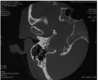
Figure 2. CT, axial image, bone window, shows osteosclerotic changes of the right petrous pyramid.
Figure 3. CT, axial image, bone window. Widening and erosion of the right jugular foramen with tumor invasion into the tympanic cavity.
sinuses [8, 17]. Paragangliomas are characterized by low signal intensity on T1-weighted images and heterogenous, hyperintense signal on T2- weighted images [9, 11]. In 19 patients of the evaluated group T1-weighted images demonstrated within the tumor mass tortuous, signal-void areas corresponding to blood vessels with high blood flow velocity (Fig. 4). Such pattern may also be seen in other tumors with abundant vascular component, e.g. juvenile angiofibromas. However, in the jugular foramen it is almost pathognomonic for paragangliomas [16, 17]. In patients with suspected paraganglioma contrast administration is recom-mended both in CT and MRI, because these highly vascu-larized tumors show quick and intensive contrast enhance-ment. It was observed in all patients in the analyzed group. Angiography is an important part of preoperative assess-ment in patients with suspected paraganglioma. Typical angiographic image of this tumour consists of a hypervas-cular mass, intensive tumor blush, enlargement of feeding
arteries and the presence of arteriovenous shunts with early draining veins [9, 16, 18]. These features were observed in all patients from the analyzed group who underwent angio-graphy (Fig. 5). Due to typical localization and characteristic presentation, angiography allows to confirm the diagnosis of paraganglioma. The highest sensitivity of detection of even small tumors makes this method the best tool for exclusion or confirmation of multiple paragangliomas. For this reason, the examination should always be performed bilaterally [15]. Jugular bulb paragangliomas derive their vascular supply primarily from the branches of the external carotid artery (the ascending pharyngeal, posterior auricular and occipital artery), meningeal branches of the vertebral artery and the internal carotid artery [6, 9, 16]. Apart from the diagnostic value, angiography has also an important role in presurgical work-up of patients with paragangliomas: facilitates intra-operative identification of misplaced blood vessels, allows assessment of their patency, determines the extent of tumor blood supply from the internal carotid artery and enables preoperative embolization of the tumor. In case of tumor encasing the internal carotid artery, when its occlusion during the surgical procedure is considered, the examina-tion should include the Mattas’s test to assess the efficiency of the circle of Willis [9, 19].
The role of ultrasound in the evaluation of jugular bulb paragangliomas is limited due to surrounding bony struc-tures and location at the skull base [20]. This technique
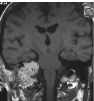
Figure 4. MR T1-weighted post-contrast coronal image shows within the right petrous bone and below the skull base strongly enhanced tumor with multiple signal-void areas.
Figure 6. Colour-coded Doppler ultrasonography, bilateral transversal sections of the carotid bifurcation area show a hypoechoic tumor (arrows) in the left carotid bifurcation – angiographically confirmed carotid body tumor.
Figure 5. Carotid angiography, lateral view. Typical picture of hypervascular tumors in a patient with three synchronous paragangliomas on one side of the head and neck: jugular bulb paraganglioma, vagal paraganglioma and carotid body tumor.
is useful for quick, noninvasive exclusion of a coincident carotid body tumor. In the reported group of 3 patients who underwent Doppler sonography, jugular bulb paraganglio-mas were not accessible to sonographic assessment. On the side of the tumor low-resistance blood flow of high velocity in the external carotid artery was observed, as well as enhanced blood flow in the jugular vein, which indicated a vascular, shunt-like character of the temporal pyramid tumor. In 2 patients coincident contralateral carotid body tumors were diagnosed (Fig. 6). In one of these patients, both paragangliomas were resected in separate surgical procedures. The other patient after surgical removal of the jugular bulb paraganglioma, is followed-up by means of ultrasonography and no signs of tumor progression are observed.
Before introduction of CT and MRI into clinical routine, the primary modalities used in the diagnostic management of jugular bulb paragangliomas were conventional radiogra-phy and angiograradiogra-phy. Technological advances in the field of imaging methods has led to increased importance of
non-invasive techniques, which have become the fundamentals of the diagnostic algorithm in patients with clinical symp-toms of paraganglioma. Carotid angiography should be per-formed as the last diagnostic step before surgery, in order to visualize tumor vascularity and allow its embolization. Angiography is not necessary in cases, where due to the extent of the tumor or other reasons not surgery but radio-therapy or clinical observation is recommended,.
Conclusions
1. CT and MRI allow best evaluation of tumor extension and demonstrate features characteristic for jugular bulb paraganglioma.
2. Carotid angiography confirms the diagnosis of a vascular tumor and allows its embolization as an adjunct to surgi-cal treatment.
3. Ultrasonography is a useful technique for exclusion of coexisting carotid body paraganglioma.
1. Kliewer KE, Wen D-R, Cancilla PA et al.: Paragangliomas: assessment of prognosis by histologic, immunohistochemical and ultrastructural techniques. Hum Pathol, 1989; 20: 29–39.
2. Megerian CA, McKenna MJ, Nadol JB Jr.: Non-paraganglioma jugular foramen lesions masqueradingas glomus jugulare tumours. Am J Otol, 1995; 16: 94–98.
3. Rao AB, Koeller KK, Adair CF: Paragangliomas of the head and neck: radiologic-pathologic correlation. Radiographics, 1999; 19: 1605–1632.
4. Lee JH, Barich F, Karnell LH et al.: National cancer data base report on malignant paragangliomas of the head and neck. Cancer, 2002; 94: 730–7.
5. Patetsios P, Gable DR, Garret WV et al.: Management of carotid body paragangliomas and review of a 30-year experience. Ann Vasc Surg, 2002; 16: 331–338.
6. Myssiorek D: Head and neck paragangliomas. Otolaryngol Clin North Am, 2001; 34 (5): 829–836.
7. Baysal B: Genetics of familial paragangliomas. Otolaryngol Clin North Am, 2001; 34 (5): 863–879.
8. Jackson CG, Kaylie DM, Coppit G et al.: Glomus jugulare tumours with intracranial extension. Neurosurg Focus, 2004; 17 (2): 45–50. 9. Van den Berg R: Imaging and management of head and neck
paragangliomas. Eur Radiol, 2005; 15: 1310–1318. 10. Som PM, Curtin HD: Tumors of the temporal bone and the
cerebellopontine angle. In: Som P.M., Cortin H.D (eds.): Head and Neck Imaging, ed 3, vol. 2. Mosby, St Louis, 1996, pp. 915–951.
11. Lustrin ES, Palestro C, Vaheesan K: Radiographic evaluation and assessment of paragangliomas. Otolaryngol Clin North Am, 2001; 34 (5): 881–906.
12. Ramina R, Maniglia JJ, Fernandes Y.B et al.: Jugular foramen tumors: diagnosis and treatment. Neurosurg Focus, 2004; 17 (2): 31–40. 13. Oldring D, Fish U: Glomus tumors of the temporal region. Arch
Otolaryngol, 1981; 107: 209.
14. Glasscock ME, Jackson CG, Harris PF: Glomus tumors: Diagnosis, classification, and management of large lesions. Arch Otolaryngol, 1982; 108: 401.
15. Boedeker CC, Ridder GJ, Schipper J: Paragangliomas of the head and neck: diagnosis and treatment. Familial Cancer, 2005; 4: 55–59. 16. Lowenheim H, Koerbel A, Ebner FH et al.: Differentiating imaging
findings in primary and secondary tumors of the jugular foramen. Neurosurg Rev, 2006; 29: 1–11.
17. Phelps DP, Cheesman AD: Imaging jugulotympanic glomus tumors. Arch Otolaryngol Head Neck Surg, 1990; 116: 940–945.
18. Weber AL, McKenna MJ: Radiologic evaluation of the jugular foramen: anatomy, vascular variants, anomalies, and tumors. Neuroimag Clin North Am, 1994; 4: 579–598.
19. Tikkakoski T, Luotonen J, Leinonen S et al.: Preoperative embolization in the management of neck paragangliomas. Laryngoscope, 1997; 107: 821–826.
20. Stoeckli SJ, Schuknecht B, Alkadhi H et al: Evaluation of
paragangliomas presenting as a cervical mass on color-coded Doppler sonography. Laryngoscope, 2002; 112: 143–146.