Mesh Repair of Hernias
of the Abdominal Wall
€ducost Publishers
Cover design: Sacha Hart-Schellart Printpartners Ipskamp B.V., Enschede
© W.W. Vrijland, Rotterdam, 2003
No part of this book may be reproduced by print, xerography or any other means without the authors permission in writing.
Mesh repair of hernias
of the abdominal wall
Correctie van buikwandhernia’s
met kunststof materiaal
Proefschrift
ter verkrijging van de graad van doctor aan de
Erasmus Universiteit Rotterdam
op gezag van de
Rector Magnificus
Prof.dr.ir. J.H. van Bemmel
en volgens besluit van het College voor Promoties.
De openbare verdediging zal plaatsvinden op
woensdag 19 maart 2003 om 15:45 uur
door
Wietske Willemijn Vrijland
geboren te Enschede
Promotiecommissie
Promotoren:
Prof.dr. J. Jeekel
Prof.dr. H.J. Bonjer
Overige leden:
Prof.dr. Th.J.M. Helmerhorst
Prof.dr. H.W. Tilanus
Prof.dr. J.B.M.Z. Trimbos
CONTENTS
1 General
Introduction
1
2
Randomized Clinical Trial of Non-Mesh versus Mesh Repair
of Primary Inguinal Hernia
17
3
Prosthetic Mesh Repair should be Used for Any Defect
in the Abdominal Wall
27
4
Introduction to Pathophysiology and Prevention of
Postoperative
Adhesions
31
5
Peritoneal Adhesions to Prosthetic Materials:
Choice of Mesh for Incisional Hernia Repair
35
6
Intraperitoneal Polypropylene Mesh Repair of Incisional Hernia
is not Associated with Entercutaneous Fistula
43
7
Fewer Intraperitoneal Adhesions with Use of Hyaluronic Acid-
Carboxymethylcellulose Membrane; A Randomized Clinical Trial
51
8 Abdominal
Adhesions:
Intestinal
Obstruction, Pain and Infertility
65
9 Discussion
75
Summary
81
Conclusions
83
Samenvatting
85
List of Publications
88
References
89
Dankwoord
101
Curriculum
Vitae
105
Introduction
CHAPTER 1
Introduction
1.1
Overview of the thesis
Hernia of the ventral abdominal wall
A hernia of the abdominal wall is a permanent or intermittent protrusion of abdominal
contents outside the abdominal cavity through a defect in the abdominal wall.
Approximately 75% of all hernias occur in the inguinal region. Other types of hernias
of the ventral abdominal wall are incisional, umbilical, epigastric and Spigelian
hernia. In chapter 1 an overview of hernias of the abdominal wall is described. The
incidence, clinical implications and treatment options and their complications are
described, based on the available literature regarding this subject.
Since there are numerous methods for abdominal wall hernia repair, without
consensus about the preferred method, we decided to perform a randomized clinical
trial to compare mesh and non-mesh repair for inguinal hernias. This randomized
clinical trial is described in chapter 2.
The preferential method of hernia repair is discussed in an editorial, not only for
inguinal hernias, but also for other types of abdominal wall hernias such as incisional
hernias and large umbilical hernias. Endoscopic hernia repair was included in this
editorial, which is described in chapter 3.
Complications of mesh repair of abdominal wall hernias
Ideally, prosthetic mesh is placed preperitoneally. If the mesh can be placed
between the abdominal wall and peritoneum or prefascially, opening of the
abdominal cavity can be avoided and the peritoneum and the abdominal contents
are not exposed to injury. For uncomplicated inguinal hernias prefascial or
preperitoneal placement of mesh is easy to perform, but for incisional and recurrent
umbilical hernias intraperitoneal placement of mesh is often unavoidable. Possible
complications of intraperitoneal mesh placement are adhesions and
entero-cutaneous fistulas.
Adhesions developing after abdominal surgery are abnormal attachments between
tissues and organs. The formation of adhesions results from peritoneal laceration
and is enhanced by the presence of foreign materials in the abdominal cavity such
as sutures and prosthetic mesh. An introduction to postoperative adhesion formation
is described in chapter 4.
An animal study on the formation of adhesions in the presence of a non-absorbable
mesh which is often used in hernia repair is described in chapter 5. This mesh is
applied with and without coverage by an absorbable mesh, which was suggested to
prevent the formation of adhesions. A newly developed mesh with an inert surface
was also tested.
Enterocutaneous fistulas are a feared complication after intraabdominal mesh
placement because their morbidity is severe and repair technically difficult. Although
only few enterocutaneous fistulas have been reported in literature, the common
opinion is that polypropylene mesh should never be in contact with intraabdominal
organs to avoid this complication [Morris-Stiff 1998]. In chapter 6 a retrospective
analysis of the outcome of incisional hernia repair with polypropylene mesh is
described to assess the risk of enterocutaneous fistula formation, combined with an
overview of literature concerning this topic.
Prevention and treatment of postoperative complications related to adhesions
To prevent the formation of adhesions, it was suggested that a mechanical barrier
could be used peroperatively to temporarily separate the intraabdominal viscera from
the abdominal wall or prosthetic mesh. A membrane containing hyaluronic acid has
been developed for this purpose, which already has been shown effective in
experimental studies. To assess the value of this material in clinical circumstances, a
randomized controlled multicenter study was performed which is described in
chapter 7.
Surgery is the only modality to confirm the presence of adhesions in the abdominal
cavity and surgical lysis is the only therapy available. The reported results of
adhesiolysis vary widely, and studies cannot be compared because the indication for
adhesiolysis and duration of follow-up differ. In Chapter 8 the indication, method and
success rate of adhesiolysis for intestinal obstruction, chronic abdominal pain and
infertility are reviewed.
Introduction
1.2 Inguinal
hernia
Definition and incidence
An inguinal hernia is defined as a protrusion of abdominal contents through a defect
in the abdominal wall of the groin.
Although no exact figures are available, a prevalence varying from 10% to 15% in
adults in the Western hemisphere has been estimated, with a male to female ratio of
12:1. Although the incidence of inguinal hernia rises with age, relatively young
people are affected: an incidence between 5 and 8 % in patient 25 to 40 years of age
has been reported [Abrahamson 1997]. Inguinal hernia repair is the most frequently
performed surgical operation. In the United States approximately 700.000
procedures are performed annually [Lichtenstein 1993], and in the Netherlands
25.000 [Health Care Information 1995]. Consequently, inguinal hernias not only
affect individual patients but also have a great impact on society. Failure of inguinal
hernia repair leads to increased patient discomfort, reoperations and sick-leave, and
therefore may result in a considerable economical burden [Liem 1997b].
Clinical picture, diagnosis and indications for inguinal hernia repair
The primary manifestation of an inguinal hernia is usually a bulge in the inguinal
region. The patient may describe minor pain or vague discomfort. Severe pain only
occurs in case of incarceration of bowel. In adults, the onset of inguinal hernia is
usually rapid. At physical examination, a hernia can be observed if standing upright,
while it may disappear in the supine position. Manipulation might be necessary to
reduce the bulge and finally some hernias are not reducible anymore, either because
of incarceration or because of adhesions. The most important tool in diagnosing an
inguinal hernia is physical examination. If there is any doubt about the nature of the
inguinal bulge ultrasonography or MRI can provide more evidence. If a hernia cannot
be diagnosed by physical examination because of the absence of a clear bulge,
herniography may be of help, although the accuracy of this procedure remains to be
assessed [Van den Berg 1993].
Strangulation is a complication mainly of longer existing inguinal hernias. Abdominal
contents become trapped in the abdominal wall defect, can not be reduced and
become ischemic which results in bowel obstruction with severe abdominal pain.
Although the incidence of incarceration is about 3 % and the incidence of
strangulation no more than 1 %, morbidity and mortality increase considerably after
emergency repair of incarcerated or strangulated hernias, and therefore it is
generally advised to perform inguinal hernia repair timely [Oishi 1991,
Kulah 2001].
The general opinion is that in case of an inguinal hernia, a repair should always be
done.
Classification
Numerous classification systems for inguinal hernias have been described. Since the
clinical significance is limited, these systems are rarely used in daily practice. The
most important distinction is between a direct and an indirect hernia. A crucial role in
this distinction is played by Hesselbach’s triangle, which is bordered on the medial
side by the rectus sheath, on the craniolateral side by the epigastric vessels and in
the inferior side by the inguinal ligament.
An indirect inguinal hernia is situated lateral of Hesselbach’s triangle and thus lateral
of the epigastric vessels. The peritoneal sac protrudes through the internal inguinal
ring and passes down the inguinal canal together with the spermatic cord. It is
suggested that lack of the obliteration of the processus vaginalis is the primary factor
leading to the development of an indirect inguinal hernia, and therefore can be
defined as a congenital disease. Inguinal hernias in children are always indirect.
A direct inguinal hernia protrudes through the floor of the inguinal canal in
Hesselbach’s triangle, medial to the epigastric vessels. It is suggested to be acquired
by repetitive straining, as with prostatism, constipation, coughing and heavy lifting,
although solid evidence lacks. Defects in collagen synthesis might predispose to this
type of hernia [Wagh 1974].
Other classifications have been described by Casten [1967],
Halverson [1970],
Gilbert [1987], Robbins [1993], Nyhus [1993] and Rutkow [1993] but since the clinical
significance is limited and surgical treatment is identical for all different types in
adults, these classifications will not be described in this thesis. The only exception
might be the indirect hernia in young adults; it is thought reposition of the peritoneal
sac and narrowing of the internal inguinal ring is a sufficient repair in these patients,
but clinical reports on this subject lack.
Treatment and outcome
Numerous methods have been described for inguinal hernia repair. These can be
divided in non-mesh or suture repairs and repairs with the use of prosthetic mesh.
Non-mesh repair
Bassini, an Italian surgeon, performed the first inguinal hernia repair with
reconstruction of the inguinal canal to preserve the functional anatomy in 1894, and
described this procedure in 1897 [Bassini 1897]. The operation involved high ligation
of the hernia sac by opening the transversalis fascia and consequently suturing the
internal oblique and transversus abdominis muscles, together with the upper leaf of
the transversalis fascia, to the inguinal or Poupart’s ligament and the lower leaf of
the transversalis fascia. Interrupted silk sutures were used. His technique
dramatically decreased postoperative mortality, morbidity and recurrence rate and
his method has been the method of choice for about a hundred years.
In 1940 McVay popularized a method first described by Lotheissen, which described
suturing the conjoint tendon to the pectineal (Cooper’s) ligament instead of to the
inguinal ligament [McVay 1981, Lotheissen 1898]. This method is based on the
observation that the conjoint tendon originally is attached to Cooper’s ligament.
Shouldice [1953] described a multi-layered repair based on Bassini’s repair, which is
probably the most successful method of non-mesh repair. Stainless steel continuous
sutures are applied. The transversalis fascia is also opened exposing the internal
ring and widely dissected from the preperitoneal fat. The first layer of the repair
involves suturing the lower flap of the transversalis fascia to the posterior side of the
upper flap of this fascia and to the posterior side of the rectus abdominis muscle and
of the aponeurosis of the transversus abdominis. The upper flap of the transversalis
fascia is sutured to the base of the lower flap and to the inguinal ligament forming the
second layer. The third layer consists of the conjoint tendon sutured to the inguinal
ligament and lower flap of the external oblique aponeurosis. For the fourth layer, the
Introduction
anterior rectus sheath and the lower aspect of the conjoint tendon from the front to
the inner surface of the lower flap of the external oblique aponeurosis are sutured.
Then the external oblique aponeurosis is closed over the spermatic cord.
This repair is technically complicated, time consuming and not always feasible,
especially in patients with large direct hernias, who do not have sufficient
transversalis fascia.
These three types of non-mesh repair represent the most widely used surgical
procedures for inguinal hernia repair without the use of prosthetic material. Although
many other methods have been described, the common problem of these
procedures is that suturing and displacement of anatomic structures may cause
excessive tension on the suture line and surrounding tissue, thus increasing the risk
of recurrence of the hernia. More elaborate descriptions of these procedures and
their modifications have been described in several textbooks [Nyhus 2002].
Recurrence rates of non-mesh repairs vary from 0.2 to 33 %, depending on the
surgical method, experience, length of follow-up and type of hospital [Beets 1997, De
Wilt 1990, Hay 1995, IJzermans 1991, Janu 1997, Kux 1994, Paul 1994, Rand
Corporation 1983, Simons 1996].
Mesh repair
Abdominal wall hernia repair with the use of polypropylene mesh was initially
described by Usher [1958]. Inguinal hernia repair employing polypropylene mesh to
achieve a so-called ‘tension-free’ repair was first described by Lichtenstein and
Shulman [1986]. This technique avoids tension on the sutured structures bordering
the defect by refraining from approximating these structures. The Lichtenstein
technique involves dissecting and inverting the hernia sac without opening it. Closure
of the hernial orifice is not attempted. The defect is covered with a polypropylene
mesh sized about 6 x 8 cm trimmed to fit the area. A non-absorbable suture is used
to fix the mesh. The mesh is fixed medially to the rectus sheath and the lacunar
ligament close to the pubic tubercle. On the inferior side the mesh is sutured to
Poupart’s ligament. A slit in the mesh on the lateral side at the internal ring allows
emergence of the spermatic cord and vessels. The two lateral tails of the mesh are
crossed to embrace the spermatic cord and vessels thus creating a new internal ring.
The superior side of the mesh is loosely sutured to the rectus sheath and conjoint
tendon. Then, the external oblique aponeurosis is closed over the mesh. This
method is associated with a recurrence rate of less than one per cent [McGillicuddy
1998, Lichtenstein 1989, Friis 1996]. Randomized clinical trials concerning this
subject have been done, and are mentioned in chapter 2 and 3 of this thesis
[McGillicuddy 1998, Friis 1996, Collaboration 2000a].
Other types of repair with prosthetic mesh are for example Gilbert’s plug and patch
repair [Gilbert 1987] which has been modified by Robbins and Rutkow [1993] and
the Rives’ repair involving placement of a larger mesh preperitoneally. [Rives 1987].
Stoppa [1987] described a repair with a very large preperitoneal mesh covering the
lower half of the parietal peritoneum, which may be used in case of multiple
recurrences. These repairs will not be discussed in this thesis.
Endoscopic repair
Endoscopic repair of inguinal hernia can be done totally extra peritoneally (TEP), a
procedure first described by McKernan and Laws [1993] and transabdominally
(TAPP), first reported by Arregui [1991].
Both procedures require the use of prosthetic mesh. It has been proven that
endo-scopic repair causes less recurrences if compared to open non-mesh repair, but if
compared to open mesh repair no differences in recurrence rate exist [Liem 1997a,
Collaboration 2000b]. The advantages of endoscopic procedures are less
post-operative pain and more rapid return to normal activities, but since endoscopic
procedures take longer to perform and may be related to rare but serious
complications, the method of choice for inguinal hernia repair remains to be
established [Collaboration 2000b]. In the Netherlands, the acceptance of endoscopic
inguinal hernia repair is still low, only 16% of surgeons applies this method on a
regular basis [Knook 2001].
Complications
The incidence of wound infections after inguinal hernia repair varies from 0.4% to 9%
[Bailey 1992, Gilbert 1993, Holmes 1994, Karran 1992, Mertens 1994]. The wide
variation of this incidence might be explained by a variation of surgical techniques
and operative measures. The administration of antibiotic prophylaxis is generally
advised if prosthetic mesh is used, although no strong evidence exists that it
decreases the incidence of wound infections and serious complications like
necrotizing fasciitis [Platt 1990]. Definitions of wound infection differ among studies
which impedes interpretation of several studies at a time. Furthermore, retrospective
studies tend to underestimate the rate of wound infection while prospective analyses
record clinical events such as wound infection more accurately. There is great
concern for infection of mesh, although it is an infrequent complication [Anonymous
2002].
Chronic pain is a common complication with an incidence of 2 to 5 per cent [Starling
2002]. However, few studies of inguinal hernia repair address chronic pain [Callesen
1999, Cunningham 1996]. Chronic pain may be related to peroperative nerve injury.
The inguinal region receives sensory innervation from the iliohypogastric, ilioinguinal,
genitofemoral and lateral femoral cutaneous nerves, all stemming from the eleventh
thoracic through second lumbar nerve. In open inguinal hernia repair, the
iliohypogastric nerve, the ilioinguinal nerve and the genital branch of the
genitofemoral nerve are at stake. In endoscopic or laparoscopic repair the femoral
branch of the genitofemoral nerve and the lateral femoral cutaneous nerves are at
risk.
Starling [2002] provides an excellent outline of this problem in Nyhus and
Condon’s Hernia. Although it has been suggested that the use of mesh and the
development of chronic pain are related, no evidence can be found in literature. The
EU Hernia Trialist Collaboration concluded that mesh appears to reduce the chance
of persisting pain rather than to increase it [Anonymous 2002].
The incidence of testicular atrophy has not been frequently described. Bendavid et
al. [1995] of the Shouldice Hospital described an incidence of 0.08 %. It was
suggested that extensive dissection of the funiculus damaging the venous blood flow
is responsible for this complication rather than the creation of a narrow internal
inguinal ring [Wantz 1995].
Introduction
General complications include pulmonary atelectasis and pneumonia which can be
prevented by early postoperative reactivation. Exact data are lacking probably due to
the low incidence of these sequelae. Urinary retention is more common, and has
been related to prostatism and regional anesthesia. Both predispose to postponed
micturition postoperatively that may evolve into urinary retention.
Non-mesh and mesh repairs have shown no significant difference in the incidence of
complications in a systematic review [Collaboration 2000a].
1.3 Incisional
hernia
Definition, incidence and risk factors
An incisional hernia is defined as a protrusion of abdominal contents through a
defect in the abdominal wall located at the site of a former incision in the abdominal
wall. The bowel contents remain covered by peritoneum and skin. Incisional hernia
has been a common complication, reported in 2 – 19 % of patients after abdominal
surgery [Mudge 1985, Bucknall 1982, Luijendijk 1997, Regnard 1988, Israelsson
1993].
Some incisional hernias develop within days after an abdominal operation while
other hernias may develop many years after primary surgery. Incisional hernias that
occur within days postoperatively have been suggested to originate from technical
failure or raised intra-abdominal pressure due to persisting ileus or chronic
pulmonary disease. In a later stage, wound healing disturbance and co-morbidity
may be responsible for incisional herniation. The evidence on this subject is still
incomplete, but it has been shown that there are several patient-related factors that
predispose to the development of an incisional hernia. Male gender [Wissing 1987],
increasing age [Viljanto 1966], pulmonary disease [Wissing 1987, Gecim 1996],
prostatism [Luijendijk 2000] diabetes mellitus [Sugerman 1996], obstructive jaundice
[Armstrong 1984] and aneurysmatic disease [Stevick 1988, Luijendijk 2000] have
been indicated as risk factors in some studies, whereas this remained unconfirmed
in others. Disturbances in collagen metabolism probably play a role in the
development of incisional hernias [Wagh 1974, Si 2002]. Closure of the abdominal
wound after surgery is a risk factor as well [Niggebrugge 1999].
Israelsson [1993] described the suture length to wound ratio as an important
parameter for healing of midline incisions closed with a continuous suture technique.
It was stated that this ratio should be ≥ 4 to reach a lower incidence of incisional
hernias.
Clinical picture, diagnosis and indications for incisional hernia repair
Incisional hernias are often asymptomatic, especially in small hernias. However, if
they cause symptoms, pain, discomfort and the presence of a bulge constitute the
clinical picture. In large hernias, cutaneous ulceration and necrosis may develop.
Strangulation of the hernia occurs in 2.4 % of patients with incisional hernia [Read
1989].
To ascertain the presence or absence of an incisional hernia, physical examination
of the abdominal wall is mandatory. A weakness in the abdominal wall at the site of a
scar with palpable fascial rims suggests the presence of a hernia. Bulging during
Valsalva’s manoeuvre or at getting up from a supine position may occur. An
incisional hernia should be distinguished from local paralysis of the abdominal
muscles which can occur postoperatively, and from diastasis of the rectus abdominis
muscle. Diastasis of the rectus abdominis muscle, or divarication, occurs when this
muscle is loosened from the linea alba. This may be related to pregnancy and
obesity. When in doubt, ultrasonography or CT scanning can be of help to detect and
locate a defect of the abdominal wall and assess the diameter of the defect.
Not all incisional hernias need to be repaired. It is generally thought safe to refrain
from operating in case of minor symptoms [Abrahamson 1997]. Hernias with a small
Introduction
fascial defect and a large protrusion and incisional hernias in patients who suffer
from recurrent bowel obstruction are at risk for strangulation, and therefore should be
considered as a definitive indication for surgery. For all other incisional hernias there
is a relative indication for surgery without international, validated guidelines. When
considering repair of incisional hernias, the benefits should be weighed against the
recurrence rate which can be as high as 49 % [Van der Linden 1988].
Treatment and outcome
Numerous methods have been described for incisional hernia repair. These can be
divided in non-mesh repairs and repairs with the use of prosthetic mesh.
Non-mesh repair
Before the introduction of prosthetic material for hernia repair, all incisional hernias
were repaired by suturing the fascial edges. In spite of varying techniques and
different suture materials, recurrence rates between 24 and 49 % were encountered
in the larger studies [Van der Linden 1988, Langer 1985, George 1986, Read 1989].
Primary closure is performed in single or multiple layers. In single layer closure, all
layers of the abdominal wall are approximated with one bite of suture. In multiple
layer closure, different layers, for example the anterior and posterior rectus sheath
are approximated and sutured separately. Both techniques are associated with high
recurrence rates in large studies with long-term follow-up [Langer 1985, George
1986, Gecim 1996].
Mayo or overlap repair provides overlap of the fascial edges and fixated suturing.
This method has shown a high recurrence rate of 31 to 78% [Paul 1998, Luijendijk
1997].
To prevent tension on the suture lines relaxing incisions have been advocated.
These incisions are made in a vertical fashion and bilateral to the incisional hernia in
the anterior sheath of the rectus abdominis muscle before closure of the defect. No
studies involving larger numbers of patients have been reported assessing the value
of this technique.
Rectus sheath techniques involve mobilizing of healthy tissue with subsequent
primary closure to cover the defect in the abdominal wall. Different techniques have
been described but no success rates have been reported. The components
separation technique of Ramirez [1990] showed recurrence rates ranging from 4.5 to
8.6% in small series of large incisional hernias [DiBello 1996, Girotto 1999, Shestak
2000]. In some cases in these series, prosthetic material was used as well to
reinforce the abdominal wall.
Mesh repair
Abdominal wall hernia repair with the use of polypropylene mesh was initially
described by Usher [1958]. Since then, mesh repair was popularized and other types
of mesh were developed. Three types of prosthetic mesh are currently used in hernia
repair: polypropylene, expanded polytetrafluoroethylene and polyester. These
meshes are all non-absorbable since the application of absorbable meshes leads to
unacceptable high recurrence rates. There is much debate about which mesh to
choose, because of different characteristics of these meshes, such as tissue
susceptibility to infection as well as disintegration of the mesh. In incisional hernia
repair, different positions of the mesh are possible: intraperitoneal, preperitoneal or
between rectus abdominis muscle and posterior fascia and the onlay method in
which the prosthesis is placed on top of the anterior rectus fascia. It remains to be
established which position is preferable. Larson
and Harrower [1978] advised to
place the mesh subfascially, but this is not supported by others.
Mesh repair is associated with lower, but still considerable recurrence rates of 4 – 17
% in different studies with a follow-up of 6 months to 7.6 years [Leber 1998, Molloy
1991, Liakakos 1994, Sugerman 1996, McCarthy 1981, Matapurkar 1991,
McLanahan 1997, Turkcapar 1998, Whiteley 1998, Ladurner 2001, Martin-Duce
2001, Schumpelick 1996]. In 2000, the first randomized clinical trial comparing
non-mesh repair and non-mesh repair was published by Luijendijk et al. [2000]. In this study,
it was concluded that mesh repair is the method of choice for all non-emergency
incisional hernia repairs, even in defects as small as 3 cm in diameter. Three year
recurrence rates were 43 and 24 per cent for non-mesh versus mesh repair
respectively.
Laparoscopic repair
Laparoscopic repair of incisional hernia with prosthetic mesh was introduced by
LeBlanc
and
Booth
[1993].
Cassar and Munro [2002] described 14 series of
laparoscopic incisional hernia repair. In all studies, the mesh was placed
intraperitoneally after installation of pneumoperitoneum, insertion of trocars as far as
possible from the defect and careful adhesiolysis to create sufficient overlap of the
mesh. It has been shown necessary to use full thickness sutures to fixate the mesh
to the abdominal wall because only tackers or hernia staples have shown to provide
inadequate fixation [Riet 2002]. Our own technique was described in the Dutch
Journal of Surgery [Vrijland 1998]. The recurrence rate with the use of this technique
varies between 0 and 9 per cent. The follow-up is still relatively short, but
comparable to that of open mesh repair. [Cassar 2002]. The only randomized clinical
trial on laparoscopic versus open incisional hernia repair shows similar recurrence
rates, less morbidity and shorter hospital stay [Carbajo 1999]. Since this study is
relatively small and other comparative studies can not confirm these results, more
studies are necessary.
Complications
Since incisional hernia repair requires rather extensive dissection, postoperative
bleeding and hematoma formation occurs in approximately 10% [Luijendijk 2000]. It
is assumed that hematomas predispose to wound infections and since the use of
drains does not reduce the incidence of hematomas, the only way to prevent
hematoma formation and related wound complications is meticulous hemostasis and
obliteration of dead space [White 1998]. A seroma is defined as a collection of
serous fluid in the subcutaneous space which is related to extensive dissection as
well. The incidence of seroma is 1 – 15 % in different studies [Cassar 2002]. This
figure is not influenced by the placement of drains and therefore, there is still
discussion about the use of drains in incisional hernia repair [White 1998].
Wound infection is a serious complication of incisional hernia repair which eventually
may lead to recurrence of the incisional hernia [Bucknall 1982, Luijendijk 2000].
Bucknall et al. showed that an infected wound has a fivefold increased risk for
Introduction
developing a ventral hernia. It is thought that recurrence of incisional hernia after
incisional hernia repair occurs more frequently if infection occurs. Wound infection
has been documented in 4 – 15 % after incisional hernia repair when mesh was
used [Cassar 2002, Houck 1989, White 1998]. Details about the incidence of
hematomas and seromas after non-mesh repair are lacking, but Luijendijk et al.
[2000] showed no differences in complication rate between non-mesh and mesh
repair in a randomized clinical trial. However, Korenkov et al. [2002] described a high
infection rate after polypropylene mesh repair.
In mesh repair, wound infection may lead to an infection of the mesh. This is a
serious complication, because sometimes removal of the mesh is required. Evidence
exists that a polytetrafluoroethylene mesh requires removal more often than a
polypropylene mesh in case of infection [Cassar 2002]. Some have suggested that
the small pore size in polytetrafluoroethylene mesh enhances bacterial binding and
therefore promotes chronic mesh infection. Another wound complication is wound
sinus formation which was described to occur in 4 % of patients in one study
[Liakakos 1994] and in 12 % of patients in another [Molloy 1991].
Antibiotic prophylaxis before surgery seems necessary to prevent wound
complications, but a comparative study regarding this subject has never been
executed.
Enterocutaneous fistula formation after mesh repair was first described by Kaufman
et al. [1981]. The incidence of this complication is low, but it is a very serious
complication requiring surgery and usually removal of the mesh [Cassar 2002].
Intraperitoneal placement of the mesh possibly increases the risk for
enterocutaneous fistulas. In chapter 5 this complication is discussed more
extensively.
Chronic pain is an issue in incisional hernia as well as in inguinal hernia.
Martin-Duce et al. [2001] reported chronic pain in 28 per cent of patients after mesh repair
of incisional hernias. In most studies chronic pain has not been reported. The origin
and possible treatment of chronic pain after incisional hernia repair remain unclear.
General complications like pneumonia and urinary tract infections appear to occur at
similar rates in mesh and non-mesh groups Luijendijk et al. [2000].
Carbajo et al. [1999] reported less complications after laparoscopic repair of
incisional hernia in a small randomized clinical study which was confirmed by
Goodney et al. [2002] in a meta-analysis. Other studies showed a higher
complication rate after laparoscopic repair [Cassar 2002]. More clinical studies
randomizing patients with incisional hernias for either open or laparoscopic surgery
are necessary to establish the value of laparoscopic hernia repair. By the Erasmus
Medical Center Rotterdam such a randomized clinical trial has been started.
1.4
Other types of ventral abdominal wall hernia
Umbilical hernia
Umbilical hernias occur when the fascia at the abdominal entry of the umbilical cord
does not close completely. Hernias just below or just above the umbilicus in the
midline are called paraumbilical hernias and are usually included in the group of
umbilical hernias.
The incidence of umbilical hernias in children is high, but decreases with age
because of spontaneous closure of the defect. The incidence of umbilical hernias in
adults is unknown. Little is known about the cause of umbilical hernia in adults, but
umbilical hernia in childhood is a risk factor [Jackson 1970]. Middle aged obese
women with multiple pregnancies are at risk. Adults have a considerable risk of
incarceration with associated morbidity and mortality, but detailed information on this
subject lacks.
Non-mesh repair with non-absorbable sutures has been the method of choice
[Abrahamson 1997]. Recently Arroyo et al. [2001] published a randomized clinical
trial comparing non-mesh and mesh repair for umbilical hernias in adults with
recurrence rates of 11 versus 1 per cent respectively. The mean follow-up was 64
months. The complication rate was comparable between both groups. The authors
state that umbilical hernias should be treated with a mesh repair, regardless of the
size of the hernia.
Epigastric hernia
An epigastric hernia may be defined as a fascial defect in the linea alba between the
xyphoid process and the umbilicus. The prevalence of this hernia is between 0.5 and
10 per cent, as concluded from autopsy studies. Males are predominantly affected
[Abrahamson 1997]. Probably the epigastric hernia is acquired, and results from
excessive straining [Askar 1978, Lang 2002].
The majority of epigastric hernias, up to 75 per cent, is asymptomatic. Related
symptoms are epigastric pain, abdominal distention, dyspepsia, nausea and
vomiting. Incarceration, usually of the omentum, is common, but strangulation is
rare.
The presence of epigastric hernia can be confirmed by clinical examination, although
obesity can obscure epigastric hernia. In case of doubt, ultrasonography or
CT-scanning may be of help.
Non-mesh repair is still advised, but recent randomized trials comparing different
treatment modalities lack. Probably it is wise to consider mesh repair, especially in
larger defects [Abrahamson 1997].
Spigelian hernia
The Spigelian hernia is called after Adriaan van der Spieghel, a Belgian anatomist
and surgeon, who discovered the linea semilunaris. The linea semilunaris is the
course of the lateral border of the rectus abdominis muscle, and in the muscular gap
between the linea semilunaris and the medial borders of the oblique and transversus
abdominis muscles the Spigelian aponeurosis is located, which is broadest just
Introduction
caudal to the umbilicus. Spigelian hernias protrude through the Spigelian
aponeurosis and usually in the lower abdomen [Abrahamson 1997]. They are rare
and usually difficult to diagnose because of their intramural location and unspecific
symptoms. An intermittent mass and local pain are the common symptoms of a
Spigelian hernia [Larson 2002]. Clinical diagnosis can be confirmed by ultrasound,
CT-scanning and ultimately herniography.
Generally, it is advised to perform non-mesh repair, but in larger hernias mesh repair
should be considered. No comparative studies are available [Larson 2002].
Non-Mesh vs. Mesh Repair
CHAPTER 2
RANDOMIZED CLINICAL TRIAL OF NON-MESH VERSUS
MESH REPAIR OF PRIMARY INGUINAL HERNIA
Abstract
Background
: The optimum method for inguinal hernia repair has not yet been
determined. The recurrence rate for non-mesh methods varies between 0.2 and
33%. The value of tension-free repair with prosthetic mesh remains to be confirmed.
The aim of this study was to compare mesh and non-mesh suture repair of primary
inguinal hernias with respect to clinical outcome, quality of life and cost in a
multicentre randomized trial in general hospitals.
Methods
: Between September 1993 and January 1996, all patients scheduled for
repair of a unilateral primary inguinal hernia, were randomized to non-mesh or mesh
repair. The patients were followed up at 1 week and at 1, 6, 12, 18, 24 and 36
months. Clinical outcome, quality of life and costs were registered.
Results
: Three hundred patients were randomized of whom 11 were excluded.
Three-year recurrence rates differed significantly: 7 per cent for non-mesh repair
(n=143) and 1 per cent for mesh repair (n=146). There were no differences in clinical
variables, quality of life and costs.
Conclusions
: Mesh repair of primary inguinal hernia is superior to non-mesh repair
with regard to hernia recurrence and is cost-effective. Postoperative complications,
pain, and quality of life did not differ between groups.
Non-Mesh vs. Mesh Repair
2.1 Introduction
Inguinal hernia repair is the most frequently performed operation in The Netherlands
[
Health Care Information 1995
]. Consequently, failure of inguinal hernia repair not
only affects individual patients but also has a great influence on society. Failure
leads to increased patient discomfort, reoperations and sick-leave, and thus may
result in a considerable economical burden [
Liem 1997b
]. No consensus has been
reached yet about the best operation of inguinal hernia repair, which should show
good cost-effective clinical results [
MRC 1999, Barth 1998, McGillycuddy 1998
].
Recurrence rates after non-mesh suture repair of inguinal hernia vary between 0.2%
and 33%, depending on the surgical method, experience, length of follow-up and
type of hospital [
Beets 1997, De Wilt 1990, Hay 1995, IJzermans 1991, Janu 1997,
Kux 1994, Paul 1994, Rand Corp. 1983, Simons 1996
]. Tension-free hernia repair,
or repair with the use of mesh, was popularized by
Lichtenstein
and
Schulman
[
1986
]. This method was associated with a lower recurrence rate than suture repair
in a non-randomized study of primary inguinal hernia repair [
Lichtenstein 1989
], and
in two randomized studies [
McGillicuddy 1998, Friis 1996
].
The aim of this study was to establish the value of open mesh hernia repair for
primary inguinal hernia in the general hospital setting, not only with respect to clinical
outcome but also quality of life and cost. A multicentre randomized trial with
long-term follow-up was conducted.
2.2
Patients and methods
Between September 1993 and January 1996, patients older than 18 years scheduled
for repair of a primary unilateral inguinal hernia were randomized to non-mesh or
mesh repair. Patients could only be enrolled once and were not included if they
suffered from bilateral inguinal hernia. Patients were informed about the trial both
verbally and in writing. Six hospitals participated in the study.
Randomization was achieved by calling an independent randomization centre, where
computer-generated lists were available, stratified by hospital. The protocol was
approved by the ethics committees of all participating hospitals.
Age, obesity, intermittent high abdominal pressure (cough, constipation, prostatism)
and factors that may interfere with wound healing (diabetes, use of steroid
medication, smoking) were noted. Obesity was defined as a Body Mass Index of 30
kg/m
2or more. The type of inguinal hernia was also noted.
Evaluation of operation-related factors included surgical technique, type of
anaesthesia, clinical setting or day-care, and whether the operation was performed
by a surgeon or by a surgical resident. Drainage, wound hematoma, seroma, wound
dehiscence and wound infection were also recorded. Wound infection was defined
as discharge of pus from the wound.
At the induction of anaesthesia, a single dose of intravenous broad spectrum
antibiotics was administered according to hospital protocol. Non-mesh repair was
performed according to the surgeons' method of choice, provided that 2/0
polypropylene (Prolene, Ethicon, Johnson & Johnson, NJ, USA) sutures were used.
Mesh repair was performed according to a strict protocol as described by
Lichtenstein and Shulman
[
1986
]
using a polypropylene prosthetic mesh (Prolene,
Ethicon, Johnson & Johnson, NJ, USA or Marlex,
C.R. Bard, Inc., Billerica, MA, USA)
of 7.5 x 15 cm to avoid tension on the suture lines. The duration of surgery (from first
incision to last skin suture), hospital stay and time off work were noted. Patients were
followed up at 1 week and at intervals of 1, 6, 12, 18, 24 and 36 months. Awareness
of hernia recurrence and complaints about the groin were noted and the groin was
examined physically for recurrence of inguinal hernia. Hernia recurrence was defined
as a bulge or weakness in the operative area exacerbated by a Valsalva manoeuvre
and palpable outside the external ring. Hernia recurrence and death were the study
endpoints. Patients who did not visit the outpatient department for follow-up at 36
months, were asked to complete a questionnaire, and were visited at home by a
physician who was not aware of the method used for inguinal hernia repair. If
recurrences were found after follow-up had terminated, they were not included in the
statistical analysis in accordance with the protocol.
To assess quality of life (or current health state) before and after surgery, the Dutch
version of the EuroQol EQ-5D and the EuroQol Visual Analogue Scale [
Brooks 1996,
Kind 1999
] was administered for self-completion by patients before operation and 1
week, 1 month and 6 months after operation.
To determine the cost-effectiveness of both methods of inguinal hernia repair, a
questionnaire about costs was completed 1 month and 6 months after surgery.
This included questions about the need for help from a general practitioner, nurse or
housekeeper, the need for pain medication and the duration of sick-leave.
Cost-effectiveness also involved quality of life- and operation-related factors, such as
duration of surgery, duration of hospital stay and time off work.
Statistical analysis was done with the Statistical Product and Service Solutions
software (SPSS, Chicago, Illinois, USA). Percentages and continuous variables were
compared using Fisher's exact test and Mann-Whitney
U
test respectively.
Cumulative recurrence rates were calculated and compared using Kaplan-Meier
curves and the log rank test. P-values given are two-sided; P = 0.05 was considered
the limit of significance. The primary analysis was by intention to treat. A univariate
regression analysis for the non-mesh repair group was performed.
Non-Mesh vs. Mesh Repair
2.3 Results
Three hundred patients were randomized. Eleven patients were excluded. In four
patients another type of hernia was demonstrated at operation. One patient needed
bilateral repair. The operation was cancelled for three patients. In spite of inclusion in
the trial two patients underwent laparoscopic inguinal hernia repair and one patient
withdrew consent before operation. Preoperative characteristics were well matched
between the two groups (
Table 2.1
). Eight patients (3 per cent) were women.
Table
2.1
Comparison of study groups
Non-mesh repair group Mesh repair group
Variable Age1 [y] 53 (19-85) 58 (24-83) BMI1 [kg/m2] 25 (19-34) 25 (18-34) Prostatism2 [n] 14/140 12/141 Constipation [n] 11/143 10/146 Coughing [n] 22/143 25/146 Diabetes [n] 8/143 2/146 Use of steroids [n] 2/143 4/146 Type of hernia Indirect [n] 67 75 Direct [n] 45 37 Combined [n] 27 29 Not described [n] 4 5
1) Values are: median (range). 2) Men only.
Intention to treat analysis.
Of the remaining 289 patients, 143 had been randomized to non-mesh repair and
146 to mesh repair. The type of inguinal hernia repair in the non-mesh repair group
was Bassini-McVay in 75 patients (53 per cent), Shouldice in 36 (25 per cent),
Bassini in 26 (18 per cent) and McVay in three (2%). Three patients received a mesh
because the surgeon decided at operation that a mesh repair was preferable. These
procedures were marked as conversions. In the mesh repair group, 125 patients
received a
Prolene
mesh, whereas
Marlex
was used in 13 cases. In one occasion a
resorbable polyglactin 910 mesh (Vicryl, Ethicon, Johnson & Johnson, NJ, USA) was
used in error. Seven patients did not receive a mesh repair and these operations
were marked as conversions.
Thirteen patients (4 per cent) died within the follow-up period from causes unrelated
to inguinal hernia and more than 1 month after hernia repair.
Follow-up was complete for 254 patients (88 per cent). Thirty-five patients (12 per
cent) were lost to follow-up: twelve patients withdrew from follow-up, twelve patients
could not be traced, and eleven patients were followed up in writing at 36 months but
were not physically examinated at this time. All patients were included in the analysis
with their follow-up censored at the time of last physical examination.
Recurrences
During the 3-year follow-up, nine recurrences were found in the non-mesh repair
group and one in the mesh repair group. The only recurrence in the mesh group
occurred in the patient who received a resorbable mesh in error. The 3-year
cumulative recurrence rates in the non-mesh and mesh repair were 7 and 1 per cent
respectively (P = 0.009,
Table 2.2
).
Table
2.2
Cumulative recurrence rates 1-36 months of follow up after primary inguinal
hernia repair
Months after operation [mo]
Number at risk for recurrence [n]
Cumulative recurrence rate [%]1 Non-mesh repair 1 143 0 6 137 1 (1) 12 131 1 (1) 18 127 1 (1) 24 125 3 (2) 36 119 7 (2) Mesh repair 1 146 0 6 138 0 12 138 0 18 133 0 24 131 0 36 122 1 (1)
1) Values in parentheses are s.e.
Exclusion of the patient who received a resorbable mesh from the analysis (major
trial violation) decreased the 3-year cumulative recurrence rate from 1 per cent to
zero, increasing the difference between groups (P = 0.002). There were no
recurrences after inguinal hernia operations that were converted peroperatively.
Univariate analysis
The non-mesh repair group was associated with a significantly higher recurrence
rate. Risk factors were evaluated within this group. The recurrence rate was higher
for older patients; 3-year recurrence rates for patients younger than 65 years of age
and older patients were 3 and 16 per cent respectively (P = 0.01). Other patient
characteristics and wound complications were not identified as significant risk
factors.
Non-Mesh vs. Mesh Repair
Operation-related factors
Median duration of surgery was 45 min for both non-mesh repair (range 19-105) and
mesh repair (range 20-120). Seventy-nine percent of patients were treated in a
clinical setting and 21 percent in day-care; there was no difference between
treatment groups. Median hospital stay was 2 days in both groups, with a range of
0-14 and 0-11 days respectively. Median time off work was 17 (range 0-56) days after
non-mesh repair and 19 (range 2-113) days after mesh repair.
The type of anaesthesia did not differ between the groups (general 62 percent,
epidural 23 percent, spinal 15 percent). The type of hernia encountered at operation
was comparable between the two groups (
Table 2.1
). In the non-mesh group there
was no recurrence of an indirect hernia, four recurrences of direct hernias and five
recurrences of combined hernias. Surgeons and residents assisted by a surgeon
operated on comparable numbers of patients (68 versus 78 and 78 versus 66
respectively). Of the ten patients with a recurrence, six were primarily treated by a
surgeon and four by a resident (P not significant).
Complications
There was no significant difference between the non-mesh and the mesh repair
group regarding postoperative wound infection (none of 143 versus one of 146; p =
0.32), wound dehiscence (none of 143 versus one of 146; p = 0.32), hematoma (17
of 143 versus 15 of 146, p = 0.66) and seroma (none of 143 versus 4 of 146, p =
0.62). In the non-mesh group one patient suffered from urinary retention and one
patient from pneumonia. Apart from recurrences, there were no long-term
complications.
Postoperative pain (week 1: 45 of 140 versus 58 of 140 (p = 0.11); 36 months: 9 of
125 versus 8 of 129 (p = 0.73)) and discomfort (week 1: 78 of 140 versus 72 of 140
(p = 0.42); 36 months: 13 of 125 versus 11 of 129, (p = 0.6)) were similar at all
timepoints.
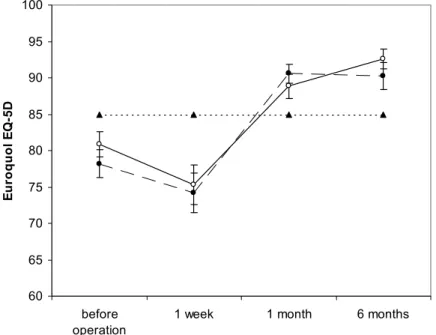
Quality of life
The response rate for the Euroqol questionnaire and VAS ranged from 49 to 74
percent for the non-mesh repair group and from 56 to 79 percent in the mesh group,
varying between timepoints. The quality of life did not differ significantly between
groups at any timepoint. There were no significant differences between mean (s.d.)
values for EuroQol EQ-5D or EuroQol VAS measured in the general population
(85(8) (
Kind 1999
) and 81(14) (
Van Agt 1994
) respectively) and either study group.
(
Figures 2.1
and
2.2
).
Number of respondents per time point ranges from 90 to 111 per treatment group.
Figure 2.1
Quality of life measured by Current Health State,
EuroQol EQ-5D (minimum score 0, maximum score 100).
Number of respondents per time point ranges from 101 to 119 per treatment group.
Figure 2.2
Quality of life measured EuroQol Visual Analogue Scale (VAS)
(minimum score 0, maximum score 100).
60 65 70 75 80 85 90 95 100 before operation
1 week 1 month 6 months
E u roqu ol E Q -5 D
Non-mesh repair group Mesh group General population
60 65 70 75 80 85 90 95 100 before operation
1 week 1 month 6 months
E u roqu ol V A S