0099-2240/03/$08.00⫹0 DOI: 10.1128/AEM.69.11.6908–6922.2003
Copyright © 2003, American Society for Microbiology. All Rights Reserved.
Analysis, Characterization, and Loci of the
tuf
Genes in
Lactobacillus
and
Bifidobacterium
Species and Their Direct Application for
Species Identification
Marco Ventura,
1* Carlos Canchaya,
1Vale
`rie Meylan,
1Todd R. Klaenhammer,
2and Ralf Zink
1†
Nestle´ Research Center, 1000 Lausanne 26, Switzerland,1and Department of Food Science, College of Agricultureand Life Sciences, North Carolina State University, Raleigh, North Carolina 27695-76242
Received 16 June 2003/Accepted 19 August 2003
We analyzed the tuf gene, encoding elongation factor Tu, from 33 strains representing 17 Lactobacillus
species and 8Bifidobacteriumspecies. Thetufsequences were aligned and used to infer phylogenesis among
species of lactobacilli and bifidobacteria. We demonstrated that the synonymous substitution affecting this gene renders elongation factor Tu a reliable molecular clockfor investigating evolutionary distances of
lactobacilli and bifidobacteria. In fact, the phylogeny generated by thesetufsequences is consistent with that
derived from 16S rRNA analysis. The investigation of a multiple alignment oftufsequences revealed regions
conserved among strains belonging to the same species but distinct from those of other species. PCR primers complementary to these regions allowed species-specific identification of closely related species, such as Lactobacillus caseigroup members. Thesetufgene-based assays developed in this study provide an alternative
to present methods for the identification for lactic acid bacterial species. Since a variable number oftufgenes
have been described for bacteria, the presence of multiple genes was examined. Southern analysis revealed one
tuf gene in the genomes of lactobacilli and bifidobacteria, but thetuf gene was arranged differently in the
genomes of these two taxa. Our results revealed that thetufgene in bifidobacteria is flanked by the same gene
constellation as the str operon, as originally reported for Escherichia coli. In contrast, bioinformatic and
transcriptional analyses of the DNA region flanking thetufgene in fourLactobacillusspecies indicated the same
four-gene unit and suggested a noveltufoperon specific for the genusLactobacillus.
The members of the genera Lactobacillusand Bifidobacte-riumare gram-positive organisms considered to belong to the general category of lactic acid bacteria (LAB), even though the genusBifidobacteriumis phylogenetically unrelated and has a unique mode of sugar fermentation (44). These organisms are inhabitants of a wide range of environments, including the gastrointestinal and urogenital tracts of humans and animals. Many LAB strains have a worldwide industrial use as starters in the manufacture of fermented foods. Moreover, some Lac-tobacillusandBifidobacteriumstrains have been shown to have beneficial effects on human and animal health (45).
The evolutionary relationships among LAB have been de-termined by comparing rRNA gene sequences (mainly 16S rRNA) because of their ubiquity and their resistance to evo-lutionary changes. Several new genetic approaches for the identification ofLactobacillusandBifidobacteriumspecies have been used in recent years, including the sequencing of rRNA genes (2, 46, 49, 50, 51, 53), restriction endonuclease finger-printing (51, 52), analysis with oligonucleotide probes (13, 33, 35), analysis of plasmid content (41), analysis of sodium dode-cyl sulfate (SDS)-polyacrylamide gel electrophoresis patterns of whole-cell proteins (13, 33), and comparisons of tuf se-quences (4, 26, 27). Now, with the advent of the genomics era, this rRNA-based view of bacterial phylogeny is being critically
examined. Indeed, many microbial genome sequencing projects are providing phylogenetic markers that supply alter-natives for the widely accepted small-subunit rRNA marker.
Many studies emphasize that the present LAB phylogeny, deriving almost entirely from the analysis of only a single gene, may be unsatisfactory; a critical reevaluation of phylogenetic relationships is needed (11, 25). A highly conserved protein, such as RecA, was proposed as an alternative phylogenetic marker for comparative phylogenetic analysis of the genus
Bifidobacterium (22) and the Lactobacillus plantarum group (47). Alternative molecules, such as 23S rRNA (26), ATPase subunits (26), RNA polymerase (25), and other proteins (16, 36), recently were used to examine whether phylogenies de-rived from comparative analysis of 16S rRNA reflect the evo-lution of microorganisms in general or only their own history. In addition, the significance of 16S rRNA genes as molecular markers sometimes has been questioned, as in the genus Hel-icobacter, where a large insertion of DNA could change the overall evolutionary scenario. The low rate of 16S rRNA evo-lution is responsible for the failure of this molecule to provide multiple diagnostic sites for closely related but ecologically distinct taxa. Rates of evolutionary substitution in protein-coding genes are 1 order of magnitude greater than those for 16S rRNA genes. Thus, some pairs of ecologically distinct taxa may have had time to accumulate neutral sequence divergence at rapidly evolving loci but not yet at the 16S rRNA level (11, 30). The highly conserved function and ubiquitous distribution of the gene encoding elongation factor Tu (EF-Tu) may render this gene a valuable phylogenetic marker for eubacteria; this
* Corresponding author. Mailing address: Department of Microbi-ology, National University of Ireland, Cork, Western Road, Cork, Ireland. Phone: 353 21 4901365. Fax: 353 21 4903101. E-mail: [email protected].
† Present address: Cognis, Dusseldorf, Germany.
gene already has given satisfying results for enterococcal spe-cies (18, 19) and some eubacterial spespe-cies (27).
EF-Tu is a GTP binding protein playing a central role in protein synthesis. It loads the amino-acyl tRNA molecule onto the ribosome during the translation process. The EF-Tu pro-tein is encoded by thetufgene in eubacteria and is present in various copy numbers per bacterial genome. Thetufgene be-longs to a large transcriptional unit, the str operon, which encodes many ribosomal proteins and related regulatory pro-teins (5, 21). Thestroperon ofEscherichia coliis composed of four genes:rpsL(coding for ribosomal protein S12),rpsG (ri-bosomal protein S7),fus(elongation factor G), andtufA (EF-Tu). The order of these genes in this transcriptional unit is similar to that described for many species, including Entero-coccusspp.,Bacillus subtilis, andNeisseria meningitidis(24). In myxobacteria, EF-Tu is genetically organized in the tRNA-tufB
operon, where thetufgene is preceded by four tRNA genes which are cotranscribed with thetufgene (3).
In this study, short tuf gene sequences of different LAB strains were obtained and used to analyze the phylogeny of manyLactobacillusandBifidobacteriumspecies. We also de-scribe the genomic locations of thetufgenes in some Lacto-bacillus and Bifidobacterium species and their transcription patterns. Moreover, species-specific primers for the identifica-tion of members of theL.caseigroup were designed based on available genome sequences and used successfully in a multi-plex PCR assay.
MATERIALS AND METHODS
Bacterial strains and culture conditions.The bacterial strains and their origins
are summarized in Table 1. AllBifidobacteriumstrains were grown anaerobically
in MRS medium (Difco, Detroit, Mich.) supplemented with 0.05%L-cysteine–
HCl and incubated at 37°C for 16 h.Lactobacillusstrains were grown aerobically
in MRS medium and incubated at 37°C for 16 h.
DNA amplification and cloning of thetufgene and its locus.PCR was used to
amplify thetufgene in all investigatedLactobacillusstrains. A DNA fragment
corresponding to thetufgene was amplified by using oligonucleotides TUF-1
(5⬘-GATGCTGCTCCAGAAGA-3⬘) and TUF-2 (5⬘-ACCTTCTGGCAATTCA
ATC-3⬘). Thetuffragment sequence ofBifidobacteriumstrains was amplified by
using oligonucleotides BIF-1 (5⬘-GAGTACGACTTCAACCAG-3⬘) and BIF-2
(5⬘-CAGGCGAGGATCTTGGT-3⬘). In order to amplify DNA sequences
lo-cated upstream of thetufgene inL.delbrueckiisubsp.bulgaricusATCC
BAA-365, we used primers rp (5⬘-ATAAGACCTTTAGAAGCAGC-3⬘) and Tu-inv
(5⬘-CACGAGTTTGTGGCATAG-3⬘), targeting therpsTgene and the 5⬘end of
thetufgene, respectively.
Each PCR mixture (50l) contained a reaction cocktail of 20 mM Tris-HCl,
50 mM KCl, 200M each deoxynucleoside triphosphate (dNTP), 50 pmol of
each primer, 1.5 mM MgCl2, and 1 U ofTaqDNA polymerase (Gibco BRL,
Paisley, United Kingdom). Each PCR cycling profile consisted of an initial denaturation step (3 min at 95°C), followed by amplification for 30 cycles as follows: denaturation for 30 s at 95°C, annealing for 30 s at 52°C, and extension for 2 min at 72°C. PCR was completed with an elongation phase (10 min at 72°C). The resulting amplicons were separated on 1% agarose gels, followed by ethidium bromide staining. PCR fragments were purified by using a PCR puri-fication kit (Qiagen, West Sussex, United Kingdom) and then cloned in the pGEM-T Easy plasmid vector (Promega, Southampton, United Kingdom) by following the supplier’s instructions.
DNA sequencing and phylogeny study.Nucleotide sequencing of both strands from cloned DNA was performed by using a fluorescence-labeled primer cycle sequencing kit (Amersham Buchler, Braunschweig, Germany) by following the supplier’s instructions. The primers used were TUF-1, TUF-2, BIF-1, and BIF-2 labeled with IRD800 (MWG Biotech, Ebersberg, Germany). The sequences
determined for thetufgenes of allLactobacillusandBifidobacteriumstrains used
in this study and those available in the GenBank database were compared. Sequence alignments were done by using the MultiAlign program and the Clustal-W package. Phylogenetic trees were constructed by using the
neighbor-joining program from the PHYLIP software package, version 3.5c (10). Calcu-lation of distance matrices was carried out by using the DNADIST and PROTDIST programs (10) for nucleotide and putative amino acid sequences, respectively, and by using the default models. Dendrograms from gene sequences were also drawn by using the Clustal-X, DNAML (maximum likelihood), and DNAPARS (parsimony) programs (10). The numbers of synonymous
substitu-tions between all possible pairs oftufgenes were determined by applying the
method of Nei and Gojobori (29) and by using the MEGA computer program (23). The correction for multiple substitutions was made by using the Jukes-Cantor formula (17).
Reference sequences used.tufgene sequences from the following bacteria
(GenBank accession numbers) were used for our phylogenetic analysis:L.
hel-veticus ATCC 15009 (AJ418903),L.acidophilusATCC 4356 (AJ418902), L.
amylovorus DSM 20531 (AJ418904), L. delbrueckii subsp. bulgaricusATCC
11842 (AJ418910),L.delbrueckiisubsp.delbrueckiiATCC 9649 (AJ418911),L.
delbrueckiisubsp.lactis(ATCC 12315),L.reuteriATCC 23272 (AJ418925),L.
fermentumATCC 14931 (AJ418939),L.rhamnosusATCC 11443 (AJ459828),L.
rhamnosusATCC 11981 (AJ459829),L.caseiNCDO 173 (AJ459390),L. para-caseisubsp.paracaseiATCC 27216 (AJ418937),L.paracaseisubsp.paracasei
ATCC 335 (AJ459399),L.lactisATCC 11154 (AF274745),Enterococcus faecalis
ATCC 29212 (AF124221),E.gallinarumATCC 49573 (tufA) (AF124223),E.
gallinarum ATCC 49573 (tufB) (AF274725),E.faeciumATCC 19434 (tufA)
(AF124222),E.faeciumATCC 19434 (tufB) (AF274724),Streptococcus pyogenes
ATCC 19615 (AF274743), andS.mutansATCC 25175 (AF274741).
We extracted the genes surrounding thetufgene from theBifidobacterium
longumNCC 2705 genome (GenBank accession number NC004307) and from theL.plantarumWCFS1 genome (GenBank accession number AL935263).
Preliminary sequence data for theL.gasseriATCC 33323 genome (Genbank
accession number NZAAAB00000000), theL.caseiATCC 334genome, and the
L.delbueckiisubsp.bulgaricusATCC BAA-365 genome were obtained from the U.S. Department of Energy Joint Genome Institute at http://www.jgi.doe.gov/ JGI_microbial/html/index.html.
Southern hybridization.Ten micrograms of bacterial DNA was digested to
completion with restriction endonucleaseHindIII as recommended by the
sup-plier (Roche, Sussex, United Kingdom). This restriction enzyme was chosen
because no restriction sites were observed within the amplifiedtufgene
frag-ments. Southern blots of agarose gels were performed with Hybond N⫹
mem-branes (Amersham, Little Chalfont, United Kingdom) as described by Sambrook and Russell (37). The filters were hybridized with a PCR-generated probe
ob-tained with primer pairs TUF-1–TUF-2 and BIF-1–BIF-2 and labeled with␣-32P
by using a random-primer DNA labeling system (Roche) (37) and DNA
tem-plates extracted fromB.longumNCC 2705 andL.johnsoniiNCC 533.
Subse-quent prehybridization, hybridization, and autoradiography were carried out as described by Sambrook and Russell (37).
RNA isolation and Northern blot analysis.Total RNA was isolated by
resus-pending bacterial cell pellets in TRIzol (Gibco BRL), adding 106-m glass beads
(Sigma), and shearing the slurry with a Mini-Beadbeater cell disruptor (Biospec Products) as described by Walker et al. (55). An initial Northern blot analysis of
thetufactivity of lactobacilli was carried out with 15-g aliquots of RNA isolated
from 10 ml ofLactobacillusstrains collected after 8 or 18 h under the growth
conditions described above. The RNA was separated in 1.5% agarose–formal-dehyde denaturing gels, transferred to Zeta-Probe blotting membranes (Bio-Rad, Hemel Hempstead, United Kingdom) as described by Sambrook and Rus-sell (37), and fixed by UV cross-linking with a Stratalinker 1800 (Stratagene). PCR amplicons obtained with primers TUF-1 and TUF-2 were radiolabeled (37). Prehybridization and hybridization were carried out at 65°C with 0.5 M
NaHPO4(pH 7.2)–1.0 mM EDTA–7.0% SDS. Following 18 h of hybridization,
the membranes were rinsed twice for 30 min each time at 65°C in 0.1 M NaHPO4
(pH 7.2)–1.0 mM EDTA–1% SDS and twice for 30 min each time at 65°C in 0.1
mM NaHPO4(pH 7.2)–1.0 mM EDTA–0.1% SDS and then exposed to X-Omat
autoradiography film (Eastman-Kodak). The sizes of the transcripts were esti-mated by direct comparison to a molecular RNA ladder (Life Technologies).
Primer extension analysis.The 5⬘ends of the RNA transcripts were deter-mined in the following manner. Separate primer extension reactions were
con-ducted with 15-g aliquots of RNA isolated as described above and mixed with
1 pmol of primer (IRD800 labeled) and 2l of buffer H (2 M NaCl, 50 mM
PIPES [pH 6.4]). The mixture was denaturated at 90°C for 5 min and then
hybridized for 60 min at 42°C. After the addition of 5l of 1 M Tris-HCl (pH
8.2), 10l of 0.1 M dithiothreitol, 5l of 0.12 M MgCl2, 20l of 2.5 mM dNTP
mixture, 0.4l (5 U) of reverse transcriptase (Sigma), and 49.6l of
double-distilled water, the enzymatic reaction mixture was incubated at 42°C for 2 h. The
reaction was stopped by the addition of 250l of ethanol-acetone (1:1), and the
mixture was incubated at⫺70°C for 15 min and centrifuged at 12,000⫻gfor 15
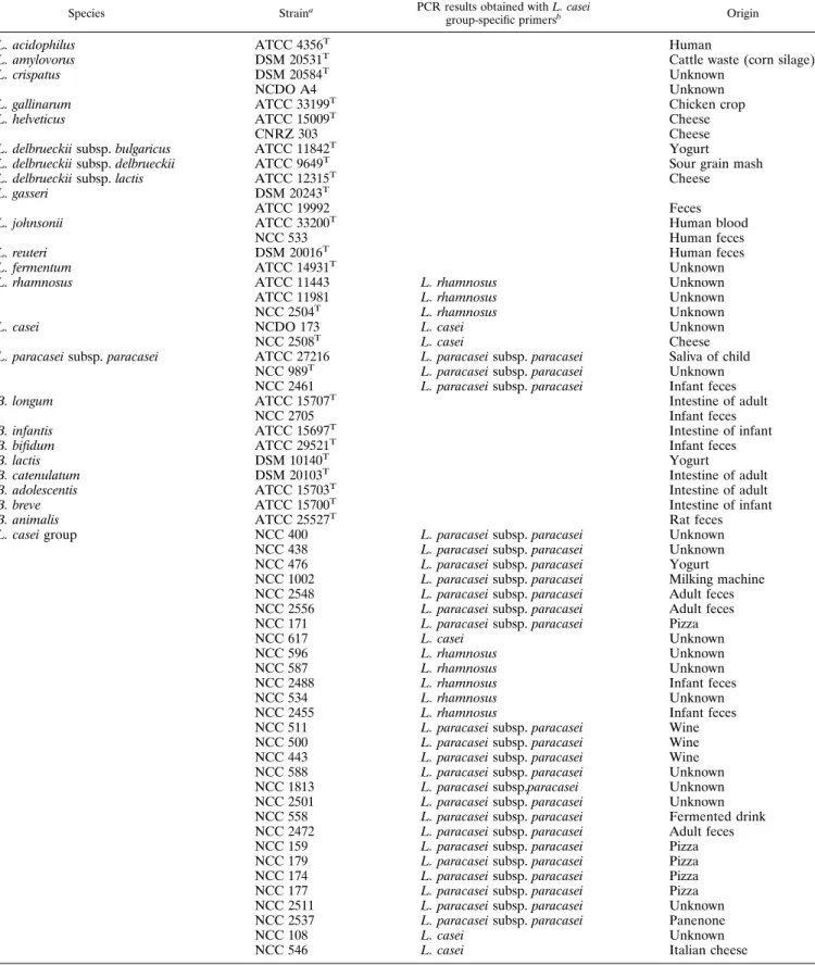
TABLE 1. Bacterial strains used
Species Straina PCR results obtained withL. casei
group-specific primersb Origin
L. acidophilus ATCC 4356T Human
L. amylovorus DSM 20531T
Cattle waste (corn silage) L. crispatus DSM 20584T
Unknown NCDO A4Unknown L. gallinarum ATCC 33199T
Chicken crop
L. helveticus ATCC 15009T Cheese
CNRZ 303 Cheese
L. delbrueckiisubsp.bulgaricus ATCC 11842T
Yogurt L. delbrueckiisubsp.delbrueckii ATCC 9649T Sour grain mash L. delbrueckiisubsp.lactis ATCC 12315T
Cheese L. gasseri DSM 20243T ATCC 19992 Feces L. johnsonii ATCC 33200T Human blood NCC 533 Human feces
L. reuteri DSM 20016T Human feces
L. fermentum ATCC 14931T
Unknown
L. rhamnosus ATCC 11443 L. rhamnosus Unknown
ATCC 11981 L. rhamnosus Unknown
NCC 2504T
L. rhamnosus Unknown
L. casei NCDO 173 L. casei Unknown
NCC 2508T L. casei Cheese
L. paracaseisubsp.paracasei ATCC 27216 L. paracaseisubsp.paracasei Saliva of child NCC 989T
L. paracaseisubsp.paracasei Unknown NCC 2461 L. paracaseisubsp.paracasei Infant feces
B. longum ATCC 15707T
Intestine of adult
NCC 2705 Infant feces
B. infantis ATCC 15697T Intestine of infant
B. bifidum ATCC 29521T
Infant feces
B. lactis DSM 10140T
Yogurt
B. catenulatum DSM 20103T Intestine of adult
B. adolescentis ATCC 15703T
Intestine of adult
B. breve ATCC 15700T
Intestine of infant
B. animalis ATCC 25527T Rat feces
L. caseigroup NCC 400 L. paracaseisubsp.paracasei Unknown
NCC 438 L. paracaseisubsp.paracasei Unknown
NCC 476 L. paracaseisubsp.paracasei Yogurt
NCC 1002 L. paracaseisubsp.paracasei Milking machine NCC 2548 L. paracaseisubsp.paracasei Adult feces NCC 2556 L. paracaseisubsp.paracasei Adult feces
NCC 171 L. paracaseisubsp.paracasei Pizza
NCC 617 L. casei Unknown
NCC 596 L. rhamnosus Unknown
NCC 587 L. rhamnosus Unknown
NCC 2488 L. rhamnosus Infant feces
NCC 534 L. rhamnosus Unknown
NCC 2455 L. rhamnosus Infant feces
NCC 511 L. paracaseisubsp.paracasei Wine
NCC 500 L. paracaseisubsp.paracasei Wine
NCC 443 L. paracaseisubsp.paracasei Wine
NCC 588 L. paracaseisubsp.paracasei Unknown
NCC 1813 L. paracaseisubsp.paracasei Unknown
NCC 2501 L. paracaseisubsp.paracasei Unknown
NCC 558 L. paracaseisubsp.paracasei Fermented drink NCC 2472 L. paracaseisubsp.paracasei Adult feces
NCC 159 L. paracaseisubsp.paracasei Pizza
NCC 179 L. paracaseisubsp.paracasei Pizza
NCC 174 L. paracaseisubsp.paracasei Pizza
NCC 177 L. paracaseisubsp.paracasei Pizza
NCC 2511 L. paracaseisubsp.paracasei Unknown
NCC 2537 L. paracaseisubsp.paracasei Panenone
NCC 108 L. casei Unknown
NCC 546 L. casei Italian cheese
aATCC, American Type Culture Collection; DSM, Deutsche Sammlung von Mikroorganismen; NCDO, National Collection of Dairy Organisms; CNRZ, Centre
National de Recherches Zootechniques; NCC, Nestle´ Culture Collection.L. caseigroup strains were used for species-specific detection.
min. The pellets were dissolved in 4l of distilled water and mixed with 2.4l of loading buffer from the sequencing kit (Thermosequenase, fluorescence la-beled). The cDNA was separated on 8% polyacrylamide–urea gels. Sequencing reactions were conducted with the same primers as those used for the primer extension reactions and detected by using a LiCor sequencer (MWG Biotech).
The synthetic oligonucleotides used (designed in this study) were tuf-a (5⬘-CA
AAACAGTAGTAATAGCTGC-3⬘) and tgf-1 (5⬘-CGAGAAACGTGACCTTT
AC-3⬘).
Amplification with species-specific primers.Amplification reactions were
per-formed with a 50-l (total volume) solution containing 10 mM Tris-HCl, 50 mM
KCl, 1.5 mM MgCl2, 200M each dNTP (Gibco BRL), 10 pmol each of primers
PAR (5⬘-GACGGTTAAGATTGGTGAC-3⬘), CAS (5⬘-ACTGAAGGCGACA
AGGA-3⬘), and RHA (5⬘-GCGTCAGGTTGGTGTTG-3⬘), 50 pmol of primer
CPR (5⬘-CAANTGGATNGAACCTGGCTTT-3⬘), 25 ng of template DNA, and
2.5 U ofTaqDNA polymerase. Amplification reactions were performed by using
a thermocycler (Perkin-Elmer Cetus 9700) with the following temperature pro-files: 1 cycle at 95°C for 5 min; 30 cycles at 95°C for 30 s, 54°C for 1 min, and 72°C for 1.5 min; and 1 cycle at 72°C for 7 min. Primers CAS, PAR, RHA, and CPR were all designed in this study. For routine identification, cells were lysed by using a rapid DNA extraction protocol and were used as direct PCR templates. PCR amplicons were analyzed by 2% (wt/vol) agarose gel electrophoresis in Tris-acetate-EDTA buffer at a constant voltage of 7 V/cm, visualized with
ethidium bromide (0.5g/ml), and photographed under UV light at 260 nm.
Nucleotide sequence accession numbers.The GenBank accession numbers for
the partialtufgene sequences generated in this study are as follows:L.gallinarum
ATCC 33199 (AY372032),L.helveticusCNRZ 303 (AY372033),L.crispatus
DSM 20584(AY372034),L.crispatusNCDO 4(AY373256),L.gasseriATCC
19992 (AY372035),L.johnsoniiATCC 33200 (AY372036),L.johnsoniiNCC
533 (AY372049),L.rhamnosus NCC 2504(AY372037),L.caseiNCC 2508
(AY372038),L.paracaseisubsp.paracaseiNCC 989 (AY372039),L.paracasei
subsp.paracaseiNCC 2461 (AY372040),B.bifidumATCC 29521 (AY372041),
B.longumATCC 15707 (AY372042),B.infantisATCC 15697 (AY372043),B.
catenulatumDSM 20103 (AY372044),B.adolescentisATCC 15703 (AY372045),
B.breveATCC 15700 (AY372046),B.animalisATCC 25527 (AY370920), andB.
lactisDSM 10140 (AY370919). Since theL.gasseri tufsequence extracted from
the ongoing genome sequencing ofL.gasseriATCC 33323 (NZAAAB00000000)
contained various reading errors, we decided to sequence thistufgene again and
deposited it in GenBank under accession number AY372047). The DNA region
located upstream of thetufgene ofL.delbueckiisubsp.bulgaricusATCC
BAA-365 and reported here was deposited in GenBank under accession number
AY372048. The GenBank accession number for thetuflocus sequence ofL.
johnsoniiNCC 533 is AY372049.
RESULTS
Identification and alignment oftuf sequences. The tuf
quences from selected bacterial species for which genome se-quences are publically available were aligned and compared. Four conserved regions were identified, and two pairs of prim-ers (BIF-1–BIF-2 and TUF-1–TUF-2) for amplifying regions of 800 bp were designed. These primers allowed the amplifi-cation oftufsequences from differentBifidobacteriumand Lac-tobacillusspecies. All PCR products were cloned into the vec-tor system pGEMT-Easy. Subsequently, the nucleotide sequence of the inserted DNA fragment was determined by sequencing of three randomly selected clones on both strands for each bacterial species.
A multiple alignment of thetufsequences determined in our laboratory with those retrieved from databases revealed re-gions which were conserved in all strains from the same species but which were variable in other species. A similarity compar-ison of thetufsequences for lactobacilli and for bifidobacteria demonstrated that thetufgenes were highly conserved among allLactobacillusspecies investigated here, with identities rang-ing from 78 to 98% for DNA (reachrang-ing a value of 100% between strains belonging to the same species) and from 76 to 100% for translated gene products (Table 2). Identities among the tuf genes of the bifidobacteria ranged from 89 to 97% (reaching a value of 100% for strains belonging to the same species) for DNA and from 91 to 99% for amino acid se-quences (Table 3). Many of the differences observed in DNA
TABLE 2. Comparison of nucleotide and amino acid sequence identities for EF-Tu among differentLactobacillusstrainsa
Strain
no. Strain
% Sequence identity for strain no.:
1 2 3 45 6 7 8 9 10 11 12 13 1415 16 17 18 19 20 21 22 23 24 1 ATCC 11443 80 79 100 79 93 82 82 79 80 81 81 79 100 96 81 80 93 96 79 93 79 79 78 2 ATCC 15009 87 80 80 96 79 89 89 95 99 8488 95 80 80 88 97 79 80 96 79 88 88 87 3 ATCC 14931 86 83 79 80 80 82 82 80 80 89 82 81 79 81 82 80 80 81 81 80 80 80 80 4ATCC 11981 108 87 86 79 93 82 82 79 80 81 81 79 100 96 81 80 93 96 79 93 79 79 78 5 ATCC 33199 78 90 76 80 78 88 88 95 98 83 88 96 79 78 87 96 78 78 96 78 87 87 87 6 ATCC 27216 98 85 86 96 76 82 82 79 79 80 81 78 93 92 81 80 100 92 78 100 79 79 79 7 ATCC 19992 87 91 8487 8486 99 88 88 87 97 88 82 81 97 88 82 81 89 82 85 85 85 8 DSM 20243 88 91 8488 8487 100 88 88 87 97 89 82 81 97 89 82 81 89 82 85 85 85 9 ATCC 4356 85 97 83 85 89 85 91 92 95 8487 9479 79 87 96 79 79 9479 87 87 87 10 CNRZ 303 87 100 83 87 90 85 91 91 97 8488 95 80 80 88 96 79 80 96 79 87 87 87 11 DSM 20016 88 85 9488 78 86 86 85 8485 87 8481 81 87 8480 81 8480 81 81 81 12 ATCC 33200 85 87 82 85 82 8495 9487 87 88 88 81 81 100 88 81 81 88 81 86 86 85 13 DSM 20584100 87 86 100 78 98 87 88 85 87 88 89 79 79 88 95 78 79 100 78 87 87 87 14NCC 2504 85 96 82 85 90 83 90 90 95 96 8485 85 96 81 80 93 96 79 93 79 79 78 15 NCC 2508 100 88 85 100 78 97 87 87 86 87 88 84100 97 81 80 92 100 79 92 79 79 79 16 NCC 533 88 90 85 88 82 87 98 98 90 90 87 97 88 89 87 88 81 81 88 81 85 85 85 17 DSM 20531 85 97 83 85 90 85 91 92 98 97 85 87 85 96 86 90 80 80 96 79 88 88 88 18 NCC 2461 98 85 86 98 76 100 86 87 85 85 86 84 98 83 97 87 85 92 78 100 79 79 79 19 NCDO 173 100 88 85 100 78 97 87 87 86 87 88 84100 85 100 87 86 97 79 92 79 79 79 20 NCDO A480 91 77 80 89 78 85 85 89 91 79 8480 92 80 8491 78 80 78 87 87 87 21 NCC 989 98 85 86 98 76 100 86 87 85 85 86 8498 83 97 87 85 100 97 79 79 79 79 22 ATCC 9649 85 91 83 85 848489 89 91 91 85 86 85 91 85 89 91 8485 85 84 99 99 23 ATCC 12315 85 91 8485 848488 88 91 91 85 85 85 90 85 89 91 8485 85 84100 99 24ATCC 11842 85 90 83 85 83 8488 88 91 90 85 85 85 90 85 88 91 8485 848499 100
aData in the upper right triangle represent DNA sequence identities of thetufgenes inLactobacillusstrains, while data in the lower left triangle represent deduced
amino acid sequence identities of the corresponding EF-Tu proteins.
sequences among species were silent in terms of their effects on the encoded amino acid sequences. Thus, there were many identical protein sequences, even though their encoding DNAs exhibited substantial divergence.
Alignment of the amino acid sequences deduced from the
tufgenes of lactobacilli and bifidobacteria with other EF-Tu sequences available in databases demonstrated that their gene products are conserved and carry conserved amino acid resi-dues typically found in prokaryotic EF-Tu (18). The portion of thetufgenes of lactobacilli and bifidobacteria described in this study encodes the portion of the EF-Tu protein from residues 104to 335, according to the numbering for E. coli (42). A secondary structure prediction for this portion included the last four ␣helices and two  sheets of domain I, the entire domain II, and the N-terminal portion of domain III, on the basis of the experimentally determined structure of EF-Tu of
E. coli(42). These domains have been determined to play a crucial role in the correct folding of the protein (42); conse-quently, the corresponding sequences have remained highly conserved among eubacterial species.
Phylogenetic analysis.Phylogenetic analysis of thetufDNA
sequences within the generaLactobacillusandBifidobacterium
by neighbor-joining and maximum-parsimony methods showed clear distinct positions of the two genera (Fig. 1). These data were supported by the reported bootstrap values. For com-pleteness, we included in our analysis thetufDNA sequences of other strains belonging to different genera representing the LAB group (e.g., Lactococcus, Streptococcus, and Enterococ-cus). The tree shows two major clusters representing the low-G⫹C-content gram-positive bacteria (genera Lactobacillus,
Lactococcus, Streptococcus, and Enterococcus) and the high-G⫹C-content gram-positive bacteria (genusBifidobacterium). Moreover, a further subdivision into three groups correspond-ing toLactobacillus,Lactococcus-Streptococcus, and Enterococ-cuswas identified.
In order to improve the accuracy of our phylogenetic esti-mation, we traced trees using different methods. The tree to-pologies obtained showed similar hierarchical arrangements (data not shown).
The differentLactobacillusandBifidobacteriumspecies un-der investigation were unambiguously differentiated by a com-parative sequence analysis of a fragment of thetuf gene, as indicated by the phylogenetic tree in Fig. 1. A phylogenetic tree was also constructed on the basis of 16S ribosomal DNA
(rDNA) sequences available in databases. The tree topologies obtained with the 16S rDNA sequences showed a phylogenetic arrangement very similar to that of thetuf-based tree (data not shown). A striking feature of thetufphylogeny is thatL. del-brueckii tuf sequences clustered closely with L. acidophilus
group A tuf sequences, while L. acidophilus group B tuf se-quences clustered more distantly. Interestingly, closely related strains with nearly identical 16S rRNA sequences, e.g., theL.
casei group (L. casei, L. paracasei subsp. paracasei, and L.
rhamnosus), the L. acidophilus B group (L. gasseri and L.
johnsonii), B. animalis-B. lactis, and B. longum-B. infantis, clearly branched separately in thetuf-based tree (Fig. 1).
Most of the base substitutions in thetufgenes were synon-ymous, i.e., did not result in amino acid changes. The synony-mous distances calculated from the nucleotide substitution ra-tios at synonymous positions in the tufgenes were examined for all possible combinations ofLactobacillusand Bifidobacte-rium tuf genes. The relationships between the pairwise dis-tances for the 16S rRNAs and the synonymous disdis-tances for the tuf genes are shown in Fig. 2. There was a significant correlation between the genetic distances for the16S rDNA sequences and those for thetufsequences. In fact, lactobacilli and bifidobacteria showed a correlation coefficient (r) of 0.94 (Fig. 2). The two groups of dots depicted in Fig. 2 represent the
Lactobacillus andBifidobacteriumspecies in accordance with the different G⫹C contents of their 16S rRNAs andtufgenes. Therefore, it can be concluded that the base substitutions oc-curring in the tuf sequences during the evolutionary process render thetufgene a reliable molecular evolutionary clock.
Presence oftufgenes in theLactobacillusandBifidobacterium
genomes.We surveyed all available genomic data for the
pres-ence of thetufgene and its genomic location in various Lac-tobacillusandBifidobacteriumspecies and strains (Fig. 3). InB.
longumNCC 2705, thetufgene is directly downstream of the
fusA gene (translation elongation factor G), the rpsG gene (30S ribosomal protein S7), and therpsLgene (30S ribosomal protein S12) and directly upstream of an unidentified open reading frame (Fig. 3a). PCR amplification with a primer tar-geting a conserved region of the fus gene and the tuf gene yielded the expected amplicons for all nine bifidobacteria tested, indicating the presence of the conservedfus-tuf orga-nization in the Bifidobacteriumspecies tested here (data not shown). The overall organization of thetufgene of
bifidobac-TABLE 3. Comparison of nucleotide and amino acid sequence identities for EF-Tu among differentBifidobacteriumstrainsa
Strain
no. Strain
% Sequence identity for strain no.:
1 2 3 4 5 6 7 8 9 1 DSM 10140 90 90 97 91 91 95 89 91 2 ATCC 15697 92 97 90 95 91 91 96 09 3 ATCC 15707 92 99 90 9490 91 100 89 4ATCC 25527 99 93 93 91 91 95 89 91 5 ATCC 29521 95 96 96 95 90 91 9489 6 ATCC 15703 93 93 93 93 91 91 90 94 7 ATCC 15700 97 95 9498 93 94 91 91 8 NCC 2705 93 99 100 93 96 93 9489 9 DSM 20103 93 91 91 93 92 93 93 91
aData in the upper right triangle represent DNA sequence identities of the
tufgenes inBifidobacteriumstrains, while data in the lower left triangle represent deduced
FIG. 1 . Phylogenetic tree of Lactobacillus EF-Tu and Bifidobacterium EF-Tu based on nucleotide sequence homology. The phylogenetic tree was constructed by the neighbor-joining method. The scale bar represents sequence divergence. In this tree topology, the phylogenetic distance between organisms is the sum of the horizontal segments. L . acidophilus group A includes the species L . acidophilus , L . amylovorus , L . gallinarum , and L . crispatus ; L . acidophilus group B includes only L . gasseri and L . johnsonii . Bootstrap values are reported for a total of 1,000 replicates. tufA and tufB indicate the tufA and tufB genes, respectively. 6913
teria displayed extensive homology with that of thestroperon ofE.coli(15) and enterobacteria (19).
Screening of the sequence data from the ongoing genome sequencing projects forL.gasseriATCC 33323,L.caseiATCC 334, L. johnsonii NCC 533, L. delbrueckii subsp. bulgaricus
ATCC BAA-365, andL.plantarumWCFS1 revealed a similar
tufgenomic location (Fig. 3). Surprisingly, thistufarrangement does not resemble any other so far described fortufloci (3, 5, 6, 14, 15, 19, 20, 21, 38, 54). InL.gasseriATCC 33323 andL.
johnsoniiNCC 533, the tuf gene is located downstream of a metallo-beta-lactamase gene, the rpsO gene (30S ribosomal protein S15), and therpsTgene (30S ribosomal protein S20), while directly downstream of thetuf gene is a transcription regulator trigger factor gene (tig gene), which is followed by genes encoding a Clp protease (clp gene), a GTP binding protein, and a phosphotyrosine protein phosphatase. Both strains showed high sequence identity (from 82 to 96%) within a 9-kb genome fragment. The consensus nucleotide binding domain, Walker motif A (GXXXXGKT), was conserved in the deduced sequences of thetufandclpgenes. An examination of the immediate neighborhood of the Walker sequence indicates that this region is preceded by astrand and followed by an␣
helix, an arrangement which complies with the rules for Walker motif A (34). A comparison with various other Lacto-bacillusspecies showed a very similar genetic organization of thetufregion. Analysis of thetufregion ofL.caseiATCC 334 revealed similar corresponding genes (Fig. 3b). Interestingly, between thetufgene and thetiggene is an insertion of a 4-kb DNA segment that bears a high resemblance to a mobile ele-ment. It contains four genes, the first of which encodes a predicted protein sharing 51% identity with a transposase of
Leuconostoc mesenteroides. The next gene encodes a protein which matches an ATP binding cassette transporter. The fol-lowing genes encode proteins that are identical to a putative transposase ofL.rhamnosusand to an ATP binding protein.
Since the ongoing genome sequencing project for L. del-brueckii subsp. bulgaricus ATCC BAA-365 provided incom-plete DNA sequences for thetufgene, these sequences were completed for this study. The DNA sequence located upstream of thetufgene ofL.delbrueckiisubsp.bulgaricusATCC BAA-365 was generated by PCR with two primers targeting con-served regions in thetufandrpsTgenes. Thetufregion ofL.
delbrueckiisubsp.bulgaricusATCC BAA-365 showed the same gene order as that identified for theL.johnsonii,L.gasseri, and
FIG. 2. Comparison between genetic distances estimated from 16S rRNA and synonymous distances estimated fromtufgenes. The synonymous distances were obtained from the nucleotide sequences of thetufgenes by applying the method of Nei and Gojobori (29). r, coefficient of correlation.
FIG. 3 . Comparison of the organization of the tuf loci in bifidobacteria (a) and in lactobacilli (b). (a) B . longum NCC 2705. The DNA sequence fragments of Bifidobacterium represented here are 4.8 kb. (b) L . casei ATCC 334, L . gasseri ATCC 33323, L . johnsonii NCC 533, L . delbrueckii subsp. bulgaricus ATCC BAA-365, and L . plantarum WCFS1. The putative function of a protein is indicated above or below each arrow. Related proteins are linked by blue shading ( ⱖ 80%) and pink shading ( ⬍ 80%) according to dif ferent amino acid similarities; percent amino acid identities are shown. The length of an arrow is proportional to the length of the predicted open reading frame. Corresponding genes are indicated by the same colors of arrows.
L.caseistrains examined (Fig. 3b). InL. plantarumWCFS1, except for two gene insertions (pmrBgene, encoding a multi-drug resistance efflux pump, and dapAgene, encoding dihy-drodipicolinate synthase) upstream of the open reading frame encoding the metallo-beta-lactamase, the gene order sur-rounding thetufgene was conserved (Fig. 3b). The degree of sequence conservation in thetufregion ofLactobacillusspecies reflects the evolutionary distance separating these different species. Bioinformatic analysis suggested a highly conserved DNA module among the Lactobacillus strains investigated here, consisting of thetuf, tig, clp, and GTP binding protein genes.
Estimation of the numbers oftufgenes in theLactobacillus
and Bifidobacterium genomes. In a Southern hybridization analysis,HindIII-digested genomic DNAs from 13 Lactobacil-lus species and from 8 Bifidobacterium species were probed with thetufgene (Fig. 4a). All investigated strains of lactoba-cilli and bifidobacteria yielded single bands of different sizes (ranging from 1,500 to 8,600 bp in lactobacilli and from 1,100 to 2,100 bp in bifidobacteria), suggesting that only onetufgene is present in all of the genomes. All bacterial DNAs were also digested withBamHI, and the resulting hybridization patterns again yielded only one band for each bacterial species, con-firming the presence of a singletufgene(data not shown). This result was also confirmed by analysis of the incomplete lacto-bacillus and bifidobacterium genomes. Sequence analysis of the entire genomes of L. gasseri ATCC 33323, L. johnsonii
NCC 533, andB.longumNCC 2705 (39) reveals a unique copy of thetufgene, whereas other eubacterial taxa (e.g., enterobac-teria) have a duplication of EF-Tu.
tuf transcription analysis. Northern hybridization
experi-ments were performed in order to determine whether thetuf
gene is cotranscribed with its flanking genes. Total RNA was extracted from L. johnsonii NCC 533 and L. gasseri ATCC 33323 in the exponential and stationary growth phases. A probe corresponding to thetufgene hybridized to transcripts of 4.7, 2.7, and 1.1 kb (Fig. 4b). A second probe, overlapping a gene next to thetufgene that encodes a trigger factor, hybrid-ized to 4.7-, 2.7-, and 1.3-kb transcripts (Fig. 4b). It is highly unlikely that the 1.3-kb transcripts are merely processed prod-ucts of the 4.7- and 2.7-kb transcripts, since only one band was systematically found with theclp gene probe. In fact, a third probe targeting the gene encoding the Clp protease revealed only one transcript, of 4.7 kb, as did a probe targeting the gene encoding the GTP binding protein (data not shown). These results led us to conclude that the transcripts of 4.7 kb corre-spond to thetufgene cotranscribed with thetigandclpgenes
and with the gene encoding the GTP binding protein and that the transcript of 2.7 kb includes thetufandtiggenes.
The genes located upstream of thetuf gene showed tran-scription patterns independent of those of thetufgene. In fact, two probes corresponding to the genes encoding the metallo-beta-lactamase and a hypothetical protein hybridized to a 2.2-kb transcript (Fig. 4b and data not shown). Moreover, these transcripts were present in both exponential and latent growth phases, while thetuf,tig, andclpgenes followed differ-ent kinetics and appeared to be transcribed only during the exponential growth phase. The amount of transcript corre-sponding only to thetufgene is larger than that of the largest transcript species covering the entire tuf gene. These results confirmed what was described earlier for thestroperon ofB.
stearothermophilusandB. subtilis(20). The hybridization sig-nals corresponding to lanes loaded with RNA samples ex-tracted from L. gasseri ATCC 33323 were absent or were weaker than those ofL.johnsoniiNCC 533 because we used probes for the DNA of L. johnsonii NCC 533 which shared variable degrees of similarity with the DNA ofL.gasseri (rang-ing from 70 to 80%) (Fig. 3b).
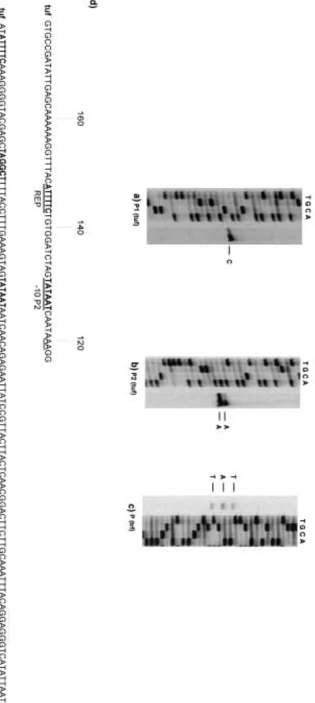
Analysis of the nucleotide sequence of thetuf operon re-vealed several notable features (Fig. 4b). Thetuf operon was delimited at the border by two strong terminator sequences, one located at the 5⬘end of a gene upstream of thetufgene and a second one located at the 5⬘end of the GTP binding protein gene. To map precisely the transcription start sites directly upstream of the tuf gene, primer extension experi-ments were carried out with RNA isolated from exponentially growing L. johnsonii NCC 533 (Fig. 5). Multiple promoter structures have been found preceding thetufgene. In fact, two transcription start sites were identified at ⫺64bp (putative promoter P1) and at⫺119 bp (putative promoter P2) relative to the start site of the coding sequence (Fig. 5a and b). Putative promoter P1 had a⫺10 region (TATAAT) and a⫺35 region (TAGGCT), while putative promoter P2 had a⫺10 box iden-tical to that of putative promoter P1, but no consensus⫺35 sequences were found (Fig. 5d). Notably, two direct repeats (ATTTTC) were detected in the region upstream of the⫺10 box for both start sites and could play a role in the recognition of the RNA polymerase. Primer extension experiments con-firmed that the gene encoding the trigger factor not only is cotranscribed with the tuf gene but also possesses its own promoter. Primer extension experiments located the 5⬘end 47 bp upstream of the start codon of thetig gene (Fig. 5c). An analysis of the putative promoter regions revealed a potential promoter-like sequence having a putative ⫺10 hexamer (TA
FIG. 4. Southern and Northern hybridization analyses. (a) Southern hybridization analysis ofHindIII-digested genomic DNAs of 13 Lacto-bacillus and 8 Bifidobacterium species with the tuf gene fragment as a probe. Northern hybridization analysis of Lactobacillus RNA and transcription unit mapping of thetuflocus. In panel a, the sizes of hybridizing fragments are shown. The tested strain was the type strain (Table 1). In the top portion of panel b, all predicted open reading frames are indicated and are annotated with their database matches. The positions of the probes used in Northern blot experiments are indicated under the gene map. The transcripts are depicted as arrows; the arrows point to the 3⬘end of the mRNA. The length of the arrow is proportional to the length of the mRNA derived from the Northern blots. The estimated sizes of the mRNAs are indicated. Hairpins indicate possible rho-independent terminators. The transcripts are positioned with respect to the gene map. The width of an arrow indicates the relative abundance of the mRNA species. bind., binding. The bottom portion of panel b shows Northern blot hybridization of RNA isolated from lactobacilli with probes corresponding to the open reading frames in the gene map. The sizes calculated for the hybridization signals are provided.
FIG. 5. Primer extension analysis and comparison of the putative promoter sequences of the tuf and trigger factor genes. (a and b) Results of primer extension experiments with oligonucleotides the 5 ⬘ end of the tuf gene. P1, putative promoter P1; P2, putative promoter P2. (c) Results of primer extension experiments with an oligonucleotide targeting the 5 ⬘ end of the trigger factor The lanes in panels a, b, and c indicate the sequencing products for the tuf and trigger factor genes. (d) Overview of experimentally determined 5 ⬘ ends of the tuf gene ( tuf ) and the trigger factor ( trf ). The endpoints of the primer extension products are underlined. Underlined bold type indicates the ⫺ 10 and ⫺ 35 boxes and direct repeats (REP); RBS, Shine-Dalgarno sequences. The start is given at the right end of the sequence. 6918
FIG. 6. Amplification products obtained from thetufmultiplex assay. Lane M, 50-bp DNA molecular marker (Sigma); lane m, 1-kb DNA ladder (Gibco BRL). Lane 1,L.caseiNCC 2508; lane 2,L.paracaseisubsp.paracaseiNCC 989; lane 3,L.rhamnosusNCC 2504; lane 4,L.paracasei subsp.paracaseiNCC 2461; lane 5,L.rhamnosusATCC 11443; lane 6,L.reuteriDSM 2006; lane 7,L.fermentumATCC 14931; lane 8,L.casei NCC 617; lane 9,L.paracaseisubsp.paracaseiNCC 438; lane 10,L.paracaseisubsp.paracaseiNCC 476; lane 11,L.paracaseisubsp.paracaseiNCC 400; lane 12,L.rhamnosusNCC 596; lane 13,L.rhamnosusNCC 587; lane 14,L.rhamnosusNCC 546; lane 15,L.rhamnosusNCC 2488; lane 16,L.rhamnosusNCC 2455; lane 17,L.paracaseisubsp.paracaseiNCC 1002; lane 18,L.paracaseisubsp.paracaseiNCC 2548; lane 19,L.paracasei subsp.paracaseiNCC 2556; lane 20,L.paracaseisubsp.paracaseiNCC 171; lane 21, negative control.
AGAT) and ⫺35 box (TTGTGT) (Fig. 5d). The promoter sequences comply with all requirements ofLactobacillus pro-moter sequences necessary for efficient recognition by the subunit of the RNA polymerase involved in the transcription of housekeeping genes (7).
Primer design and PCR assay forLactobacillusspecies
iden-tification.We designed a single reverse primer (CPR) and three
forward primers (PAR, CAS, and RHA) for the specific detection ofL.paracaseisubsp.paracasei,L.casei, andL.rhamnosus. Ap-plication of the CPR-PAR-CAS-RHA oligonucleotide mixture (Fig. 6) resulted in PCR amplicons of 700, 540, and 350 bp with DNA extracted fromL.caseiNCC 2508, amplicons of 540 and 240 bp with DNA derived fromL.paracaseisubsp.paracaseiNCC 989, but only one amplicon of 540 bp with DNA isolated fromL.
rhamnosusNCC 2504. No PCR product of the above expected sizes was detectable with these primers for any otherLactobacillus
orBifidobacteriumstrains listed in Table 1. The amplicon sizes were in agreement with those expected from the analysis of thetuf
sequences. In fact, the CPR-PAR, CPR-CAS, and CPR-RHA primer pairs must generate amplicons of 240, 350, and 520 bp, respectively. Multiple products are explained by the fact thatL.
paracaseisubsp. paracaseishould generate only two amplicons (240 and 520 bp),L.caseishould produce two PCR products (350 and 520 bp), andL.rhamnosusshould generate only one ampli-con (520 bp).
The identities of the PCR fragments were confirmed by sequence analysis (data not shown). The species-specific primer sets based on the tufgene were also extended to an additional 29 lactobacillus strains (L. casei group strains in Table 1). These strains were originally allocated within theL.
caseigroup on the basis of their fermentative properties and the results of amplified rDNA restriction analysis for Lactoba-cillusspecies identification (51). As shown in Table 1 (L.casei
group strains) and Fig. 5, 21 strains were clearly allocated within the speciesL.paracaseisubsp.paracasei, 2 strains were identified as belonging to the speciesL.paracaseisubsp.casei, while the remaining 6 strains were found to belong to the speciesL.rhamnosus. All strains had been previously charac-terized by ribotyping-hybridization, which produced individual and repeatable profiles for each strain. The heterogeneity among all ribotyping-hybridization patterns clearly demon-strated that all strains investigated with species-specific tuf -based primers were different (data not shown).
DISCUSSION
Significant changes have occurred in bacterial taxonomy since the introduction of molecular techniques. The accurate identification of many bacterial species can be accomplished by reference to rRNA gene sequences (mainly the 16S rRNA gene), which is considered an important molecular marker of modern bacterial taxonomy. The use of other highly conserved macromolecules as evolutionary chronometers might have strong applications in the identification, differentiation, and tracing of bacterial species.
In this study, we have investigated the occurrence of the gene encoding EF-Tu in different species of the genera Bi-fidobacteriumandLactobacillus. The tufgene product brings aminoacylated tRNA molecules to the ribosome. This gene represents an ideal target candidate for diagnostic purposes
because it is highly conserved and ubiquitous in bacteria (26, 27). It has been already applied to infer phylogeny in the generaEnterococcus(18),Mycoplasma(1), andStaphylococcus
(28). In addition, in a very recent study, a comparative analysis of partialtufsequences was evaluated for the differentiation of some Lactobacillus species (4). It fulfills all prerequisites to server as a suitable phylogenetic marker, such as very high genetic stability and a wide distribution (25). This alternative molecular marker might corroborate and help to complete the evolutionary history of various LAB species. In this report, we demonstrated that there is a high correlation between 16S rDNA sequences and the tuf genes of lactobacilli and bi-fidobacteria. The use oftufgenes in LAB species as an alter-native or complement to the 16S rRNA marker mainly sup-ports the phylogenetic relationships that are revealed by the 16S rRNA-based determination of bacterial phylogeny but also provides more detail that can be used to distinguish closely related species and that can be helpful for inferring phylogeny in closely related species (e.g., B. animalis-B. lactis, B. lon-gum-B.infantis, andL.johnsonii-L.gasseri).
Recently, polyphasic taxonomy (48) was recognized by the International Committee on Systematic Bacteriology as a new tool for the description of species and for the revision of the present nomenclature of some bacterial groups. In view of its demonstrated effectiveness, sequence analysis of protein-cod-ing genes (e.g.,tufgenes) as alternative phylogenetic markers could be added to the arsenal of rRNA sequence databases and to the relatively small groEL (16) and recA (9, 22) se-quence databases. It has been shown that species having 70% or greater DNA similarity (at the DNA-DNA hybridization or reassociation level) possess in fact more than 97% 16S rDNA sequence identity (43). Consequently, 16S rDNA sequence analysis might not be an appropriate replacement for DNA reassociation to define closely related taxa. Our results and those of previous studies (4, 18, 19, 26, 27) suggested thattuf
gene analysis also could be a valid tool for inferring relation-ships among closely related bacterial species. The use of thetuf
gene, as well as the recAgene, as a phylogenetic marker for LAB has the advantage that the amino acid sequences from these genes can be used to infer bacterial phylogenies, avoiding the problems associated with rRNAs and the likely overesti-mation of the relatedness of taxa with similar nucleotide dif-ferences, nonindependence of substitution patterns at different sites, and bias derived from different G⫹C contents of micro-organisms (8). Moreover, at the nucleotide level, EF-Tu can tolerate mutations that do not or only slightly alter it. These mutations can provide information about recent evolutionary history which is too recent to be fixed in slowly diverging sequences such as 16S rRNAs (31).
In this study, we demonstrated that the tuf sequence is a valid molecular marker for inferring interspecies relationships. However, the lack oftufsequence variations in strains within the same species showed its inadequancy for any intraspecific relationship analysis (e.g., as a typing tool at the strain level). The analysis of variable regions within the tuf genes of the formerL.caseigroup led us to design a set of species-specific PCR primers. We focused our attention on the establishment of a tufPCR-based assay for the identification of closely re-lated microorganims (e.g., within theL.caseigroup), which so
far cannot be differentiated in a reliable manner by traditional approaches (9).
Thetufgenes have been described to be present in the bacterial genome in various copy numbers. Most gram-positive bacteria carry only onetufgene, and only a few exceptions to this rule have been described (e.g., someEnterococcus[19] andClostridium[40] species). Since it has been postulated that the second copy of the
tufgene (tufB) in enterococci has been generated from a hori-zontal gene transfer event (19), caution should be exercised in the interpretation of bacterial evolution when such events occurred. This is not the case forLactobacillusandBifidobacteriumstrains. We have determined that both the low-G⫹C-content gram-pos-itive bacteria (lactobacilli) and the high-G⫹C-content gram-pos-itive bacteria (bifidobacteria) investigated here contain only one copy of thetufgene.
The tuf genes usually are associated with characteristic flanking genes (5, 24). EF-Tu has been described to be part of either the bacterialstroperon (20) or the tRNA-tufBoperon (3, 5). The arrangement of thetufgene in thestroperon has been described for a variety of bacteria, such asE. coli(15),
Bacillus(20, 21),Streptomyces(54), andEnterococcus(19). The arrangement of thetufgene in the tRNA-tufBoperon has been described forChlamydia trachomatis(5),Thermus thermophilus
(38), andAquifex aeolicus (6), as has that of thetufBgene ofE.
coli (14). It has been postulated that the widespread EF-Tu gene arrangements might argue in favor of their ancient origins (5). All sequenced gram-positive bacteria with a high G⫹C content (e.g., bifidobacteria) contain only a single copy of the
tufgene arranged in a manner similar to that of thestroperon ofE.coli(14). This is the case forB.longum,B.lactis(M. Laloi, personal communication), and all Bifidobacterium species tested in this study.
However, in the available Lactobacillus genomes, the se-quences flanking thetufgenes differ from those of any othertuf
locus described so far. We have found a common genetic map of thetuf region in all five investigatedLactobacillusspecies, includingrpsT,rpsO, metallo-beta-lactamase,tig,clp, and GTP binding protein genes. However, only the EF-Tu-, trigger fac-tor-, Clp protease-, and GTP binding protein-encoding genes seem to constitute a highly conserved module. Functional anal-ysis of these genes seems to corroborate the hypothesis that these genes constitute the same operon. In fact, the trigger factor is a ribosome-associated protein that interacts with the EF-Tu protein and with a wide variety of nascent polypeptides to catalyze their folding (32). Clp ATP-dependent proteases are stress-induced proteins acting to refold or degrade mis-folded or denatured proteins (12). The elongation phase of protein synthesis is promoted by two proteins, EF-Tu and elongation factor G, which binds to the ribosome in its GTP form, hydrolyzes GTP to drive tRNA movement on the ribo-some, and is released in its GDP form. We might speculate that the GTP binding protein following the Clp protease could play the role of elongation factor G. Thetufregion of Lacto-bacillus species displays some features that are not found throughout the bacterial world and that could be of great interest from an evolutionary point of view.
We demonstrated that the EF-Tu-, trigger factor-, Clp pro-tease-, and GTP binding protein-encoding genes are cotrans-cribed and belong to the same transcription unit. Primer ex-tension experiments precisely mapped the start of the
transcript species occurring in thetufoperon. Transcripts de-rived from thetufpromoter and the readthroughtigpromoter were present and covered the entire tuf operon. A similar situation in which the Ef-Tu gene is cotranscribed with flanking genes has been described already forBacillus(20, 21) andE.
coli(3). Thetufgene ofL.johnsoniihas a multiple-promoter structure, which has been described previously for thetufgene ofB. stearothermophilus(20, 21). Transcription directed by a multipletuf promoter structure inStreptomyces ramocissimus
has been demonstrated to be growth phase dependent. Pre-liminary results regarding the tufgene of other LAB genera (e.g.,LactococcusandStreptococcus) show a general organiza-tion common to their loci but not in common with those of the genusLactobacillus. The analysis of the flanking regions sug-gests that in general, the genes surrounding thetufgene have coevolved with EF-Tu. The still relatively small number of LABtufregions available renders attempts to understand the direction of their evolution a challenge. Analysis of a large number of LAB tuf regions may provide important clues to better understanding the biology and the evolutionary history of thetufregion and LAB phylogeny. Thetuflocus of Lacto-bacillusshould undergo complementary studies to clarify the role of the 5⬘-proximal region of the locus in the regulation of expression of the genes and the effects of other possible factors (e.g., growth rate phase) on modulation of the promoter ac-tivity of thetufgene.
In conclusion, in this study we determined the tuf gene sequences of a large number of species of lactobacilli and bifidobacteria, increasing the already existenttufsequence da-tabases of LAB species. We demonstrated a higher distinctness of thetufsequences than of the 16S rRNA sequences and offer a valid molecular marker for inferring phylogeny among closely related taxa (e.g.,L.caseigroup). Moreover, we showed for the first time the genetic organization of thetufoperon of lactobacilli, which has no counterpart in any other known bac-terial genomes so far.
ACKNOWLEDGMENTS
We thank the members of theL.delbrueckiisubsp.bulgaricusATCC BAA-365,L.caseiATCC 364, and L.gasseriATCC 33323 genome sequencing projects, funded by the U.S. Department of Energy Joint Genome Institute, for providing us the sequence of thetufloci. We also thank G. Unnlu and J. Broadbent for making raw genome data for L.delbrueckiisubsp.bulgaricusATCC BAA-365 andL.caseiATCC 334available fortuflocus comparisons (Fig. 3) prior to publication. We are indebted to D. R. Pridmore and M. Laloi (both at NRC) for helpful discussions and comments. Finally, we thank A. Mercenier and F. Arigoni (both at NRC) for constructive and critical reading of the manuscript.
REFERENCES
1. Berg, S., E. Luneberg, and M. Frosch.1996. Development of an
amplifica-tion and hybridizaamplifica-tion assay for the specific and sensitive detecamplifica-tion of
My-coplasma fermentansDNA. Mol. Cell. Probes10:7–14.
2. Bourget, N. L., H. Philippe, I. Mangin, and B. Decaris.1996. 16S rRNA and 16S to 23S internal transcribed spacer sequence analyses reveal inter- and
intraspecificBifidobacteriumphylogeny. Int. J. Syst. Bacteriol.46:102–111.
3. Bremaud, L., C. Fremaux, S. Laalami, and Y. Cenatiempo.1995. Genetic
and molecular analysis of the tRNA-tufB operon of the myxobacterium
Stigmatella aurantica. Nucleic Acids Res.23:1737–1743.
4. Chavagnat, F., M. Haueter, J. Jimeno, M. G. Casey.2002. Comparison of
partialtufgene sequences for the identification of lactobacilli. FEMS
Mi-crobiol. Lett.217:177–183.
5. Cousineau, B., C. Cerpa, J. Lefebvre, and R. Cedergren.1992. The sequence
of the gene encoding elongation factor Tu fromChlamydia trachomatis
compared with those of other organisms. Gene120:33–41.