Loma Linda University
TheScholarsRepository@LLU: Digital Archive of Research,
Scholarship & Creative Works
Loma Linda University Electronic Theses, Dissertations & Projects
3-1-2011
IGF2 Promotes Activation of Estrogen Receptors
in Basal-like Breast Cancer Cells
Angelique Richardson
Loma Linda University
Follow this and additional works at:http://scholarsrepository.llu.edu/etd
Part of theMedical Genetics Commons, and theMedical Microbiology Commons
This Dissertation is brought to you for free and open access by TheScholarsRepository@LLU: Digital Archive of Research, Scholarship & Creative Works. It has been accepted for inclusion in Loma Linda University Electronic Theses, Dissertations & Projects by an authorized administrator of TheScholarsRepository@LLU: Digital Archive of Research, Scholarship & Creative Works. For more information, please contact
Recommended Citation
Richardson, Angelique, "IGF2 Promotes Activation of Estrogen Receptors in Basal-like Breast Cancer Cells" (2011).Loma Linda University Electronic Theses, Dissertations & Projects. 60.
LOMA LINDA UNIVERSITY School of Medicine in conjunction with the Faculty of Graduate Studies
____________________
IGF2 Promotes Activation of Estrogen Receptors in Basal-like Breast Cancer Cells
by
Angelique E. Richardson
____________________
A Dissertation submitted in partial fulfillment of the requirements for the degree of
Doctor of Philosophy in Microbiology and Molecular Genetics
____________________
© 2011
Angelique E. Richardson All Rights Reserved
Each person whose signature appears below certifies that this dissertation in his/her opinion is adequate, in scope and quality, as a dissertation for the degree Doctor of Philosophy.
, Chairperson Daisy D. De León, Associate Professor of Physiology and Pharmacology
Eileen Brantley, Assistant Professor of Basic Sciences and Pharmaceutical Sciences
Carlos Casiano, Associate Professor of Biochemistry and Microbiology
Subburaman Mohan, Research Professor of Medicine, Biochemistry and Physiology
Padma Uppala, Associate Research Professor of Environmental and Occupational Health
ACKNOWLEDGEMENTS
It is my pleasure to show my gratitude to those who made the completion of this dissertation possible. I am indebted and heartily thankful to my Principal Investigator, Dr. Daisy De Leon, whose support and guidance lead me from my first experience with research in the Initiative for Maximizing Student Development (IMSD) program as a junior in college to near completion of the MD/PhD program at LLU. I would also like to thank Dr. Nalo Hamilton and Dr. Willie Davis for all of their proofreading and support in so many ways. I am also grateful to my dissertation committee members who made available their services and advice to me. I thank my parents Mr. Alvin Ellerbee I and Mrs. Audrey Ellerbee and siblings Alvina and Alvin Ellerbee II who have always been a great source of love and strength. Your prayers were invaluable. I owe my deepest gratitude to my husband, Henry Richardson, whose perpetual love and unwavering support have been a pillar of strength every step of the way.
CONTENTS
Approval Page ... iii
Acknowledgements ... iv
Table of Contents ...v
List of Tables ... vii
List of Figures ... viii
List of Abbreviations ...x
Abstract ... xi
Chapter 1. Introduction ...13
Disparities in Breast Cancer ...13
Role of IGF2 in Breast Cancer ...14
Insulin-like Growth Factor Family ...15
IGF2 ...16
IGF1R ...17
IGF2R ...18
Insulin Receptor ...19
Estrogen Receptors ...20
Estrogen Receptor in Cancer ...20
Estrogen Receptor Signaling ...22
Growth Factor Receptor and Estrogen Receptor Cross-talk ...23
Breast Cancer Subtypes ...24
Significance of Studies ...26
Approach of Studies ...27
2. IGF2 Activates ER- and ER- via the IGF1R and IR in Basal-like Breast Cancer Cells ...30
Abstract ...31
Introduction ...31
Cell Culture ...34
Quantitative Real Time PCR ...35
Subcellular Fractionation ...36
Western Blot Analysis ...36
siRNA Analysis ...38
Statistical Analysis ...38
Results ...39
ER- and ER- mRNA in Breast Cancer cells ...39
Phosphorylation and Subcellular Translocation of ER-α and ER-β ...40
IGF-2 Stimulates the Translocation of the ERs through IGF-1R and IR ...44
Discussion ...51
Declaration of Interest...55
References ...56
3. Extracellular Expression of ERs in Breast Cancer Tissues ...62
Introduction ...62
Materials and Methods ...62
Breast Cancer Tissue Specimens ...62
Immunohistochemistry Analysis ...63
Results ...64
Detection of ER-α and ER-β in Breast Cancer Tissues ...64
Discussion ...65
4. Discussion ...67
Conclusions and Future Directions ...67
TABLES
Tables Page
FIGURES
Figures Page
1. Bar Graph of Western Blot Analysis Showing IGF2 Expression in Normal
and Malignant Breast Tissue Samples ...14 2. Schematic Representation of Pre-proIGF2, proIGF2 and mature IGF2 ...17 3. ER-α and ER-β mRNA expressed as fold change of mRNA expression in
MCF-7, CRL-2335 and Hs578T cells ...39 4. Western Blot of Subcellular Localization of Total and Phosphorylated
ER- and ER- in MCF-7 cells ...40 5. Western Blot of Subcellular localization of Total and Phosphorylated
ER- and ER- in Hs578T cells in response to proIGF2 (100ng/mL) treatment ...42 6. Western Blot of Subcellular localization of Total and Phosphorylated
ER- and ER- in CRL-2335 cells in response to proIGF2 (100ng/mL)
treatment ...43 7. Western Blot of Total and Phosphorylated expression of ER- in Hs578T
cells treated with IGF-1R siRNA (A & B) ...44 Western Blot of Total and Phosphorylated expression of ER- in Hs578T
cells treated with IGF-1R siRNA (C) ...45 8. Western Blot of Total and Phosphorylated expression of ER- in Hs578T
cells treated with IR siRNA ( A & B) ...46 Western Blot of Total and Phosphorylated expression of ER- in Hs578T
cells treated with IR siRNA (C) ...47 9. Western Blot of Total and Phosphorylated expression of ER- in
CRL-2335 cells treated with IGF-1R siRNA (A & B) ...48 Western Blot of Total and Phosphorylated expression of ER- in
10.Western Blot of Total and Phosphorylated expression of ER- in
CRL-2335 cells treated with IR siRNA (A & B) ...50 Western Blot of Total and Phosphorylated expression of ER- in
CRL-2335 cells treated with IR siRNA (C) ...51 11.IHC of ERα (A) and ERβ (B) in African-American ER Negative Breast
Cancer Tissues ...64 12.IHC of ERα (A) and ERβ (B) in African-American ER Positive Breast
ABBREVIATIONS
AA African-American BLBC Basal-like Breast Cancer ER- Estrogen Receptor alpha ER- Estrogen Receptor beta ERE Estrogen Response Element FBS Fetal Bovine Serum
DMEM Dubelcco’s Modified Eagle Medium IGF Insulin-like Growth Factor
IGF1 Insulin-like Growth Factor 1 IGF2 Insulin-like Growth Factor 2
IGFBP Insulin-like Growth Factor Binding Proteins IGF1R IGF-1 Receptor
IGF2R/M-6-P IGF-2 Receptor /Mannose-6-Phosphate Receptor IR Insulin Receptor
Kb Kilobase
Kda Kilodalton ProIGF2 Precursor IGF2 MIGF2 Mature IGF2
PAGE Polyacrylamide Gel Electorphoresis PBS Phosphate Buffered Saline
ABSTRACT OF THE DISSERTATION
IGF2 Promotes Activation of Estrogen Receptors in Basal-like Breast Cancer Cells by
Angelique Ellerbee Richardson
Doctor of Philosophy, Graduate Program in Microbiology and Molecular Genetics Loma Linda University, March 2011
Daisy D. De León, Chairperson
The autocrine-paracrine effects of IGF2 are important in the growth and differentiation of normal breast. In breast cancer (BC), IGF2 is initially stimulated by estrogen, progesterone and prolactin to regulate proliferation and cancer progression. These actions are mediated by the IGF-1R and insulin receptor A (IR-A) both members of the tyrosine- kinase receptors family. The activation of Estrogen Receptor (ER) is also very important in BC growth and progression.
As BC progresses to estrogen-independent growth, the IGF-1R and the estrogen receptor (ER) interact in crosstalk mechanisms that are synergistic and results in
enhanced activation of both receptors signaling cascades. This mechanism plays a central role in the transition of estrogen-dependent to estrogen-independent breast cancer (BC) progression.
Basal-like BC (BLBC) is a sub-group of estrogen-independent tumors that have a very aggressive clinical behavior and are resistant to hormone-based therapy resulting in reduced disease-free survival period and increasing the mortality of breast cancer (BC) patients. Our BC research team has elucidated how IGF2 crosstalk signaling results in the activation of ER pathways independent of estrogen. Central to our investigation is how this mechanism is associated to the survival disparity observed among African American
(AA) BC patients. BLBC accounts for nearly 15-20% of all breast cancers, however it represents 45% of all BC observed in AA patients.
Analyses of subcellular compartments, Western-Blot and siRNA demonstrated that IGF2 activates ER-α and ER-β in ER negative BLBC cells Hs578t and CRL-2335. Our studies show that both IGF-1R and IR crosstalk with ER-α and ER-β promoting BLBC progression. This novel mechanism offers new therapeutic targets that will significantly impact treatment and diagnosis of BLBC patients.
CHAPTER ONE INTRODUCTION
Disparities in Breast Cancer
African-Americans (AA) diagnosed with all cancer types have a higher death rate and lower survival when compared to other ethnic groups in the United States (Ward et al. 2004). This health disparity is also present in breast cancer. Breast cancer is the second most common form of cancer in women, however, it is the number one diagnosed cancer in African-American (AA) women (Ward et al. 2004). AA women are more likely to have highly aggressive tumors when compared to their Caucasian (CA) counterparts (Chlebowski et al. 2005).
Reasons for this survival health disparity in AA women are complex. Some factors are social (inequalities in work, wealth, education, standard of living) and some factors are biological such as loss of tumor suppressor gene expression, loss imprinting of insulin like growth factor 2 gene, and BRCA 1 and 2 mutations (Baker, Metzler, and Galea 2005; Fine, Ibrahim, and Thomas 2005; Woolf 2004). This study will evaluate the role of biological factors in the breast cancer survival disparities seen in AA women. There are several findings which suggest that the biological factor IGF2 is a contributor in the survival health disparity seen among breast cancer patients (Elledge et al. 1994)(Fu et al. 2003) (Poola et al. 2005).
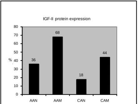
Figure 1. Bar graph of Western Blot Analysis Showing IGF2 Expression in Normal and Malignant Breast Tissue Samples.
Kalla Singh et al. 2010. Differential Insulin-like Growth Factor (IGF-II) Expression: A Potential Role for Breast Cancer Survival Disparity. Growth Hormone & IGF Research, 20:162-170.
Role of IGF-II in Breast Cancer
There is a higher expression of IGF2 in both normal and cancerous breast tissues of AA women when compared to normal and tumor breast tissues from Caucasian women (Figure 1). AA women overexpress several factors that lead to an increased expression of IGF2 which in turn promotes breast cancer cell survival (Singh, et al. 2008)(Singh, et al. 2007). One such factor is mutated p53. Mutated p53 has been shown to lead to an increase in IGF2 expression and promote the upregulation of cell survival pathways leading to increased tumor aggressiveness (Zhang, et al. 1996) (Kanashiro, et al. 2003) (Carey et al. 2006) (Carey et al. 2006). This evidence suggests that there may be a direct correlation between an increased expression of IGF2 and breast tumor
36 68 18 44 0 10 20 30 40 50 60 70 80
AAN AAM CAN CAM
% o f t o ta l
IGF-II protein expression
aggressiveness in AA women which may, in part, explain the decrease in survival seen in these women.
Multiple steps occur as a cell progresses from its normal cell phenotype to its cancerous phenotype. The cancer cell develops the ability to avoid apoptosis, maintain a limitless replicative potential, and provide its own growth signals (Hanahan and
Weinberg 2000). Interestingly, IGF2 has been shown to regulate all of the steps needed in carcinogenesis. The overexpression of IGF2 is involved in the development of various cancers, including breast cancer (Haruta et al. 2008; Honda et al. 2008; Wilkin et al. 2000)(Vu et al. 2003; Wise and Pravtcheva 2006).
Our lab has shown that IGF2 plays a role in promoting breast tumor progression towards aggressiveness. The study demonstrated that the expression of IGF-II promoted tumor growth and metastasis without the requirement of estrogen in nude and Severe Combined Immunodeficient (SCID) mice. This data suggests that IGF2 promotes tumor progression towards aggressiveness by allowing the tumor to grow independent of estrogen in these mice models.
Insulin-like Growth Factor Family
IGF2, a member of the insulin-like growth factor (IGF) family, plays an
important role in the development and maintenance of normal body function (LeRoith et al. 1995). Also, included in the IGF family is the IGF1 ligand, cell membrane receptors insulin-like growth factor receptor 1 (IGF1R), IGF2R and the Insulin Receptor (IR), as well as six IGF-binding proteins (IGFBP-1 through IGFBP-6) (Yu and Rohan 2000). Notably, the IGF family ligands are highly homologous (62%) and both IGF1 and IGF2 are able to bind to each of the IGF family receptors and promote mitogenic and
antiapoptotic actions affecting cell proliferation, differentiation, and transformation (Corps and Brown 1991; Wood et al. 2000; Yu and Rohan 2000).
IGF2
The igf2 gene is located on chromosome 11 (Brissenden, Ullrich, and Francke 1984). From four promoters (P1-P4) multiple mRNA transcripts are produced and alternatively spliced (Sara and Hall 1990)(Yu and Rohan 2000). In the adult, the P1 promoter is responsible for the transcription of IGF2 whereas in the fetus P3 and P4 regulates IGF2 transcription (Sara and Hall 1990). Importantly, P3 and P4 promoters are also activated in breast cancer (Yu and Rohan 2000). Activation of P3 and P4
promoters in cancer cells is a result of the cell reverting back to a less differentiated fetal phenotype.
IGF2 is produced as a prohormone (pre-proIGF2) and it is cleaved post-translationally by proprotein convertase 4 (PC4), a serine protease, to yield several isoforms including proIGF2 (1-156 amino acids, aa) and (1-104 aa) and mature IGF2 (1-67 aa) (Figure 2) (Qiu et al. 2005). ProIGF2 and mature IGF2 are glycosylated to yield isoforms of varying molecular weights ranging from 21 kDa (proIGF2) to 7.5 kDa (mature IGF2) (Vyas, Asmerom, and De Leon 2005). The various IGF2 isoforms have different biological activities. ProIGF2, also known as “big” IGF2, is the predominate form expressed in breast cancer and it has a different mechanism of action when compared to mature IGF2 (mIGF2) (Singh et al. 2008; Singh et al. 2007) (Ellis et al. 1998; Ishida et al. 1995; Rasmussen and Cullen 1998; Singh et al. 2007; Vyas, Asmerom, and De Leon 2005).
Figure 2. Schematic Representation of pre-proIGF2, proIGF2, and mature IGF2. This figure shows the cleavage of pre-proIGF2 into the proIGF2 (1-156 amino acids, aa) and (1-104 aa) and mature IGF2 (1-67 aa) by proprotein convertase 4 (PC4).
Adapted from Miraki-Moud F. et al. J Clin Endocrinol Metab 2005; 90:3819-3823
Insulin-like Growth Factor 1 Receptor
Insulin-like growth factor 1 Receptor (IGF1R) is activated by both IGF1 and IGF2 (Ellis et al. 1998). IGF1R is a tetrameric glycoprotein located on the cell
membrane. The expression of IGF1R is upregulated by estrogens, growth hormone (eg. IGFs), follicle-stimulating hormone (FSH) and glucocorticoids, and downregulated by wild-type p53 and the Wilms tumor protein (Sepp-Lorenzino 1998)(Werner 1998; Werner et al. 1996; Werner et al. 1993). Once IGF1 or IGF2 binds to the IGF1R a conformational change occurs resulting in its autophosphorylation at tyrosine residues in
Mature IGF-II E-domain
Mature IGF-II Mature IGF-II E-domain PC4 enzyme cleavage site PC4 enzyme cleavage site 1 24 aa 1 67 104 156 aa 1 67 aa Signal 180 aa ProIGF-II Pre-proIGF-II Mature IGF-II
the juxtamembrane and flanking regions which serve as docking sites for insulin receptor substrates (IRS) and Shc adaptor proteins. IRS and Shc recruit other proteins, such as Grb2/SOS and phosphatidyl inosital 3’kinase (PI3 Kinase), which can then activate the mitogen-activated protein kinase (MAP kinase) and PI3 kinase pathways. The MAP kinase and PI3 kinase pathways regulate transcription factors that alter gene expression, as well as, control cell proliferation, differentiation, and apoptosis (LeRoith et al. 1995; Yu and Rohan 2000).
The igf1r mRNA expression has been found to be upregulated in most breast cancer cell lines and in over 90% of human breast tumors (Papa et al. 1993; Pekonen et al. 1988). IGF1R expression is important for the inhibition of apoptosis in tumor cells and it has been postulated that the primary role of IGF1R signaling in tumor cells is to maintain tumor cell survival and protection from apoptosis (Baserga et al. 1997) (Dunn et al. 1997; Resnicoff et al. 1995).
Insulin-like Growth Factor 2 Receptor
IGF2 also binds the insulin-like growth factor 2 receptor (IGF2R), and though it is highly debated, evidence suggests that the IGF2R may mediate signaling via a G protein-coupled mechanism (Baserga 1995; LeRoith et al. 1995)(Hawkes et al. 2007). The IGF2R is monomeric and it contains three ligand binding regions in the extracellular domain: one site is for IGF2 binding and the other two sites are for the binding of mannose-6-phosphate (M-6-P) containing proteins (Brown, Jones, and Forbes 2009; Morgan et al. 1987; Oshima et al. 1988) (Oates et al. 1998) (Jones and Clemmons 1995).
Thus, the IGF2R is sometimes referred to as the IGF2R/ mannose-6-phosphate (M-6-P) receptor (Kiess et al. 1989; Vignon and Rochefort 1992).
The IGF2R regulates the bioavailability of IGF2 by leading to its degradation and thus it has been considered as a tumor suppressor (Hankins et al. 1996)(LeRoith et al. 1995; Oates et al. 1998). Thus, loss of this receptor may contribute to various aspects of tumor pathophysiology (Oates et al. 1998). Little is known about the regulation of the IGF2R but it has been suggested that IGFs, epidermal growth factor (EGF) and M-6-P may lead to an increase in its expression (Hoeflich et al. 1996; Jones and Clemmons 1995).
Insulin Receptor
The IR is a cell surface glycoprotein with a heterotetrameric structure made up of two alpha and two beta subunits. The extracellular alpha-subunits consist of the insulin binding domains, and the transmembrane beta-subunits consist of the tyrosine kinase and the phosphorylation sites. The binding of ligand to the IR activates its tyrosine kinase signaling pathway leading to autophosphorylation of the IR and its associated
endogenous substrates such as insulin receptor substrate proteins (IRS-1, -2, -3, and –4), and Shc (White 1998; White and Kahn 1994).
Insulin and IGF2 bind to and activate the insulin receptor (IR) (LeRoith et al. 1995). An IR-isoform, IR-A, has been shown to display high affinity for IGF2. IR-A is encoded by the mRNA lacking exon 11. The binding of IGF2 to IR-A mediates
isoform which includes exon 11, elicits primarily metabolic effects and cell differentiation (Frasca et al. 1999).
IR and IGF1R are structurally homologous and activated by tyrosine phosphorylation. These receptors are implicated in the regulation of cellular
differentiation, mitosis and metabolism (Moxham and Jacobs 1992; White and Kahn 1994). Moreover, the IR and IGF1R both share certain substrates such as Shc and members of the IRS family. Once IRS is phosphorylated then it forms a signaling complex with growth factor receptor binding protein (GRB2), Syp (SH PTP2)
phosphotyrosine phosphatase, and phosphatidylinositor kinase (PI3K) (Lowenstein et al. 1992; Skolnik et al. 1993). IRS-1 couples GRB2 to IR and IGF1R. Shc functions in a similar manner. The coupling of GRB2 to the IR and IGF1R leads to association of the complex with son of sevenless (SOS) Ras GFP/GTP exchanger. This leads to the activation of Ras/mitogen-activated protein kinase (MAPK) pathway. Activation of MAPK pathway regulates cell growth, differentiation, and proliferation in response to insulin, IGF1 and IGF2. The increased expression of IR in breast cancer and breast cancer cell lines has been shown to promote tumor development (Papa et al. 1990) (Milazzo et al. 1992).
Estrogen Receptors
Estrogen Receptors in Cancer
Normal estrogen receptor signaling is necessary for the development and maturation of the mammary gland in contrast aberrant ER signaling is implicated in breast carcinogenesis and progression (Huseby, Maloney, and McGrath 1984; Mueller et
al. 2002) (Herynk and Fuqua 2004). Estrogen receptors are steroid hormone receptors and members of a nuclear superfamily whose subgroup consists of the estrogen receptor alpha (ER-) and estrogen receptor beta (ER-) and its natural ligand estrogen
(Griekspoor et al. 2007)(Arpino et al. 2008; Kuiper et al. 1997; Webb et al. 1999). ER- is located on human chromosome 6 and ER- is found on human chromosome 14 (Enmark et al. 1997; Gosden, Middleton, and Rout 1986; Gustafsson 1999; Ponglikitmongkol, Green, and Chambon 1988). Though they are different gene products, ER-α and ER-β, exhibit 97% and 60% of homology in the DNA- and ligand-binding domains, respectively. Thus, ER- and ER- interact with identical DNA
estrogen response elements (EREs) and exhibit a similar binding affinity for estrogen, but they appear to respond to ligands in a receptor-specific manner (Lazennec et al.
2001)(Kuiper et al. 1997)(Driscoll et al. 1998).
Because of their high degree of homology, initially ER- was thought to represent a level of redundancy in estrogen signaling (Couse and Korach 1999; Ogawa et al. 1999; Ogawa et al. Molecular cloning and characterization of human estrogen receptor β-cx: A potential inhibitor ofestrogen action in human 1998). Current studies indicate the unique but highly controversial role of ER- in breast cancer development and progression (Flynn et al. 2008). Some studies suggest that ER- behaves like a tumor suppressor in the breast and is related to a favorable outcome (Speirs 2002). Other studies suggest that ER- expression is associated with a poorer prognosis (Iwao et al. 2000; Knowlden et al. 2000; Roger et al. 2001; Speirs et al. 1999).
The estrogen receptor subgroup also includes three orphan receptors, estrogen related receptors (ERR ), ERR , and ERR , and estrogen receptor variants
(Griekspoor et al. 2007)(Poola et al. 2005; Vladusic et al. 1998). In this dissertation, the expression and activity of the variant ER-β5 is studied because data suggests that this variant plays a significant role in the development of estrogen-independent breast cancer, and it is expressed at higher levels in African-American women. This increased
expression has been linked to an increase in tumor aggressiveness (Inoue et al. 2000;
Peng et al. 2003)(Poola et al. 2005; Vladusic et al. 1998)(Poola, Abraham, and Liu 2002). The subcellular localization of the ERs is important when considering its role in tumor progression and must be taken into account. Studies demonstrate that when ER- localizes to the mitochondria, in breast cancer cells, it then binds to the mitochondrial genome at ERE sequences leading to the regulation of estrogen’s effects at this organelle, including modulation of calcium influx, ATP production, apoptosis, and free radical species generation (Demonacos et al. 1996)(Chen et al. 2004; Demonacos et al. 1996; Nilsen and Diaz Brinton 2003; Richards et al. 1996; Sekeris 1990; Wang, Green, and Simpkins 2001; Yang et al. 2004). This dissertation shows the ability of IGF-II to promote ER-α and ER-β translocation to the mitochondria in breast cancer cells providing a mechanism in which IGF-II can regulate the mitochondria.
Estrogen Receptor Signaling
There are two signaling mechanisms for the estrogen receptor. The first mechanism discovered is called the classical ER signaling pathway or the
genomic/nuclear-initated signaling pathway (Hall, Couse, and Korach 2001) (Rosenfeld and Glass 2001) (Rosenfeld and Glass 2001). This mechanism involves the passive diffusion of estrogen into the cell and its binding to the ER, leading to receptor
dimerization and translocation into the nucleus. Once inside of the nucleus the receptor binds to the estrogen responsive element (ERE) in the promoters of target genes
(Bjornstrom and Sjoberg 2005)(Hall, Couse, and Korach 2001) (Rosenfeld and Glass 2001)(Rosenfeld and Glass 2001).
Traditionally, the genomic or nuclear-initiated ER signaling has been considered the primary action of the ERs. However, recently plasma membrane estrogen receptors have been discovered (Moriarty, Kim, and Bender 2006). These rapid, membrane-initiated signaling cascades via plasma membrane-associated ERs are referred to as nonclassical or nongenomic/membrane-initiated estrogen receptor signaling (Bjornstrom and Sjoberg 2005; Razandi et al. 2003). This alternate pathway involves the activation of cell surface receptors that lead to the rapid activation of signaling cascades resulting in the regulation of target genes (Kousteni et al. 2001; Levin 2005; Mendelsohn 2000; Xu et al. 2004; Yang et al. 2004)(Rosenfeld and Glass 2001)(Moriarty, Kim, and Bender 2006).
It is important to note that the nuclear-initiated and membrane-initiated signaling pathways do not function independently of one another. Instead, the two pathways converge in the cell to regulate ER-regulated gene expression. Therefore, the distinction between the two signaling pathways is blurred (Bjornstrom and Sjoberg 2005; Moriarty, Kim, and Bender 2006).
Growth Factor Receptor and Estrogen Receptor Cross-talk
Interactions between growth factor receptors and estrogen receptors are important in the development and progression of breast cancer (Arpino et al. 2008). Growth factor receptors have been shown to activate the estrogen receptors (ER) through a mechanism
known as cross-talk which is bidirectional (Hall, Couse, and Korach 2001)(Fagan and Yee 2008; Kahlert et al. 2000; Shou et al. 2004). There are studies showing cross-talk between the ER-α and the Human epidermal growth factor receptor (HER2/neu), the Epidermal Growth Factor Receptor (EGFR) and the IGF-1R (Kato et al. 1995; Lee et al. 1999; Richards et al. 1996; Shou et al. 2004). As the complexities of ER and growth factor signaling cross-talk are being discovered, it is becoming increasingly clear that the treatment of breast cancer will have to involve more than just endocrine therapy that only targets the ER.
Breast Cancer Subtypes
Breast cancer patients are usually grouped together based on their tumor receptor status using immunohistochemistry. Treatment is based on the presence or absence of estrogen receptor (ER) and/or human epidermal growth factor receptor (HER2) (Walker 2008). Patients who have ER/PR positive tumors receive therapy directed towards blocking the activity of estrogen (Tamoxifen and Raloxifen) or stopping estrogen production (aromatase inhibitors) (Carpenter and Miller 2005; Jordan 2007; Reis-Filho and Tutt 2008; Robson and Offit 2007). If the tumor expresses HER2 then a HER2 directed therapy (Trastuzamab/Herceptin) is given to the patient (Albanell et al. 2003; Brenton et al. 2005). Those individuals whose tumors do not express ER, PR or HER2 are usually treated with chemotherapy.
Alternatively, breast cancers may be divided into subgroups using gene
expression profiles (Brenton et al. 2005; Dawood and Cristofanilli 2007; Jumppanen et al. 2007). The subgroups are normal breast-like, luminal, basal-like, and HER2 (ERBB2)
overexpressing (Dawood and Cristofanilli 2007; Jumppanen et al. 2007). These subgroups display distinct differences in biology, behavior, prognosis and response to therapy (Brenton et al. 2005). The normal breast-like subtype consists of normal breast samples and fibroadenomas (benign tumors) (Ihemelandu et al. 2007). The luminal subgroup is mainly comprised of ER positive tumors and the ER negative tumors are in the HER2 overexpressing and basal-like subtypes (Table 1) (Brenton et al. 2005).
Basal-like breast cancers (BLBC) account for nearly 15-20% of all breast cancers (Kobayashi 2008). Fourty-five percent of those diagnosed with BLBC are young (<50 years old), African-American women (Ihemelandu et al. 2007; Reis-Filho and Tutt 2008). There appears to be a positive correlation between Breast Cancer 1 gene (BRCA 1)
mutations, increased expression of mutated p53 and the development of BLBC in AA women (Brenton et al. 2005; Sorlie et al. 2001)(Reis-Filho and Tutt 2008; Turner, Tutt, and Ashworth 2004).
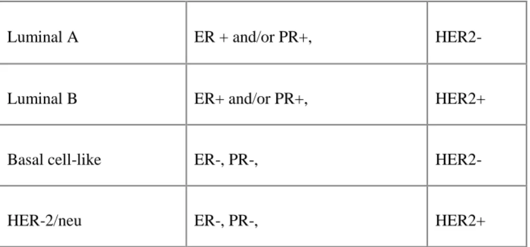
Table 1. Typical Immunohistochemical Classification of Breast Cancer Subtypes
Adapted from Ihemelandu et al. 2007. Journal of Surgical Research. Vol 143, Issue 1, p.109-118.
Luminal A ER + and/or PR+, HER2-
Luminal B ER+ and/or PR+, HER2+
Basal cell-like ER-, PR-, HER2-
The majority of BLBC present with an ER, PR and HER2/neu negative (triple negative) phenotype (Brenton et al. 2005; Jumppanen et al. 2007)(Reis-Filho and Tutt 2008). BLBC has a poor prognosis due to the absence of targeted therapies and its intrinsically aggressive biology (Turner, Tutt, and Ashworth 2004). There is a need to develop targeted therapies for women diagnosed with these tumors.
Significance of the Studies
Breast cancer is the second most common cancer in women (Ward et al. 2004). Basal-like breast cancer (BLBC) accounts for approximately 15-20% of all breast cancers in women, however 45% of these women are AA and this may partly explain why AA women overall have a higher mortality rate from breast cancer (Chlebowski et al. 2005; Finn et al. 2007; Munzone et al. 2006; Szepeshazi et al. 1992)(Ward et al. 2004).
Basal-like breast cancer has a poor prognosis due to the absence of targeted therapies and its intrinsically aggressive biology (Turner, Tutt, and Ashworth 2004). Currently, targeted therapies for breast cancer are aimed at preventing estrogen dependent growth which is not effective for the treatment of BLBC, because it has developed the ability to grow independent of estrogen. Little is known about how these cells grow independent of estrogen. This dissertation addresses this lack of knowledge by
elucidating a mechanism in which these BLBC grow independent of estrogen via growth factor and estrogen receptor cross-talk mechanisms driven by IGF-II.
This study is highly significant and transformational because several observations made in this study will lead to major changes in clinical outcomes for AA women with BLBC.
Approach of Studies
This study was designed to demonstrate the ability of IGF2 to circumvent the requirement for estrogen in breast cancer cells by acting through the IGF1R and IR to phosphorylate/activate ER- and ER-. Central to this study is the association of this mechanism with survival health disparities in AA women with breast cancer. An appropriate cell model had to be selected for demonstrating the role of IGF2 in estrogen-independent activation/phosphorylation of ER- and ER- in breast cancer cells.
The desirable cell model had to have several features important to the study. The cell model had to express ER- and ER- in order to demonstrate the ability of IGF2 to phosphorylate both receptors. The breast cancer cell line, MCF-7, was chosen. MCF-7 is derived from a Caucasian women and it is an ER positive cell line. This cell line
expresses both ER- and ER-. Hs578T cell line is also derived from a Caucasian woman, but it is an ER negative breast cancer cell line, and it was selected to serve as a control.
In addition, this study was designed to elucidate the role of IGF2 in the survival health disparity seen in AA women with breast cancer. Therefore, an ER positive and ER negative breast cancer cell line derived from AA women were selected and compared to the MCF-7 and Hs578T cell lines. CRL-2335 (ER-) and CRL-2315 (ER+) AA cell lines were choosen for this study.
The MCF-7, Hs578T, CRL-2315 and CRL-2335 cells were characterized using Real-time Polymerase Chain Reaction (RT-PCR) to determine the expression of ER- and ER-. RT-PCR revealed that the ATCC designated ER-negative breast cancer cell lines (Hs578T and CRL-2335) express ER- and ER-. Studies show that ER
designation is given to a breast cancer by immunohistochemistry. Breast tumors that express less than 10% of ER- in the nucleus are designated as ER-negative (Nadji et al. 2005; Swain 2001).
Based on these observations focus shifted to the “ER-negative” breast cancer cell lines, and the subcellular localization of ER- and ER-. First, the effect of IGF2 on the activation and subcellular localization of ER- and ER- in these breast cancer cells needed to be determined. ER activation was measured by ER phosphorylation and translocation within the subcellular compartments. In order to determine the role of IGF2 in estrogen-independent activation of ER in the CRL-2335 and Hs578T cells had to be treated with both proIGF2 and mIGF2. CRL-2335 and Hs578T cells were treated with both proIGF2 and mIGF2 at varying concentrations 25, 50 and 100 ng/mL at different time points (0, 10, 15, 20 and 30 minutes) and analyzed for ER activation by measuring ER phosphorylation and translocation. The proIGF2 (100ng/mL) treatment for 20 minutes showed the most effect in the CRL-2335 and Hs578T cell lines. The experiments in this dissertation used this time point and concentration of proIGF2.
The cell lines used for this study were basal-like breast cancer cell lines, 2335 and Hs578T. Molecular gene expression profiling data has subgrouped the CRL-2335 and Hs578T cell lines into the basal-like breast cancer subgroup (Finn et al. 2007). These cell lines were the ideal models for elucidating estrogen independent growth mechanisms in breast cancer cells. Estrogen independent growth is the primary mechanism of growth in basal-like tumors. Moreover, basal-like tumors are more common in AA women. Currently, there is little knowledge regarding how these basal-like tumors grow independent of estrogen. There is an even larger gap in knowledge
concerning what predisposes AA women to these tumors. In this study we have proposed a novel mechanism for how these basal-like breast cancers promote estrogen independent signaling mechanisms in AA women.
This dissertation study was designed to demonstrate the ability of IGF2 to activate the IGF-1R and IR promoting the phosphorylation of ER-α and ER-β via cross-talk mechanisms. This proposed mechanism was based on studies showing cross-talk between the ER-α and IGF-1R (Kato et al. 1995; Lee et al. 1999; Richards et al. 1996). Although many breast cancers express both ER-α and ER-β there is no published study demonstrating a cross-talk between ER-β and IGF-1R. Also, there are no studies in the literature showing a cross-talk between ER-α and/or ER-β and the IR. Nevertheless, we believe there is evidence supporting a crosstalk between the ER and IR based on the following observations. IGF-1R and the IR are 70% homologous, structurally and functionally, and ER may cross-talk with IR in a similar manner as IGF1R (Morrione et al. 1997). This study provides a more comprehensive understanding of this cross-talk pathway thus providing more insight into how to appropriately development more targeted therapies.
CHAPTER TWO
INSULIN-LIKE GROWTH FACTOR-2 (IGF-2) ACTIVATES ESTROGEN RECEPTOR-AND - VIA THE INSULIN-LIKE GROWTH FACTOR-1 RECEPTOR
AND THE INSULIN RECEPTOR IN BASAL-LIKE BREAST CANCER CELLS.
Richardson A. E., #Hamilton, N., *Davis, W., Brito C. and De León D.
Center for Health Disparities and Molecular Medicine Loma Linda University, School of Medicine
Loma Linda, California 92350
*Loma Linda University School of Pharmacy, Loma Linda California 92350
#
University of California Los Angeles School of Nursing, Los Angeles California 90048
Running title: proIGF-2 activates ERand ER
Key words: IGF-2, estrogen receptors, IGF-1R, IR, breast cancer
Corresponding author: Daisy D. De León, PhD
CSP 11012 Breast Cancer Laboratory Loma Linda University School of Medicine Loma Linda, CA 92350
Telephone: (909) 558-4300, ext. 42757 Fax: (909) 558-0475
E-mail: ddeleon@llu.edu Acknowledgments:
This research was supported by 5P20 MD001632, and NIGMS 5R25GM060507 No conflict of interest are declared
Abstract
The estrogen receptor (ER) is a primary target for breast cancer (BC) treatment. As BC progresses to estrogen-independent growth, the IGF-1R and the ER interact in synergistic crosstalk mechanisms which results in enhanced activation of both receptors signaling cascades. Insulin-like growth factor 2 (IGF-2) is critical in BC progression and its actions are mediated by the IGF-1R. Our previous studies showed that IGF-2 regulates survival genes that protect the mitochondria and promote chemoresistance. In this study, we analyzed BC cells by subcellular fractionation, Western-Blot, qRT-PCR and siRNA analysis. Our results demonstrate that IGF-2 activates ER-α and ER-β and modulates their translocation to the nucleus, membrane organelles and the mitochondria. IGF-2 actions are mediated by the IGF-1R and the insulin receptor (IR). This novel mechanism of IGF-2 synergistic crosstalk signaling with ER-α and ER-β can promote estrogen-independent BC progression and provides new therapeutic targets for the treatment of breast cancer patients.
Introduction
Estrogen signaling is mediated through two receptors, ER and ER (Arpino, 2008; Kuiper, 1997; Webb, 1999) When the ER- was discovered it was thought that it
represented a splice variant of ER, however current studies showed that ER- and ER- are different genes with distinct physiological functions (Flynn, 2008). While ERα mediates its effects in the nucleus, ER- is predominantly localized in the mitochondria and regulates mitochondrial gene expression and membrane potential as well as ATP production (Chen, 2004; Demonacos, 1996; Nilsen, 2003; Richards, 1996; Sekeris, 1990;
Wang, 2001; Yang, 2004). Activation of either ER- or ER-, promotes BC cell growth and survival, however, estrogen dependent tumors eventually develop mechanisms to activate ER mediated pathways without the requirement of estrogen (Szepeshazi, 1992).
The development of estrogen-independent BC involves cross-talk between several growth factors and the estrogen receptors (Gee, 2005; Shou, 2004). Insulin-like growth factor-2 (IGF-2) is a growth factor that plays a critical role in organ development, differentiation and metabolism by signaling via both the IGF-1R and the IR-A (Abbas, 2007). Signaling through both receptors also regulates the growth and differentiation of normal breast epithelial cells (Ishida, 1995; Rasmussen 1998; Singh, 2007; Vyas, 2005; Yballe, 1996).
The expression of the IGF-II gene is tightly regulated and inhibited by tumor suppressor genes such as p53, Pten, WT1, GC factor (GCF) and several other oncogenes (Zhang et al. 1996, Toretsky and Helman 1996, Kitadai et al. 1993). Mutations in tumor suppressors contribute to higher IGF-II expression not only for lack of suppression but also for gain of function as mutated p53 that stimulates IGF-II (Zhang et al., 1998). The hormonal regulation of IGF-II is positively regulated by E2, progesterone, prolactin and
GH, all important hormones in normal breast development and in breast cancer progression (Brisken, et al 2002; Goldfine et. al., 1992; Hamelers et.al., 2003). Thus, IGF-II expression is important in normal breast development and increased IGF-II in the mammary gland contributes to tumor formation. In addition to hormonal stimulation and tumor suppressor inhibition of IGF-II, there are many other critical pathways that activate IGF-II that are also important in breast cancer development. The most significant include IGF-II stimulation by oxidative stress, the Wnt-pathway (disrupts eCadherin-catenin),
integrins and mTOR (Erbay et al, 2003;Goel et.al., 2006, Morali et.al., 2001). Mutations in the II receptor (Byrd, et al., 1999) and pTEN also regulate II levels and IGF-II regulates pTEN (Sukmi Kang-Park et al, 2003, Perks et.al. 2007). The extensive regulatory mechanisms involved in IGF-2 expression also includes miRNAs,
methylation, imprinting and other epigenetic alterations present in early development of cancer (Chao et al, 2008, Ge and Chen, 2011)
IGF-2 is highly expressed in breast cancer patients and plasma levels of free IGF-2 directly correlates to BC tumor size (Singer, 2004). Furthermore, transgenic animal models expressing high levels of IGF-2 develop aggressive breast cancer tumors (Pravtcheva, 1998; Pravtcheva, 2003; Bates, 1995).
We have shown that IGF-2 also promotes proliferation, inhibit apoptosis and stimulate the transformation of breast cancer cells (Singh, 2007; Singh, 2008). Our studies also showed that IGF-2 can activate estrogen-regulated genes like survivin, BCL-xl and BCL-2 independent of estrogen through IGF-1R and Insulin receptor. Thus, our particular interest in IGF-2 effects in breast cancer is based on our original observations that IGF-1 was not present in these breast cancer cells and that published studies about IGF-1 analysis by RIA reflected interference with IGFBPs and not IGF-I (De León et al, 1988,1989, 1992). Furthermore, IGF-1 is mainly regulated by GH while IGF-2, as discussed above, is tightly regulated by a wide range of tumor suppressors and hormones that are critical for breast cancer development. Thus, IGF-2 plays a critical role in normal breast differentiation and in the development and progression of breast cancer,
demonstrating that IGF-2 have a broader range of biological functions at the cell or tumor level.
Since IGF-1 activation of the IGF-1R can cross-talk and activate the ER- signaling pathway (Fagan and Yee, 2008) we propose that likewise, IGF-2 can activate the ER-. We also propose that IGF-2 may activate ER- signaling pathway via cross-talk with the IGF-1R. Moreover, since IGF-2 not IGF-1 binds the insulin receptor-A, also a member of the IGF-1R family, we thought that IGF-2 activation of the IGF-1R and IR-A signaling pathways results in the phosphorylation/activation of ER- and ER- in BC cells. Thus, the present study aims to elucidate if IGF-2 binding to both, IGF-1 receptor and the insulin receptor-A results in activation and translocation of the estrogen receptors.
Materials and Methods
Cell Culture
CRL-2335, HS578T, and MCF-7 cell lines were obtained from the American Type Culture Collection (ATCC). The CRL-2335 cell line was derived from a 60-year old African-American (AA) woman and the HS578t cell line was derived from a 74-year old Caucasian (CA) woman and both cell lines are triple negative (ER-,PR-,Her2-). MCF-7 cells were derived from a pleural effusion of a CA woman (69 yo breast cancer patient) and it is used as a positive control for ER-α and ER-β expression. Cultures were maintained at 37C in a 5% CO2 incubator. CRL-2335 cell cultures were maintained in
RPMI 1640 media (ATCC) supplemented with 10mL of penicillin/streptomycin (10,000 units/mL penicillin and 10,000 units/mL streptomycin sulfate, Cellgro), 3ug/ml B-amphotericin, and 10% fetal bovine serum (Hyclone). HS578T cell cultures were maintained in DMEM/F12 media (Cellgro) with 4mM L-glutamine, 1.5g/L sodium bicarbonate, 4.5g/L glucose, 0.01mg/ml bovine insulin (Sigma), 10mL of
penicillin/streptomycin (10,000 units/mL penicillin and 10,000 units/mL streptomycin sulfate, Cellgro), and 10% fetal bovine serum (Hyclone). MCF-7 cells were maintained in DMEM/F12 media (Cellgro) supplemented with 10 ml of 5000 U
penicillin/streptomycin(100 U/ml penicillin and 100 U/ml streptomycin sulfate, Cellgro), 4 mM L-glutamine (Cellgro), 3 µg/ml ß-amphotericin,and 5% fetal bovine serum
(Hyclone).
Quantitative Real Time PCR (qRT-PCR)
Total RNA was extracted using the AurumTM Total RNA Mini Kit (BioRad). RNA integrity was evaluated by UV spectroscopy and RNA Quality Indicator (RQI) values were obtained using the ExperionTM Automated Electrophoresis System (BioRad). Total RNA samples were stored at -80C until assayed. Quantitative reverse-transcription PCR (qRT-PCR) was performed to determine ER- and ER- mRNA expression in MCF-7, CRL-2335 and HS578T cells.
The primer pairs used for ER- (SA Biosciences) are: ER- forward – 5’-ATCCTGATGATTGGTCTCGTCT-3’ ER- reverse - 5’-TCTGGAAGAGAAGGAACCATATCC-3’ The primer pairs used for ER- (SA Biosciences) are:
ER- forward - 5’-GCTCATCTTTGCTCCAGATCTTG-3’ ER- reverse - 5’-GATGCTTTGGTTTGGGTGATTG-3’
cDNA synthesis was performed using the iScript™ cDNA synthesis kit (Bio-Rad) with 400ng total RNA in a 20 L reaction. The reaction protocol is as follows: 5 min 25C, 30 min 42C, and 5 min 85C. Each 50 L qRT-PCR reaction contained 1 µl of
cDNA product from the first-strand synthesis, 25 µl of iQ™ SYBR® Green Supermix (Bio-Rad:100mM KCL, 6 mM MgCl2, 40mM Tris-HCL, pH 8.4, 0.4mM of dATP, dCTP, dGTP and dTTP, iTaq DNA Polymerase 50 U/mL, SYBR Green I, 20mM
Fluorescein), and 300nM of each forward and reverse primer. Before PCR amplification, iTaq DNA polymerase was activated at 95C for 10 minutes. This was followed by forty cycles of PCR amplification: 30 sec denaturation at 95C, 15 sec annealing at 57C, and 90 sec elongation at 72C. Fluorescence was detected at the end of every 72C extension phase. To confirm the presence of specific PCR products, melting curve analyses were performed on the PCR products after the cycling protocol.
Subcellular Fractionation
HS578T, CRL-2335 and MCF-7 cells were plated at a density of 1 x 10⁶ cells/ml and grown to 80% confluency in 10mm culture plates. Cultures were treated with
100ng/ml proIGF-2 (GroPep) or mIGF-2 (PeproTech) in phenol-red free and serum free media for 20 minutes. MCF-7 cells were left untreated. The Proteo-Extract Subcellular Proteome Extraction Kit (Calbiochem) was used according to manufacturer’s protocol to obtain cytosolic, membrane/organelle (mitochondria) and nuclear fractions. All fractions were stored at –80C until assayed.
Western Blot Analysis
The Coomassie Plus Protein Assay Reagent (Pierce Biotechnology) was used to assess protein concentration. Forty micrograms of each protein sample were loaded into a 4-12% polyacrylamide-SDS gradient gel then transferred onto a PVDF membrane
(Invitrogen) using an X-Cell SureLock electrophoretic transfer module (Invitrogen). Membranes were blocked for 1 hour at room temperature with 5% nonfat dry milk in 1X PBS/0.05% Tween. Phosphorylated ER-α was detected by incubating membranes at 4C overnight with either rabbit anti-human pER- (Tyr 537) (Santa Cruz Biotechnology) diluted 1:500 in 3% w/v Bovine Serum Albumin (BSA) or mouse anti-human pER (Ser 118) (Cell Signaling Technology) diluted 1:500 in 5% w/v nonfat dry milk in 1X
PBS/0.05% Tween. Phosphorylated ER-β was detected by incubating membranes with rabbit anti-human pER (Ser 87, 1:1000, Santa Cruz Biotechnology). Unphosphorylated ERs were detected by incubating membranes at 4C overnight in 3% w/v non-fat dry milk in 1X PBS/0.05% Tween with either mouse anti-human ER- (1:1000, Cell Signaling Technology) or mouse anti-human ER- (1:1000, GeneTex). Proteins specific for the membrane/organelle (mitochondrial) and nuclear fractions were used to confirm the integrity of the subcellular compartments: membrane/organelle (mitochondrial) fraction - MnSOD (1:500, BD Biosciences) and nuclear fraction- LEDGF (1:1000, BD
Biosciences). Membranes were then washed 3 times for 5 minutes in 10 ml of 1X PBS/0.05% Tween. The appropriate biotinylated secondary antibody was diluted in 1X PBS/0.05% Tween and incubated with each membrane for 1 hour (rabbit IgG & anti-mouse IgG 1:5000, Amersham), followed by streptavidin-horseradish peroxidase (HRP, 1:2000, Amersham) for 1 hour. Each membrane was washed 4 times for 10 minutes in 10ml of 1X PBS/0.05% Tween. Protein visualization was achieved using enhanced chemiluminescence (ECL) (Pierce) followed by autoradiography with Hyperfilm ECL (Amersham). Band density obtained from WB analysis was quantified using the ChemiImager 4000 (Alpha Innotech Corporation).
siRNA Analysis
CRL-2335 and HS578T cells were plated in 6-well plates at a density of 1 X 10⁵ cells/well and allowed to attach overnight in a 37C 5% CO2 incubator. Cells were grown
to 60 -80% confluency then treated with 60 pmols of either IR (sc-29370, Santa Cruz Biotechnology), IGF-1R (sc-29358, Santa Cruz Biotechnology) or scrambled (sc-Santa Cruz Biotechnology) siRNA in the supplied transfection media and incubated for 9 hours at 37C in a 5% CO2 incubator. Complete media containing 20% FBS was added to the
serum-free media so that the final serum concentration was 10% FBS. The cells were then incubated for 24 hours in a 37C 5% CO2 incubator. Media was replaced with fresh
media followed by an additional 24-hour incubation period in a 37C 5% CO2 incubator.
Afterwards, cells were incubated for 6 hours in serum-free, phenol-free media and then treated with either 100ng/ml of mIGF-2 or proIGF-2 for 20 minutes. Cells treated with scrambled siRNA did not receive treatment with IGF-2. Cellular fractions were prepared as previously described and stored at -200C until assayed.
Statistical Analysis
The data for rtPCR was analyzed by One-way ANOVA with Tukey's Multiple Comparison Post Test using GraphPad Prism software version 5.02 for Windows (GraphPad Software, San Diego, CA). *** - p <0.001, ** - p <0.01.
Statistical analysis for the Western blots scans was performed using one-way ANOVA, SPSS 11.0 software (SPSS, Inc.). Values are expressed as the mean ± SEM of three or more replicate experiments performed in triplicate. Values of p < 0.05 were considered statistically significant.
Results
ER- and ER- mRNA in Breast Cancer Cells
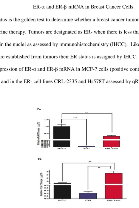
ER status is the golden test to determine whether a breast cancer tumor is sensitive to endocrine therapy. Tumors are designated as ER- when there is less than 10% staining for ER-α in the nuclei as assessed by immunohistochemistry (IHCC). Likewise, when cell lines are established from tumors their ER status is assigned by IHCC. Figure 3 depicts the expression of ER-α and ER-β mRNA in MCF-7 cells (positive control for ER-α and ER-β) and in the ER- cell lines CRL-2335 and Hs578T assessed by qRT-PCR.
Figure 3. ER-α and ER-β mRNA expressed as fold change of mRNA expression in MCF-7, CRL-2335 and Hs578T cells. Figure 3A depicts the expression of ER-α in MCF-MCF-7, CRL-2335 and Hs578T cell lines. Figure 3B shows the expression of ER-β in these same cell lines. Results are expressed as relative fold change (Ct). A p-value of less than 0.01 is represented by ** and a p value of less than 0.001 is represented by ***.
As expected, MCF-7 cells (ER+) expressed significantly higher levels of ER- mRNA (Fig. 3A) compared to the ER- cell lines CRL-2335 and Hs578T. Of note, ER- CRL-2335 cells expressed 30% of the ER-α mRNA detected in ER+ MCF-7 cells. Fig.3 B shows the expression of ER-β mRNA. The highest levels of ER-β mRNA were detected in the CRL-2335 cells (6 fold higher than MCF-7 cells) while the ER- Hs578t cells expressed the lowest levels of ER-β mRNA amongst all 3 cell lines studied.
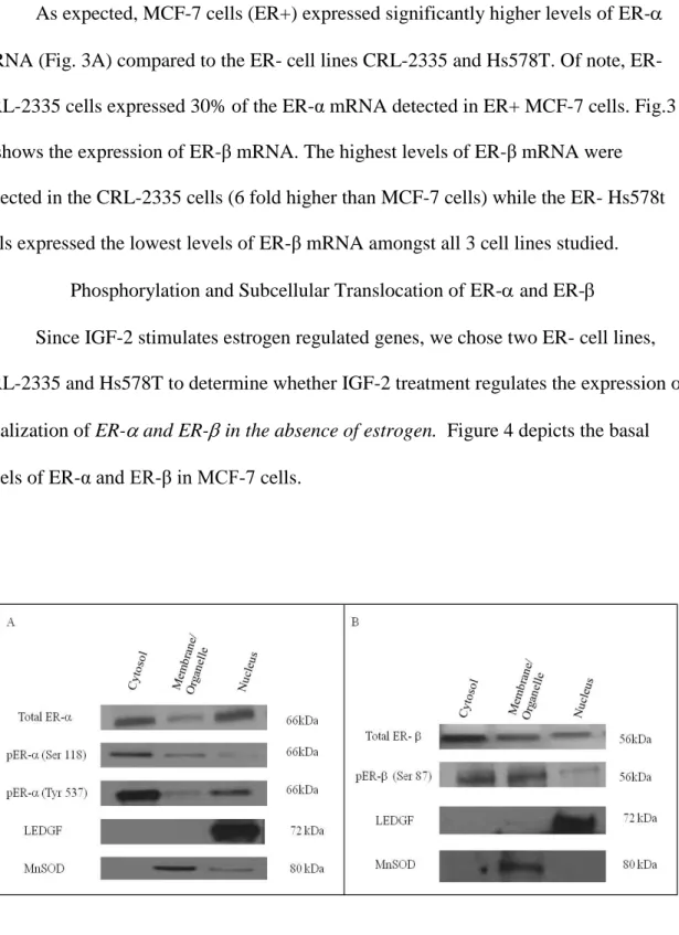
Phosphorylation and Subcellular Translocation of ER- and ER- Since IGF-2 stimulates estrogen regulated genes, we chose two ER- cell lines, CRL-2335 and Hs578T to determine whether IGF-2 treatment regulates the expression or localization of ER- and ER- in the absence of estrogen. Figure 4 depicts the basal levels of ER-α and ER-β in MCF-7 cells.
Figure 4. Western Blot of Subcellular localization of Total and Phosphorylated ER- and ER- in MCF-7 cells. The basal expression of total and phosphorylated ER- and ER- was detected in MCF-7 cells. In order to verify separation and quantify loading of the subcellular compartments LEDGF and MnSOD were used as controls. Western blot is representative of three independent experiments in triplicate.
As expected, there is significant expression of ER- and ER-β protein in the nuclear compartment of MCF-7 ER+ cells. MCF-7 cells were grown in serum-free, phenol-free media without estrogen, yet, ER-α and ER-β are present in cytosolic,
membrane/organelle (mitochondrial) and nuclear fractions. Thus, in MCF-7 cells we can detect activated nuclear and organelle ER-α and ER-β receptors without the requirement of estrogen.
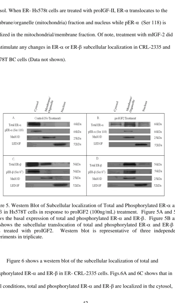
Likewise, the expression of ER- and ER-β in Hs578T and CRL-2335 cell lines was evaluated by Western blot analysis (WB). Fig. 5A and Fig. 5C shows the levels of total and phosphorylated ER- (Fig. 5A) and ER-β (Fig. 5C) in the cell compartment of ER- Hs578t cells. The basal expression of total ER- and ER-α (Ser 118) in ER- Hs578t cells is localized in the cytosol (Fig. 5A). In contrast, ER- is also detected in the membrane/organelle (mitochondrial) fraction in ER- Hs578t cells. No pER- (Ser 87) was detected in the membrane/organelle, suggesting that the ER-β present in the
organelle/mitochondrial fraction is not phosphorylated. Interestingly, when ER- Hs578t cells were treated with proIGF-2, ER- translocates from the cytosol to
membrane/organelle (mitochondrial) fraction and to the nucleus. (Fig.5B). Of great significance, proIGF-II treatment to Hs578t cells stimulates the translocation of ER-β from the cytosol to the membrane/organelle (mitochondriafraction and the nucleus (Fig.5D). ProIGF-2 also stimulated the phosphorylation of the ER- receptor present in the membrane/organelle (mitochondria) fraction pER- (Ser 87). Thus, in ER- Hs578t cells, the basal expression of total and phosphorylated ER-α (Ser 118) is localized in the
cytosol. When ER- Hs578t cells are treated with proIGF-II, ER-α translocates to the membrane/organelle (mitochondria) fraction and nucleus while pER-α (Ser 118) is localized in the mitochondrial/membrane fraction. Of note, treatment with mIGF-2 did not stimulate any changes in ER- or ER- subcellular localization in CRL-2335 and Hs578T BC cells (Data not shown).
Figure 5. Western Blot of Subcellular localization of Total and Phosphorylated ER- and ER- in Hs578T cells in response to proIGF2 (100ng/mL) treatment. Figure 5A and 5C shows the basal expression of total and phosphorylated ER- and ER-. Figure 5B and 5D shows the subcellular translocation of total and phosphorylated ER- and ER- in cells treated with proIGF2. Western blot is representative of three independent experiments in triplicate.
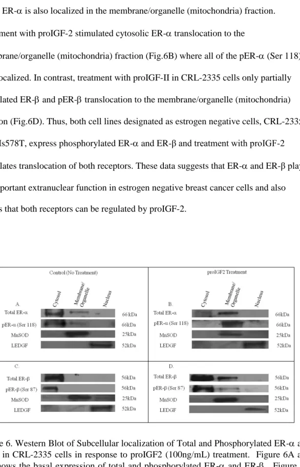
Figure 6 shows a western blot of the subcellular localization of total and phosphorylated ER- and ER-β in ER- CRL-2335 cells. Figs.6A and 6C shows that in
while ER- is also localized in the membrane/organelle (mitochondria) fraction. Treatment with proIGF-2 stimulated cytosolic ER- translocation to the
membrane/organelle (mitochondria) fraction (Fig.6B) where all of the pER- (Ser 118) was localized. In contrast, treatment with proIGF-II in CRL-2335 cells only partially stimulated ER- and pER- translocation to the membrane/organelle (mitochondria) fraction (Fig.6D). Thus, both cell lines designated as estrogen negative cells, CRL-2335 and Hs578T, express phosphorylated ER- and ER-β and treatment with proIGF-2 stimulates translocation of both receptors. These data suggests that ER- and ER-β play an important extranuclear function in estrogen negative breast cancer cells and also shows that both receptors can be regulated by proIGF-2.
Figure 6. Western Blot of Subcellular localization of Total and Phosphorylated ER- and ER- in CRL-2335 cells in response to proIGF2 (100ng/mL) treatment. Figure 6A and 6C shows the basal expression of total and phosphorylated ER- and ER-. Figure 6B and 6D shows the subcellular translocation of total and phosphorylated ER- and ER- in cells treated with proIGF2. WB is representative of at least three separate experiments.
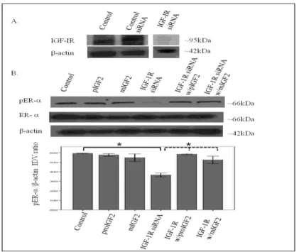
IGF-2 Stimulates the Translocation of the ERs through IGF-1R and IR Since IGF-2 actions are mediated by binding to the insulin-like growth factor-1 receptor (IGF-1R) and the insulin receptor-A (IR-A) we assessed whether proIGF-2 mediates the activation and translocation of the estrogen receptors through IGF-1R, the IR-A or both. Hs578T and CRL-2335 cell lines were treated with IGF-1R and IR-A siRNA to determine which of these receptors were mediating IGF-2 actions. Figure 7 A shows a Western Blot of Hs578t cells treated with IGF-1R siRNA, scrambled siRNA and control and demonstrates that siRNA successfully blocked the expression of the IGF-1R.
Figure 7 A&B. Western Blot of Total and Phosphorylated expression of ER- in Hs578T cells treated with IGF-1R siRNA . Figure 7A shows a WB of Hs578T cells untreated (Control), treated with “scrambled” siRNA (siRNA control) and IGF-1R siRNA. Figure 7B shows WB of ER- expression following proIGF2 and mIGF2 treatment of IGF-1R siRNA treated Hs578T cells. The bar graphs show the results of the densitometry analysis of the WBs phosphorylated ER- normalized to -actin and represent the mean +/- SE of three separate experiments. Solid brackets and * represents values significantly different from control (*p<0.05). Dashed brackets and * represents values significantly different from values between IGF-1R siRNA only treated cells and
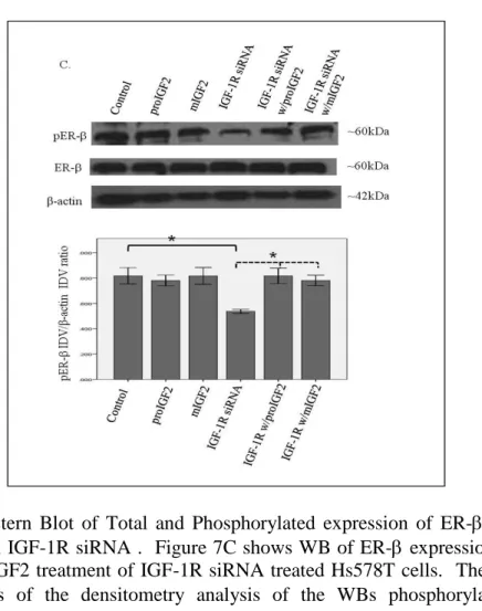
Knock-down of the IGF-1R (Fig. 7B) in the Hs578T cell line significantly (p<.05) reduced the phosphorylation of both pERα and pERβ (Fig. 7B & 7C). Interestingly, when siRNA transfected cells were treated with IGF-2 the levels of pER and pER were restored comparable to the control cells (Fig. 7B & C). Thus, IGF-2 rescued the phosphorylation of pERα and pERβ when IGF-1R was reduced or knocked-down, suggesting that another receptor(s) were mediating IGF-2 signaling in the absence of the IGF1R.
Figure 7 C. Western Blot of Total and Phosphorylated expression of ER- in Hs578T cells treated with IGF-1R siRNA . Figure 7C shows WB of ER- expression following proIGF2 and mIGF2 treatment of IGF-1R siRNA treated Hs578T cells. The bar graphs show the results of the densitometry analysis of the WBs phosphorylated ER- normalized to -actin and represent the mean +/- SE of three separate experiments. Solid brackets and * represents values significantly different from control (*p<0.05). Dashed brackets and * represents values significantly different from values between IGF-1R siRNA only treated cells and IGF-IR siRNA with proIGF2 and/ or IGF1R siRNA with mIGF2 treated cells.
In contrast, no effect in the phosphorylation of ER or ERβ was observed when the IR was successfully knocked-down in these same cells (Fig. 8A-C).
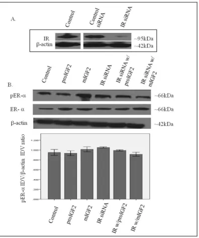
Figure 8 A & B. Western Blot of Total and Phosphorylated expression of ER- in Hs578T cells treated with IR siRNA. Figure 8A shows a WB of Hs578T cells untreated (Control), treated with “scrambled” siRNA (Control siRNA) and IR siRNA. Figure 6B shows a WB of ER- following proIGF2 and mIGF2 treatment of IR siRNA transfected Hs578T cells. The bar graphs show the results of the densitometry analysis of the WBs of phosphorylated ER- normalized to -actin and represent the mean +/- SE of three separate experiments.
These results suggest that IGF-2 mediated phosphorylation and translocation of ER and ERβ is dependent on activation of the IGF-1R but in its absence IGF-2 can activate the IR or other receptor to restore ER- and ER-β phosphorylation.
Figure 8 C. Western Blot of Total and Phosphorylated expression of ER- in Hs578T cells treated with IR siRNA. Figure 8C shows a WB of ER- following proIGF2 and mIGF2 treatment of IR siRNA transfected Hs578T cells. The bar graphs show the results of the densitometry analysis of the WBs of phosphorylated ER- normalized to -actin and represent the mean +/- SE of three separate experiments.
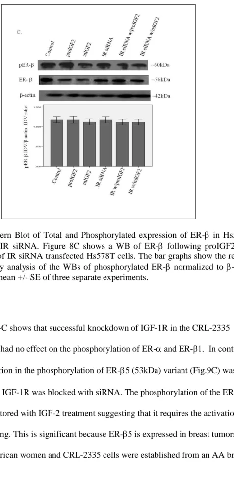
Figure 9 A-C shows that successful knockdown of IGF-1R in the CRL-2335 breast cancer cells had no effect on the phosphorylation of ER- and ER-β1. In contrast, a significant reduction in the phosphorylation of ER-5 (53kDa) variant (Fig.9C) was observed when the IGF-1R was blocked with siRNA. The phosphorylation of the ER-5 variant was not restored with IGF-2 treatment suggesting that it requires the activation of the IGF-1R signaling. This is significant because ER-5 is expressed in breast tumors from African-American women and CRL-2335 cells were established from an AA breast cancer patient.
Figure 9 A & B. Western Blot of Total and Phosphorylated expression of ER- in CRL-2335 cells treated with IGF-1R siRNA. Figure 9A shows a WB of CRL-CRL-2335 cells untreated (Control), transfected with “scrambled” siRNA (Control siRNA) and IGF-1R siRNA. Figure 9B shows a WB of ER- in CRL-2335 cells treated with IGF-1R siRNA. The bar graphs show phosphorylated ER- normalized to -actin and represent the mean +/- SE of three separate experiments.
Notably, when CRL-2335 cells were treated with IR siRNA a significant
reduction in the phosphorylation of ER-, ER-1 and ER- (Fig.10 A-C) was observed. In contrast to the Hs578t BC cells, phosphorylation of ER- and ER-β in the CRL-2335 cells is more dependent on the activation of the IR signaling. Also distinct in the CRL-2335 cells is the expression of the ER- variant which is not detected in the Hs578t BC
Figure 9 C. Western Blot of Total and Phosphorylated expression of ER- in CRL-2335 cells treated with IGF-1R siRNA . Figure 9C shows a WB of ER- in CRL-2335 cells treated with IGF-1R siRNA. The bar graphs show phosphorylated ER- normalized to -actin and represent the mean +/- SE of three separate experiments. Bar graphs with solid brackets and * represents values significantly different from control (*p<0.05). Bar graphs with dashed brackets and * show significantly different (*p<0.05) values between IGF-1R siRNA only treated cells and IGF-IR siRNA with proIGF2 and/ or IGF1R siRNA with mIGF2 treated cells.
Treatment with IGF-2 restored phosphorylation of ER-, ER-1 and ER- in the CRL-2335 BC cells (Fig. 10 B&C). Simultaneous knockdown of IR and IGF-1R was lethal (Data not shown).
Figure 10 A & B. Western Blot of Total and Phosphorylated expression of ER- in CRL-2335 cells treated with IR siRNA. Figure 10A shows a WB of CRL-2335 cells untreated (Control), transfected with “scrambled” siRNA (Control siRNA) and IR siRNA. Figure 10 B shows a WB of total and phosphorylated ER- following proIGF2 and mIGF2 treatment and/or IR siRNA treatment in CRL-2335 cells. The bar graphs show phosphorylated ER- normalized to -actin and represent the mean +/- SE of three separate experiments. Bar graphs with solid brackets and * represents values significantly different from control (*p<0.05). Bar graphs with dashed brackets and * significantly different (*p<0.05) values between IR siRNA only treated cells and IR siRNA with proIGF2 and/ or IGF1R siRNA with mIGF2 treated cells.
Figure 10 C. Western Blot of Total and Phosphorylated expression of ER- in CRL-2335 cells treated with IR siRNA. Figure 10C shows a WB of total and phosphorylated ER- following proIGF2 and mIGF2 treatment and/or IR siRNA treatment in CRL-2335 cells. The bar graphs show phosphorylated ER- normalized to -actin and represent the mean +/- SE of three separate experiments. Bar graphs with solid brackets and * represents values significantly different from control (*p<0.05). Bar graphs with dashed brackets and * significantly different (*p<0.05) values between IR siRNA only treated cells and IR siRNA with proIGF2 and/ or IGF1R siRNA with mIGF2 treated cells.
Discussion
The acquired ability of hormone refractory breast cancer cells to avoid cell death in the presence of anti-estrogen therapy means that the cells have developed the ability to maintain cell survival signaling pathways without the requirement of estrogen. The
mechanism(s) used by breast cancer to acquire this ability are not well-defined. A general consensus in the field is that “cross-talk” mechanisms between growth factor receptors (IGF-1R and EGFR) and estrogen receptors (ER-α) facilitate the progression of breast cancer tumors that become hormone insensitive and refractory (Schiff, 2004). In fact, activation of the IGF-1R (Kato, 1995; Lannigan, 2003) can lead to the activation of ER-α through activation of the mitogen-activated protein (MAP) kinase pathway resulting in the phosphorylation of ER- at Ser 118. Since IGF-2, not IGF-I, is the growth factor expressed in breast cancer cells (Pezzino, 1996), we propose that IGF-2 can maintain survival signals in an autocrine fashion by binding to the IGF-1R and possibly to the IR (LeRoith, 1995; Sciacca, 1999). Furthermore, since many breast cancers express both ER-α and ER-β, we deduced that IGF-2 can bind and activate the IGF-1R and IR leading to the activation of both ER-α and ER-β in BC cells. This IGF-1R and IR/ER cross-talk allows the cells to activate/phosphorylate the ERs without the need of estrogen. Thus, current anti-estrogen therapies would not be able to effectively prevent ER activation in these BC cells.
Binding of IGF-2 to the IGF-1R and IR activates different signaling pathways (Valentinis, 2001; Chen, 2009). Our study suggests that the activation of these different signaling pathways give each receptor a unique role in the phosphorylation of ER-α and ER-β. In ER- Hs578T cells, IGF-1R knockdown decreased the phosphorylation of both ER-α and ER-β. However, treatment with IGF-2 was able to increase the
phosphorylation of both, ER-α and ER-β, back to control levels in IGF-1R siRNA transfected Hs578T cells. Thus, IGF-1R appears to be important in the phosphorylation of ER-α and ER-β in Hs578T cells, however IGF-2 treatment was able to restore ER
phosphorylation presumably by acting through the IR-A. Indeed, Hs578t was the first breast cancer cell line shown to express high levels of IR-A (Sciacca, 1999). In contrast, knocked down expression of the IGF-1R in the ER- CRL-2335 cells had no effect on the phosphorylation of ER-α or ER-β1 but it decreased the activation of the ER-β5 variant. The ER-β5 variant is overexpressed in African-American (AA) women with aggressive BC (Poola, 2005) and this study shows that ER-β5 variant is expressed in the ER- CRL-2335 cells derived from an AA breast cancer patient (Gazdar, 1998). Treatment with IGF-2 was unable to restore ER-β5 phosphorylation in the IGF-1R siRNA transfected ER- CRL-2335 cells. These results suggest that IGF-1R expression is required for the phosphorylation of the ER-β5 variant. In contrast, ER-β5 variant is not expressed in the Hs578T cell line derived from a Caucasian breast cancer patient (Hackett, 1977).
IR knockdown further demonstrated the different roles of IGF-1R and IR in the phosphorylation of ER-α and ER-β. In contrast to the inhibition of the IGF-1R, IR knockdown in Hs578T cells had no effect on the phosphorylation of either estrogen receptor. Of note, IR siRNA transfected CRL-2335 cells showed a decrease in the phosphorylation of ER-α, ER-β and ER-β5. IGF-2 treatment restored the phosphorylation of all, ER-α, ER-β and ER-β5, possibly thorough the IGF-1R. These findings are very significant because they show the unique ability of IGF-2 to activate both the IGF-1R and IR to enhance ER activation and signaling in BC cells. Thus, both, IGF-1 and IR are important targets in the treatment of estrogen independent breast cancers.
Our study also shows that IGF-2 treatment not only phosphorylated α and ER-β but it also stimulated the sub-cellular translocation of both estrogen receptors from the cytosol to the organelles/membrane (mitochondria) fraction. The organelles/membrane