Genome-wide epigenetic profiling of B cell
leukemia and lymphoma
Mohammad Hamdy Abdelrazak Morsy
Department of Clinical Chemistry and Transfusion Medicine
Institute of Biomedicine
Sahlgrenska Academy, University of Gothenburg
Genome-wide epigenetic profiling of B cell leukemia and lymphoma © Mohammad Hamdy Abelrazak Morsy, 2020
Mohammad.hamdy@gu.se
ISBN 978-91-7833-734-7 (PRINT) ISBN 978-91-7833-735-4 (PDF) Printed in Gothenburg, Sweden 2019 Printed by BrandFactory
Dedicated to my beloved mother, my supportive brother and my father’s soul
& to the souls of uncle Magdi and uncle Atef who will always be remembered
lymphoma
Mohammad Hamdy Abdelrazak Morsy
Department of Clinical Chemistry and Transfusion Medicine, Institute of Biomedicine Sahlgrenska Academy, University of Gothenburg
Gothenburg, Sweden
ABSTRACT
Epigenetic modifications, at the level of DNA methylation and post-translational modifications of histone tails cooperatively function in the organization of the genome, and thereby establish the gene expression profiles, phenotypes, and cellular fates. In this work, we investigated the aberrant epigenome in chronic lymphocytic leukemia (CLL) which is one of the most frequent lymphoid malignancies in the west including the Nordic countries. The overall aim of this work is to address the impact of altered epigenetic patterns in CLL on the disease progression with respect to gene expression profile and gain mechanistic insights on the interplay between the different epigenetic mechanisms, such as DNA methylation and histone modifications, in regulating the expression of CLL signature genes. The first study in this thesis aims to investigate the impact of gene body hypermethylation on transcriptional activation which was not completely understood then. Based on our previous MBD seq data (Methyl-CpG-Binding Domain based next generation Sequencing) datasets on CLL samples, of the top differentially methylated genes in CLL compared to normal B cells, we nominated Ten-eleven translocation (TET1) which was shown to harbor hypermethylation at CpG islands within gene body. We found that gene body of TET1 harbors an overlapping cryptic promoter, the transcript of which attenuates the corresponding gene transcription when unmethylated and its hypermethylation in CLL was found to be associated with the overexpression of TET1. The second study aimed at globally mapping the genomic targets of enhancer of zeste homolog 2 (EZH2) the catalytic subunit of Polycomb repressive complex 2 (PRC2) in CLL by chromatin immunoprecipitation followed by sequencing (ChIP-seq) along with its prototypical repressive chromatin feature (H3K27me3). The findings of this study unraveled a non-canonical implication of EZH2 in transcriptional activation apart from PRC2. We show a mechanism by which EZH2 transactivates IGF1R gene in the more adverse CLL subgroups with IGHV mutations (mutated CLL) and how it contributes to activating PI3K/AKT pathway through IGF1R signaling. The third project is somehow pertinent to the aforementioned first study and aims at drawing a more detailed mechanistic link between CpG methylation and transcriptional regulation in terms of the residence of PRC2, as it preferentially locates GC-rich elements. Integration of our previous global methylome datasets in CLL patients and transcriptome analysis by RNA-seq after induction of global demethylation in CLL cell lines has revealed a set of genes that are supposedly prone to hypermethylation within their intragenic regions in CLL, and such hypermethyation is found to be positively correlated with their overexpression in CLL. Out of the top significant genes, MNX1 was selected to probe the mutual exclusivity of PRC2 and intragenic CpG islands and the possible implication of gene body hypermethylation in upregulating MNX1 in CLL through impeding the PRC2-mediated
detailed investigations that look forward to improve the therapy options and accordingly the clinical outcomes in CLL.
Keywords: CLL, PRC2, EZH2, Epigenetics, CpG islands, DNA methylation, ChIP-seq, RNA-seq ISBN 978-91-7833-734-7 (PRINT)
Förändringar i arvsmassan som leder till ändrat uttryck av gener utan att DNA-sekvensen ändras kallas epigenetik och inkluderar metylering av DNA och post-translationella modifieringar av histonernas svansar. Dessa samverkar med varandra och kan leda till ändrat genuttryck och därigenom också en ändrad fenotyp hos celler, utan att DNA-sekvensen påverkats. I denna studie undersökte vi det avvikande epigenetiska mönster som ses vid kronisk lymfocytisk leukemi (KLL), som är den vanligaste lymfoida neoplasi i västvärlden inklusive de nordiska länderna. Det övergripande syftet med denna studie är att avgöra betydelsen av dessa epigenetiska förändringar och deras relation till förändrat genuttryck och sjukdomsprogress som ses vid KLL samt förstå samspelet mellan olika epigenetiska mekanismer, så som DNA-metylering och modifieringar av histoner, och genuttryck.
Den första studien syftade till att undersöka hypermetylerings betydelse vid aktivering av genen Ten eleven translocation 1 (TET1), där våra tidigare resultat från kartläggning av den globala DNA-metylering vid KLL visat att genen var differentiellt metylerad vid jämförelse mellan KLL-celler och normala B-celler. Genen för TET1 visade sig ha en överlappande kryptisk promotor, vars transkript dämpar transkriptionen av TET1 när den inte är metylerad.
Den andra studien syftade till att globalt kartlägga vilka gener som Enhancer of zeste homolg 2 (EZH2), den katalytisk enheten i polycomb repressive complex 2 (PRC2), reglerar genom att analysera väl karakteriserade KLL prover genom användning av kromatin-immunprecipitering riktade mot H3K27me3, följt av sekvensering (s.k. ChIP-seq). På detta sätt identifierade vi en PCR2 oberoende icke-kanonisk funktion hos EZH2 vid transkriptionell aktivering. Sammantaget visar våra resultat att EZH2 transaktiverar IGF1R-genen vilket bidrar till aktivering av PI3K / AKT-signalvägen i den prognostiskt mer ogynnsamma subgruppen av KLL.
Vårt tredje projekt syftade till att ge en mer detaljerad mekanistisk förklaring mellan CpG-metylering och transkriptionell reglering av MNX1-genen och dess roll för bindning av PRC2 till GC-rika element och aktivering av promotorer.
KLL är en obotlig sjukdom men med mycket varierande kliniskt förlopp. Ökad förståelse för de epigenetiska förändringarna som ligger bakom en mer ogynnsam sjukdomsprogression kan bana väg för utveckling av mer riktade behandlingsstrategier med avsikt att reversera dessa förändringar för att förbättra de kliniska resultaten vid KLL.
i
This thesis is based on the following studies, referred to in the text by their Roman numerals.
I. Pradeep Kumar Kopparapu*, Mohammad Hamdy Abdelrazak
Morsy*,Chandrasekhar Kanduri and Meena Kanduri.Gene-body hypermethylation controlled cryptic promoter and miR26A1-dependent EZH2 regulation of TET1 gene activity in chronic lymphocytic leukemia.Oncotarget ,2017, Sep 29; 8(44): 77595-77608.
* Equally contributing first author.
II. Kosalai ST, Morsy MHA, Papakonstantinou N, Mansouri L, Stavroyianni N, Kanduri C, Stamatopoulos K, Rosenquist R, Kanduri M: EZH2 upregulates the PI3K/AKT pathway through IGF1R and MYC in clinically aggressive chronic lymphocytic leukaemia. Epigenetics 2019:1-16.
III. Mohammad Hamdy Abdelrazak Morsy, Mohamad Moustafa Ali, Chandrasehkar Kanduri and Meena Kanduri. DNA methylation at intragenic CpG islands controls PRC2-mediated transcriptional regulation of MNX1 in Chronic lymphocytic leukemia. (Manuscript)
1 INTRODUCTION ... 1
1.1 Genome: The pivotal dimensions of complexity in normal development and diseases 1 1.1.1 Genome and complexity of living organisms ... 1
1.1.2 The problem of cancer ... 2
1.1.3 Cancer epigenome ... 3
1.2 Epigenetics: the flexible mediators of genome dynamicity ... 5
1.2.1 Epigenetics at a glance ... 5
1.2.2 Epigenetics: what stands behind genome’s function? ... 5
1.2.3 Epigenetic patterns establishment ... 6
1.2.4 DNA methylation ... 7
1.2.5 Histone modifications ... 14
1.2.6 Polycomb repressive complex 2 (PRC2) ... 18
1.3 Chronic lymphocytic leukemia (CLL) ... 25
1.3.1 CLL overview ... 25
1.3.2 CLL pathobiology ... 25
1.3.3 CLL epigenome ... 29
2 AIM ... 32
2.1 Specific aims ... 32
3 PATIENTS AND METHODS ... 33
3.1 Patients cohort ... 33
3.2 Methods ... 33
3.3 Laboratory assays ... 36
3.4 Statistical analysis ... 38
4 RESULTS ANDDISCUSSION ... 39
4.1 Paper 1 ... 39 4.2 Paper 2 ... 41 4.3 Paper 3 (manuscript) ... 43 5 FUTURE PERSPECTIVES ... 45 ACKNOWLEDGEMENT ... 46 REFERENCES ... 48
iii
5-cC 5-carboxy Cytosine
5-fC 5-formyl Cytosine
5-mC 5-methyl Cytosine
AID Activation induced deaminase
BER Base excision repair
CATCH-IT Covalent attachment of tags to capture histones and identify turnover
CGI CpG island
CIMP CpG methylator phenotype
CLL Chronic lymphocytic leukemia
cPRC1 Canonical polycomb repressive complex1
DNMT DNA methyl transferase
dsDNA Double stranded DNA
EED Embryonic ectoderm development
EZH1 Enhancer of zeste homolog 1 EZH2 Enhancer of zeste homolog 2
GEP Gene expression profile
GRO-seq Global Run on-sequencing
HCP High CGI-promoter
HDAC Histone deacetylase
HMT Histone methyl transferase
HP1 Heterochromatin protein 1
HSC Hematopoietic stem cell
IGHV Immunoglobulin heavy chain variable gene JARID2 Jumonji and ARID domain containing protein 2
JmjC Jumonji C domain
M-CLL IGHV-mutated CLL
mESC Mouse embryonic stem cell
MLL Mixed lineage leukemia
ncPRC1 Non-canonical polycomb repressive complex1
PcG Polycomb group protein
PCGF Polycomb group ring-finger domain proteins
PCL Polycomb like protein
PHD Plant Homeodomain
PRC1 Polycomb repressive complex PRC2 Polycomb repressive complex 2
PRE Polycomb response elements
PWWP Pro-Trp-Trp-Pro domain
RNAPII RNA polymerase II
RYBP Zinc finger and YY1 binding protein
SAM S-adenosyl methionine
SET Su(var)3-9, Enhancer-of-zeste and Trithorax SNV single nucleotide variation
SUZ12 Suppressor of zeste 12
SWI/SNF Switch/ sucrose non-fermentable
TDG Thymine DNA glycosylase
TET Ten eleven translocation
TrxG Trithorax group protein TSS Transcriptional start site
U-CLL IGHV-unmutated CLL
1
1 INTRODUCTION
The rise of high throughput and massively parallel technologies has revolutionized the field of genomics and epigenomics. This revolution has in turn enhanced understanding of genome organization and the mechanisms orchestrating the interplay between the genome and epigenome towards driving both normal and diseased phenotypes.
The emergence of regulatory genomic elements and the expansion of the haploid genome size in higher organisms entail highly sophisticated mechanisms, so that they govern and regulate the genome function in establishing gene expression profiles in response to either normal cellular conditions or stochastic environmental circumstances. Along with the sequence-based intrinsic features of the genomic domains, epigenetic mechanisms serve an integral part in this regard without altering the genomic sequences. Epigenetic mechanisms involve chemical modification of DNA bases such as cytosine methylation and histone post-translational modifications, which in harmony with long non-coding RNAs (LncRNAs) organize the eukaryotic, particularly the mammalian genome into functionally distinct compartments, thereby control gene expression in a spatial and temporal manner.
Alterations that encounter any of these mechanisms have extensively been reported in many diseases including cancer. Aberrant epigenetic patterns have been shown to adopt the mutational load and encompass many notorious cytogenetic lesions that arise with the progression of malignant diseases, suggesting that cancer is more than a disease of mutations. Altered DNA methylation has been well addressed in cancers and viewed to cooperate with abnormal histone modification pattern to foster the acquisition of the malignant hallmarks.
By means of high through-put sequencing techniques and with integration of current along with previously published global datasets by our team, this work mainly aims at identification of the altered epigenome in terms of DNA methylation and polycomb repressive complex 2 (PRC2) deregulation in chronic lymphocytic leukemia (CLL). In addition, we aimed at dissecting the crosstalk between these epigenetic mechanisms in reprogramming and altering gene expression. More specifically, the main concern of this work is drawing more detailed mechanistic links between DNA methylation and PRC2 in regulating sets of biologically relevant genes that are related to the proliferative pathways such as IGF1R signaling, PI3K/AKT pathway as well as homeobox-related genes and how that contributes to CLL pathogenesis.
Comprehensive understanding of the altered aspects of epigenetic mechanisms in CLL and other cancers in general, along with translational medicine may pave the way for improving and elaborating promising and more efficient lines of therapy and improve the patients’ prognosis.
1.1 Genome: The pivotal dimensions of complexity in normal
development and diseases
1.1.1 Genome and complexity of living organisms
The last two decades following the completion of Human Genome and ENCODE projects, have witnessed a stunning paradigm shift. It was previously believed that the living organisms are hard-wired by their genes, as implied by the central dogma of molecular biology.[1] The central dogma defines life or a biological phenomenon as a flow of genetic information embedded in the DNA into a messenger RNA (mRNA), which is in turn translated into a biologically functional protein that contributes to manifestation of phenotypes.
Throughout the evolution of the living organisms, a wide range of biological diversity and scalable broad spectrum of organismal complexities are observed from symbiotic bacteria to humans.[2] The intriguing question is that, what stands behind and what confers such diverse biological complexities?
It was believed until the early 1970s of the last century that the organismal complexity is scaled by the cellular contents of DNA (the C-value).[3] The notion that the C-value failed to show a
significantly consistent relationship to biological complexity raised the C-value paradox.[2, 4-7] The C-value paradox was rationally resolved by the fact that the C-value is not more than a crude measure of cellular DNA content and is not normalized to varying degrees of ploidy and the emergence of non-protein coding elements into the genomes of higher organisms.[8, 9]
Another intriguing paradox in molecular biology is the inconsistent relationship between developmental complexity and the number of protein-coding genes, referred to as the G-value paradox.[10] According to the central dogma, one would expect that the organismal complexity is supposedly scaled up by the increase of the protein-coding sequences. However, one striking example amidst myriads, has casted a remarkable skepticism on this tenet; the mean number of protein-coding genes in human and in the microscopic nematode C.elegans is 20,000 genes.[2] Moreover, the human genome project revealed that the sequences of DNA that are synonymous with functional proteins represent less than 2% of the whole haploid genome, while the rest was termed as “Junk DNA” or “Selfish DNA”.[8, 9] After the revolution in the field of genomics, the Junk DNA turned out to be biologically relevant to the development of the higher organisms. This 98% of the human genome is broadly partitioned into cis-acting regulatory elements and non-protein-coding sequence that are not inert and code for trans-acting non-coding RNAs (ncRNAs), which in turn fulfil a broad spectrum of cellular and developmental functions. Together, these elements cooperatively contribute to the establishment of the highly sophisticated architecture and dynamic organization of the genome, and thus control gene expression in a spatial and temporal manner and account for the gene product diversity throughout the multicellular ontogeny.[2] The non-random overrepresentation of non-genic elements including introns and intergenic elements would evoke our thinking of the mechanisms beyond the intrinsic sequence properties and the information stored in our genomes, and how these mechanisms shape our genomes and accordingly control the flow of genetic information towards a stable phenotype.[2, 11] These mechanisms are known as epigenetic mechanisms which control, influence and get influenced by the genome without altering the DNA sequence, and thus account for cellular memory upon reaching the terminal stage of differentiation.[12]
By virtue of next generation sequencing and massively-parallel technologies, our comprehension of biological and physiological processes and the development of diseases such as cancer, has experienced a transition from a gene-centered to a genome-wide approach. Also, as best expressed by Adrian Bird[12], the revolution in the field of epigenetics has offered an antidote to the tenet that a living organism is equal to the summation of its genes; an organism is rather a function of its genome.
1.1.2 The problem of cancer
During the multistep process of cancer development from normal cellular incipient, new biological capabilities are acquired, following the cumulative disruptions to normal growth controls. These new traits comprise the hallmarks of cancer and confer the survival and aggressiveness of cancer through providing the greatest clonal advantages, that in turn qualify the progression of the neoplastic cells that are ultimately becoming malignant tumor.[13] These hallmarks were first introduced in the year 2000 by Douglas Hanahan and Robert A.Weinberg.[14] This proposition constituted an organizing principle and a conceptual framework for understanding the diversity and complexity of human neoplasms.[15]
These hallmarks include six biological traits: 1. Sustainable proliferative
signaling
2. Evading growth suppressors 3. Resistance to cellular death
4. Enabling replicative immor-tality
5. Induction of angiogenesis 6. Activation of invasion and
3
A decade later and with the conceptual progress and accumulation of a wealth of knowledge, Douglas and Weinberg introduced an ancillary proposition which implied additional two hallmark traits. Reprogramming of energy metabolism and evasion of immune-mediated destruction are two emerging capabilities have been added to the list of cancer hallmarks.
The fairly intriguing question is: what underlies the acquisition, preservation and fostering of these traits? And which physiological niche would embrace the clonal selection of the competent neoplastic cells?
Genomic instabilities and inflammation underlie these hallmarks through generating genetic diversity, thereby expedite their acquisition and foster their functions. Both are considered as enabling characteristics that account for the Darwinian selection of these new traits, thus provide the greatest clonal advantages for survival and generation of macroscopic tumors. The dimensions of complexity of cancers is superimposed by their ability to recruit apparently normal cells that help in establishing the so called tumor microenvironment which constitutes as a physiological niche encompassing the acquisition and contribution of the hallmarks traits in tumor progression.[16, 17]
Throughout the course of tumorigenesis, the cells destined to become malignant experience suc-cessions of drastic conditions elicited by the natural barriers which are hard wired into the cells to impede outgrowth of either pre-neoplasms or frank neoplastic lesions and functions in various combinations of tumor suppressive modes of action.[15] Neoplastic cells relentlessly invest the malignant capabilities they have been acquiring to circumvent such anticancer defense activities. Therefore, the developing neoplastic cells can overcome such bottle neck by compromising the gene expression profile (GEP) for the tumor advantage.[18, 19]
By means of epigenetic mechanisms, the expression of the components of surveillance system that ensure genetic and genomic integrity is altered [20-22]. Most importantly gate-keepers and care-takers that evoke genomic maintenance and DNA repair or otherwise trigger senescence and/or apoptosis of genetically damaged cells are compromised; thereby cancer increases the mutational load and guarantees the accumulation of genetic aberration and cytogenetic lesions that are well suited as vehicles of persistent phenotypical oncogenic changes and candidates for clonal selection.
The dynamic reprogramming of gene expression profile and the shaping of malignant phenotype is a culminate result of cooperative integration between altered genetic and epigenetic mechanisms like methylation of DNA or post-translational modification of histones that foster acquisition of hallmark traits.[13, 23, 24] Here comes the significance of epigenetic mechanism in providing alternative and flexible, yet persistent ways to regulate and acquire stable oncogenic traits throughout multiple cellular division cycles that would potentially boost the neoplastic phenotypes.[25]
1.1.3 Cancer epigenome
During the last century, cancer has generally been viewed as a genetic disease; the general tenet that was adopted then implied that mutations are the driving forces that initiate the neoplastic transformation.[13] However, it has become clear that rate of mutations, copy number alterations and insertion/deletions take place at relatively low frequencies; thus rendering them inefficient means of initially driving neoplastic transformation.
Considering the local influence of base composition on single nucleotide variation (SNV), chromatin structure, replication timing and regional effects of sequence composition and mutation rates across the genome vary markedly.[26] Notably, the mutational load in cancer can by no means be inferred directly from the number or frequency of observed mutations, without taking the number of cell divisions that have occurred or the influence of epigenetic mechanism on the rate of generation and repair of such altered genetic lesions into account.
Epigenetic mechanisms have an influential role in mutational rate in several ways. Most importantly, certain subsets of colorectal cancers and glioblastomas show epigenetically-addictive phenotypes, wherein the incipient pre-neoplastic cells exhibit a predisposition towards
exceptionally high frequency of cytosine methylation at CpG-rich promoter of certain genes the silencing of which is of a molecular and biological relevance to the respective malignancies. This phenomenon is referred to as CpG island methylator phenotype (CIMP) [27, 28], where the exceeding frequency of promoter methylation is neither stochastic nor spontaneous, but instead an epigenetically attributed and highly coordinated event. Such predisposition towards epigenetic addicted phenotypes may be a consequence of germline variation that increases the likelihood of cancer development.[29, 30] Another facet of epigenetic addition is exhibited in aberrant chromatin organization that is attributed to gain-of function of chromatin modifiers as evident in Polycomb repressive complex addiction in diffuse large B cell lymphoma (DLBCL) [31-33] and DOT1L-addiction in subsets of mixed lineage leukemia (MLL) which is associated to altered regulation of Homoebox-related genes.[34-36]
Also, cytosine methylation in the context of CIMP has been shown to play a crucial role in the boosting the rate of C-to-T mutation within CpG islands up to tens of folds of magnitudes.[26] The C-to-T mutations during DNA replication in highly proliferative cells result in T:A substitution lesion which are not recognized by the DNA repair machinery. These kind epigenetically-attributed mutations constitute about 25% of all TP53 mutations in human cancers.[37]
Altogether, the aforementioned facts support the contribution of epigenetics to cancer mutational load and that epigenetic alterations might act upstream to foster the acquisition of the cytogenetic lesions throughout the course of tumorigenesis and maintain them to boost the malignant phenotypes and achieve the best clonal traits.[13, 38]
It has become evident that both genome and epigenome influence each other as a mean of high-fidelity and tight control of gene expression profile and fate specification. The next chapter explains the different epigenetic mechanisms at the levels both DNA and histone modification, the regulation of epigenetic marks at the levels of writing and erasure, and the interaction between the different epigenetic patterns and show that they are not operating in isolation, but rather within a an integrative network of mutually exclusive, reinforcing and counteracting signals.
5
1.2 Epigenetics: the flexible mediators of genome dynamicity
1.2.1 Epigenetics at a glance
The term “Epigenetics” was first coined in 1939 by Conrad Hal Waddington, nevertheless, so many definitions have been suggested for epigenetics thereafter.[12] According to Waddington, and apart from classical genetics, epigenetics concerns about how genotypes manifest phenotypes. In his own words, epigenetics is defined as “the branch of biology which studies the causal interactions between genes and their products, which bring the phenotype into being”.[39, 40] The definition of epigenetics remained subject of debate, until the mid-1970s, when the field of epigenetics was revived once again by Arthur Riggs and coworkers.[41] They could reformulate the definition and reintroduce the biological importance of epigenetics, so that the term refers to the study of mitotically and/or meiotically heritable changes in genetic functions beyond sequence alteration of DNA itself. More specifically, epigenetics describes the interplay between the reversible chemical modifications of histone tails and/or DNA without changing the genetic information, and chromatin-associated proteins, in regulating chromatin structure and transcriptional programs of the cells.[42]
In brief, epigenetics is inheritance, but not as we know. It is suggested to be described as “the soul” of the genome that organizes its function in response to the environmental conditions or developmental demands, thereby coins the cellular identity and guarantees the phenotypic distinctiveness of each type of the genetically identical cells that comprise a multicellular organism.
1.2.2 Epigenetics: what stands behind genome’s function?
The succession of the technological advances in the post-sequencing era and the rise of the high throughput technologies have revolutionized the field of epigenetics and offered several genome-wide-based lines of investigations. By virtue of such technological revolution, the functions and distribution of several epigenetic marks over the genome have been accurately mapped and comprehensively understood. The different epigenetic modifications at the levels of DNA and histones modification are considered as surrogates of the functional genomic elements, structural organization of chromatin domains and transcriptional potentials across the chromatin domains.[43]
As explained in section1.1, the evolution of the higher organisms had experienced genomic size expansion, as a consequence of the emergence of non-protein-coding elements including repetitive elements which are mainly transposons-derived, intergenic regulatory elements and intragenic introns. This latter set of elements largely accounts for the expansion of eukaryotic proteomes by alternative splicing.[44] Such complexity of the eukaryotic genomes, in particular the mammalian genomes, entails highly sophisticated patterns of organization, that aim at not only structural packaging of the genome within the nuclear vicinity, but also functional arraying of the genomic elements and delineating the distinct chromatin domains. The mammalian genome is viewed as a series of superimposed organizational layers rooted in the double stranded DNA (dsDNA).[13, 45, 46] Approximately 147 bp of DNA is wrapped twice around histone octamer that is composed of dimers of H2A, H2B, H3 and H4 core histones, comprising a nucleosome, the building unit of chromatin.[47-49]
The chromatin is compartmentalized into structurally and functionally distinct domains that are established and demarcated by epigenetic modifications. These domains are principally classified into euchromatin regions, the conformation of which is accessible to the transcriptional machinery, and heterochromatin, which is on the contrary, closed and is transcriptionally inactive.[50] This latter is further classified based on several factors, most importantly the intrinsic sequence-based criteria and their replicative timing of the genomic sequences that
comprise these domains, into facultative heterochromatin and constitutive heterochromatin.[51-53]
This organization is highly context-dependent, in the sense that chromatin is not static, but rather dynamically varies across the cellular conditions, either during development and lineage specification, or upon cellular reprogramming and neoplastic transformation.[13, 43] Also, the higher order of chromatin organization is cell-type specific and is established by interplay between the genomic features and epigenetic machinery. Thus, epigenetic patterns elicit a “memory” for the cell, that they mediate and implement the decisions that has been taken by the cell during development from totipotent towards its terminally differentiated state, through shaping the gene expression profile of the cell and manifesting the unique phenotype. Perhaps, the key fundamental facet of epigenetic patterns is that they themselves are faithfully maintained throughout the successive cell division cycles, and in turn reciprocate by stabilizing the phenotype of the terminally differentiated cell and maintaining its identity.[54] Accordingly, the cellular epigenome seems more likely to be the “mind” of the cell.
1.2.3 Epigenetic patterns establishment
Establishing the epigenome demands reversible chemical modification either at the DNA level; most famously, DNA methylation which is achieved by adding methyl group on the fifth carbon atom of cytosine bases forming 5-methyl cytosine (5-mC), or by post translational modification of histone proteins by acetylation, phosphorylation, sumoylation, ubiquitinylation or most prominently methylation. Such chemical modifications are referred to as “epigenetic marks”. These marks are either surrogates of open chromatin structures and active transcription, or prototypes of closed conformation and diminished transcriptional potential. Each of these marks is regulated by counterbalancing activities of “writers” and “erasers”. The writers catalyze the establishment of the chemical modification at DNA or histones levels, while the erasers catalyze the removal of the epigenetic mark.[55] For example, a histone lysine methylation mark is catalyzed by a dedicated histone methyl transferase and erased by a demethylase. The balance between writers and erasers is tightly controlled based on the cellular conditions and developmental needs. As pointed out above, epigenetic patterns are not insulated from each other and they are operating within an integrative network governed by a collaboration of multiple regulatory mediators including:
1. Readers: Readers are proteins with featured domains by which they recognize specific epigenetic marks. These domains include PHD fingers, WD40, Ankyrin, PWWP, MBT, Chromodomains, Tudor domains and others.[55] Upon recognizing a certain epigenetic feature, readers can either convey signals to downstream effectors that implement a dedicated epigenetic function in establishing local chromatin structure (e.g. ATP-dependent chromatin remodelers), or recruit other epigenetic regulators and moderate the crosstalk between the mutually reinforcing epigenetic machineries. Also, some readers of certain epigenetic marks function as erasers of another antagonizing epigenetic feature. For example, PHF8 reads the active mark H3K4me3 via its PHD domain in the euchromatic contexts, and under certain circumstances it serves in erasure of the repressive H3K27me3 mark from the same context by its Jumonji C (JmjC) domain.[56, 57]
2. Long non-coding RNAs (LncRNAs): LncRNAs are also known as “initiators” that serve in recognizing the chromatin status in terms of conformation and transcriptional activity and recruiting the epigenetic machineries to their targets in a spatial and temporal manner. Many lncRNAs have been shown to recruit polycomb repressive complex 2 (PRC2) to their targets over the genome.[58-61]
3. Genomic sequence: DNA sequences in part, play a role for directing and shaping epigenetic patterns. Among several examples, DNA methylation is highly dependent on and influenced by the genomic context. For instance, CpG islands (CGIs) are by default unmethylated and are rendered protected from methylation, in spite of the
7
methylation attrition, by several factors, most prominently, the strand asymmetry at the GC-rich regions which is in turn associated with R-loop formation.[62]
On the other hand, CpG dinucleotides in the context of CpG oceans are more prone to methylation, as explained in details below (section1.2.4).[63-65] It is also noteworthy that the functionally distinct chromatin domains that are studded by counteracting epigenetic marks are delineated, in a way that prevents the spreading of the repressive regions or the firing of unintended promoters or enhancers activities. This is mainly accomplished by the binding of CTCF proteins to insulator elements, so that the different chromatin domains are stably demarcated.[66-68]
4. Nucleosome positioning and turnover: Nucleosome turnover has been suggested to play a role in regulating the pattern of histone methylation, in terms of influencing the processive kinetics of the corresponding histone methyl transferases and the valence of methylation mark on histone lysine residues. For instance H3K79 methylation is known to be written by DOT1L, while no dedicated eraser has been characterized so far. Recently, it was found that the rate of nucleosomal turnover preferentially associate with the lower states of H3K79 methylation valence and in turn influence the dynamicity of the local chromatin and its transcriptional potential as well.[69] Also, it was shown that the rate of nucleosomal turnover affect the inheritance of repressive chromatin regions that are marked by H3K9me3, in a way that entails suppression of the turnover rates by SMARCAD1 of the SNF2 family proteins to faithfully maintain the transmission of such epigenetic feature to the progeny cell.[70] In brief, the rate of nucleosome turnover is likely to be negatively correlated with suppressive epigenetic features, while directly proportional to active chromatin marks.
5. DNA replication timing: The maintenance and faithful copying of epigenetic patterns is influenced by the replication timing, in the sense that the epigenetic features on the early replicated regions are rapidly restored on the daughter DNA strand compared to the late replicating regions that are mainly decorated with repressive chromatin marks like H3K9/27 methylation which might take longer time that exceeds the time of the cell division, so that the restoration completes after the entry into G1 phase of the next cycle.[54, 71-73]
1.2.4 DNA methylation
DNA methylation has been viewed as an epigenetic prototype of transcriptional silencing that characterizes long-term stable repression.[74] In contrast to histone modification, DNA methylation is less dynamic and not easily reversed, consistent to its implication in long-range silencing that is most evident in X-chromosome inactivation and genomic imprinting.[75, 76] Methylation of cytosine bases within the genomic DNA is the most prominent methylation mark that has been a matter of deep interrogation, and the succession of the findings that have come out since the mid-1970s raised remarkable debate regarding the relationship between DNA methylation and transcriptional silencing.[41, 77]
To accurately describe the functional link between DNA methylation and transcriptional regulation, the distribution of methylation across the different genomic elements and its location within the transcriptional unit should be considered.[64, 74, 78] DNA methylation takes place to modify either Cytosine or Guanine residues yielding 5-methyl Cytosine (5-mC or O6-methyl Guanine, respectively.[79, 80]
I hereby in this thesis focus on the methylation at cytosine and its implication in establishing chromatin structure and regulation of gene expression.
DNA methylation machinery: Writing, reading and erasing
The deposition of a methyl group at the fifth carbon atom of cytosine bases is catalyzed by a set of evolutionarily conserved DNA methyl transferases (DNMTs).[81, 82] More tellingly, the DNA methylation patterns are established de novo during the early embryonic stages by DNMT3A, DNMT3B, and a non-catalytic DNMT3L, the function of which is to recognize chromatin status in terms of histone modification (discussed below) and accordingly direct the de novo methyl transferases DNMT3A/B to the target genomic sequence.[83, 84] On the other hand, the maintenance of DNA methylation is carried out principally by DNMT1 along with the E3 ubiquitin-protein ligase (UHRF1).[85] Together, DNMT1 and UHRF1 associate with the replication machinery at the hemi-methylated parental DNA strand and faithfully transmit the DNA methylation pattern to the newly synthesized DNA.[86, 87] It was previously believed that DNMT1 alone can copy DNA methylation pattern and pass it across the successive cellular generations. However, it turned out that the maintenance of DNA methylation upon cellular division is cooperatively achieved by both the de novo DNMT3A/B and DNMT1.[78, 88, 89] The implication of DNA methylation in establishing chromatin conformation and regulating transcription cannot be explained by the conviction that the presence of 5-mC per se controls the access of either transcriptional machinery or chromatin modifiers. One would rather consider the crosstalk between such an epigenetic mark with the rest of epigenetic mechanisms including histone modifiers and chromatin remodelers. Here comes the importance of the reader proteins that recognize methylated CpG sites and accordingly mediate the function of DNA methylation. Two families of proteins read methylated CpGs, namely, the MBD family (MeCP2 and MBD1-4) and the BTB/POZ zinc finger containing family (ZBTB 4, 33 and 38 and Kaiso).[90, 91] These reader proteins implement the crosstalk between DNA methylation and histone modifications that are known to establish local or long-range heterochromatin structure. For instance, upon reading 5-mC in the CpG contexts, MeCP2 recruits Suv39h1/2 which in turn catalyzes the methylation on lysine 9 on histone H3 tail, thus influences heterochromatinization and reinforces silencing.[92-94]
Despite the fact that DNA methylation is stable and not easily reversed, DNA methylation can be erased either passively during DNA replication or actively by dedicated demethylases including Ten Elven translocation family (TET1-3), activation induced deaminase (AID) and thymine DNA glycosylase (TDG).[95-97] The TETs belong to the Fe2+ and α-ketoglutarate (α-KG)-dependent dioxygenases, they catalyzes oxidative deamination of 5-mC, yielding intermediate products starting from 5-hydroxymethyl cytosine (5-hmC), which is further oxidized into 5-formyl cytosine (5-fC) and eventually into 5-carboxy cytosine (5-cC). Both 5-fC and 5-cC are subject to excision by TDG giving rise to (abasic) positions at the sites that were marked by methylation. Base excision repair (BER) is then elicited to restore cytosine residues in their original positions.[97, 98]
9
Establishment of DNA methylation pattern is not merely a culminate consequence of the balance between the DNMTs and the TETs. It is rather governed by other factors. Perhaps, the bimodal distribution of DNA methylation at CpG sites across the different genomic context suggests an impact of intrinsic sequence-based features on setting the global pattern of DNA methylation.[64, 86, 99] Moreover, with the attrition of indiscriminate methylation during the early stages of embryonic development, CpG dinucleotides in the context of CpG islands (CGIs) show diminished degrees of methylation, if any; which would support the aforementioned suggestion that DNA methylation, in the sense of its establishment and function, is first and foremost dependent on the genomic context. (Explained in the next section)
Establishing bimodal pattern of DNA methylation:
During the pre-implantation stage of the mammalian embryo’s life, the genome is subjected to a massive DNA methylation at CpG dinucleotides. Upon implantation, the differential DNA methylation pattern is established over the genome, by two counteracting waves: [63, 64, 74, 99] • An indiscriminate surge of de novo methylation of the majority, if not all CpG
dinucleotides, that are sparsely distributed in a context called CpG oceans.
• A mechanism comprised of a group of reinforcing loops that keep CpG dinucleotides within the context of CpG islands (CGIs) unmethylated. The protection of CGIs from methylation could be a consequence of either spontaneous or enzymatic demethylation. However, the shared features of CGIs better explain this predominant deficiency of methylated CpG dinucleotides within CGIs context.
As pointed out above, it is very unlikely to accurately explain DNA methylation in terms of writers/erasers balance, without considering the genomic targets for methylation and how their intrinsic characteristics direct the bimodal DNA methylation profiles. Since DNA methylation takes place on CpG dinucleotides, it is of a prime importance to understand the distribution of these CpG dinucleotides over the mammalian genome, and to hyperlink this understanding to DNA methylation.
The majority of the mammalian genomic elements are deprived of CpG dinucleotides; most probably because they are subject to intensive methylation during the early primordial stages of embryonic development. As a consequence of that, the majority of the methylated cytosines in CpG context undergo spontaneous deamination into thymine, thus explains the lack of CpG dinucleotides over the mammalian genomes.[64] The persistent CpG dinucleotides are scattered within long genomic elements (CpG oceans). On the other hand, CpG islands are relatively short (up to 1 KB), interspersed and both CpG and GC-rich genomic elements, that are punctuated by the CpG oceans. Such genomic elements harbor some intrinsic criteria that supposedly stand behind the predominant lack of methylation within their vicinities and place them apart from the rest of the bulky genomic elements. CGIs share general sequence-based features, in spite of their sequence heterogeneity; they are adapted for a transcriptionally permissive and nucleosome-depleted chromatin structure.[100, 101] It is suggested that the open structure of CGI regions is by default and is transcription-independent, evidenced by the finding that CGIs showed reluctance to assemble into nucleosomes in vitro.[102, 103] Consistently, CGIs have been shown to be well suited for promoter activity, even if they are remotely located with respect to any of the currently annotated promoters.[76]
In embryonic stem cells and during pluripotency, CGIs have been shown to recruit transcription factors and RNA polymerase II (RNAPII), consistent to the fact that the genome of pluripotent cells is more permissive.[104] RNA polymerase then recruits the writers of the active histone mark H3K4me3, for example SETD1A/B, which in turn catalyzes the deposition of such histone mark that features active transcription. The CGIs are almost nucleosome depleted, yet flanked by nucleosomes that are embellished by H3K4me3. Methylation at any level of valence on H3K4 impedes the binding of DNMT3L to the target CGI, thus prohibiting the assembly of the de novo methylation machinery that is comprised of DNTM3A/B and the non-catalytic DNMT3L.[105-107] This in part explains the diminished level of CpG methylation within the CGI contexts and the bimodal patterns of DNA methylation. (Figure.2)
Figure 2.The establishment of bimodal DNA methylation pattern during pluripotency in CpG islands (top panel) and in CpG ocean context (bottom panel).
DNA methylation and transcriptional regulation:
DNA methylation as an epigenetic mark was first introduced in 1948 by R.Hotchiss; however, the functions of DNA methylation was kind of obscure until the mid-1970s. The year 1975 witnessed two featured and independent publications by Arthur Riggs and Ryan Holliday; these key papers
11
suggested for the first time the direct implication of DNA methylation at CpG context in transcriptional silencing.[41, 77]
What has been proposed by the 1970s epigenesists, has actually offered a principal conceptual frame for our understanding of epigenetic mechanisms. Nevertheless some of the concepts that were adopted that time became debatable. For instance, the relationship between DNA methylation and transcriptional regulation has raised remarkable controversies.
Since the distribution of CpG dinucleotides and CGIs is bimodal, it is plausible enough to think of the influence of such genomic elements on DNA methylation and how the distribution of DNA methylation itself in the transcriptional unit affects gene expression.
About 50% of the GCIs in human genome coincide with transcriptional start sites (TSS), while the rest is distributed between intergenic and intragenic regions.[64] The annotated promoters are classified generally into high-CGIs promoters (HCP) which accounts for 70% of human promoters and the rest is comprised of low to intermediate CGI contents. The former set of promoters is annotated to crucial genes that code for proteins that are involved in development and lineage specification, and require tightly controlled modes of regulation.[78]
It is presumable that methylation at CGIs within TSS is associated to silencing of transcriptional initiation. However, the timing of DNA methylation with respect silencing is disputable and has not been clearly affirmed. Here is raised the intriguing question below:
Does DNA methylation precede or pursue silencing of transcriptional initiation?
Notably, based on the perceptions of how de novo methylation is established, it has been realized that nucleosomal DNA is a perfect substrate for the de novo methylation machinery comprised of DNMTs. Moreover, the de novo DNMTs are influenced by the epigenetic marks decorating the target nucleosomes; for instance DNMT3L binds to DNA wrapped on nucleosome lacking H3K4me3( Figure.2), emphasizing the adversarial relationship between DNA methylation and gene expression.
This would raise the speculation that DNA methylation directly induces silencing of transcription. DNA methylation is rather supposed to be an additional regulatory layer that reinforces the long-term silencing, evidenced by the finding that the promoters of the genes on the inactive X chromosomes become methylated after they are already silenced.[108]
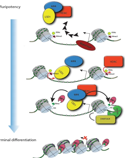
It was found that during differentiation, the pluripotency-related genes that are destined for silencing are silenced by certain repressors which in turn recruit a complex comprised of G9a, LSD1 and HDAC1 (Figure.3). Each component in this complex contribute to local heterochromatinization that is eventually mediated by heterochromatin protein (HP1), then de novo DNMTs are recruited to establish DNA methylation that serves as a “Lock” to reinforce heterochromatinization, and long-term silencing.[109]
Nevertheless, the findings that knocking de novo DNMTS out is associated with impaired differentiation of hematopoietic stem cells suggest the indispensability of DNA methylation for silencing pluripotency-associated genes and proper development and that in turn favors the postulation that DNA methylation acts upstream to silencing of transcriptional initiation.
Bottom line, the relationship between DNA methylation and silencing seems to be bi-directional, it can proceed in either direction; however, no universal conceptual conclusion has come to affirm the issue of the timing of methylation with respect to silencing so far.
Figure 3. DNA methylation in silencing of pluripotency-genes
Does DNA methylation involve in active gene expression?
Interestingly, DNA methylation in the intragenic CGIs (also called Orphan GCIs) has been shown to be associated with active gene expression. As mentioned above, the location of methylated CpG dinucleotides in the transcriptional unit is a determining factor that directs the functionality of DNA methylation. Consistent to the bimodal distribution of CGIs, methylation of CpG in their vicinities show a differential behavior regarding the regulation of gene expression.
Several functional lines have explained how CpG methylation marks the gene body regions of actively transcribed genes.[110-113] Perhaps, the most prominent mechanism is that intragenic CpG methylation enhances transcriptional elongation through inhibiting the cryptic intragenic transcription that overlap with the corresponding transcription.[114, 115] In line with the notion that CGIs are adapted for promoter activity, integrative analyses of Global Run on (GRO-seq) and transcriptome datasets have characterized many intragenic CGIs that code for non-coding or anti-sense transcripts. These transcripts have been shown to attenuate the transcriptional elongation of the corresponding protein coding transcript.[116, 117] It is most likely that methylation of these cryptic promoters, functions in the suppression of their transcriptional potentials, thereby boosting
13
the recruitment of DNMT3 at gene bodies is dependent on SETD2-mediated H3K36me3 deposition, the intragenic mark of active transcription.[118, 119]
Thus, despite the universality of the de novo methylation enzymes, the mechanism of establishing DNA methylation, in the sense of the factors that recruit DNMTs, is differential between promoter/TSS and gene bodies. This suggests that DNA methylation is not functioning separately from the genomic contexts and other epigenetic mechanisms, and casts doubts on the hypothesis that DNA methylation is the direct cause of transcriptional silencing, or transactivation.
DNA methylation in Cancer:
Altered methylome is considered as one of the most notorious features of human malignant diseases. Aberrant DNA methylation pattern has been found to contribute to the neoplastic transformation, as it is followed by altered gene expression profile which in turn expedites the acquisition of cancer hallmarks, boost mutational rate, and implement the phenotypic transformation of incipient cell into neoplastic cell with the greatest clonal advantages and eventually form a macroscopic tumor.
In most solid as well as hematopoietic malignancies, a global hypomethylation, in particular in the intergenic CGIs has been observed.[120-124] In addition to global loss of methylation, regional hypermethylation is also a feature of human neoplasms. The exceptionally high frequency of hypermethylation at certain CpG-rich promoters of tumor suppressor genes in cancer is not taking place spontaneously or stochastically as was thought previously. This has turned out to be a rather highly coordinated and epigenetically attributed event referred to as CpG islands methylator phenotype (CIMP).[28] CIMP is a phenomenon of an epigenetically addictive phenotype, where regional hypermethylation at certain CpG contexts has been found to be of a significant biological relevance to certain subsets of colorectal cancer and glioblastoma; meaning that the genes that are prone to hypermethylation are not the same in all cancer types or subsets.[27, 119, 125-127] Recently, Pan-cancer analysis including whole-genome bisulfite sequencing data from normal tissues and their respective solid tumors showed a strong correlation between gene body hypermethylation and increased expression of 43% of homeobox genes in several cancers, supporting the idea that aberrant DNA methylation in cancer is not random.[128] The integrative Pan-cancer analysis revealed that a subset of under-methylated regions (> 3.5 kb) in normal tissues and are mostly enriched in proto-oncogenes involved in transcriptional regulation and control of differentiation. These regions are called (Methylation Canyons) and are prone to hypermethylation in cancers; in particular at the intragenic locations of signature oncogenes, thereby increase their expression in cancer.[128-130]
It was found that certain sets of genes in embryonic stem cells that are silenced by PRC2-mediated H3K27 trimethylation during pluripotency are prone to methylation in a cancer-type dependent manner
.[131-133]
Upon normal differentiation and lineage specification, PRC2 is released from the promoters CGIs, so that these genes are actively transcribed. Interestingly, with the development of neoplasms, the regions that were pre-marked with PRC2 during pluripotency become methylated and silenced[133, 134]; indicating that DNA methylation mirrors the pattern of PRC2-mediated silencing in stem cells, and thus establishes a state of pseudo-pluripotency that favors the unrestrained proliferation and cellular plasticity.[132, 134-136]In conclusion, DNA methylation, in terms of its functions, establishment and crosstalk to other epigenetic mechanisms, is highly influenced by genomic context, as well as both normal cellular conditions and disease types, and thus reciprocates to cellular programming in either situation.
1.2.5 Histone modifications
Another facet of epigenetic mechanisms is the post-translational modification of certain amino acid residues on histone tails, most prominent candidate is lysine modification. Several chemical modifications of histones take place and involve in establishing structurally and functionally distinct chromatin domains.[43] Notably, post-translational modification is accomplished by adding a group or moiety to the side chain of the amino acid residue. These moieties could be phosphate, sumoyl, ubiquitinyl, acetyl, or methyl groups.[137-140] Similar to DNA methylation, a histone modification is established by a “writer” and removed by an “eraser” and is also recognized by a reader via a special domain and function in recruiting downstream effectors, thus local chromatin features are established.[55, 141] Hereby, I focus mainly on histone lysine methylation marks, in particular, methylation of lysine 27 on histone 3 (H3K27me) the repressive prototype of PRC2.
What makes histone lysine methylation distinct amid the rest of post-translational modification features, for instance lysine acetylation?
It is actually an intriguing question that comes to the minds of those who are into the field of epigenetics and chromatin. Several approaches can offer explanation to the distinctiveness of histone lysine methylation; the chemistry of lysine side chain, the functionalities of histone methylation marks and their crosstalk with the other epigenetic modifications, and the influence of the level of methylation in terms of valence on the local chromatin conformation and transcriptional potentials.
Based on the chemical structure, lysine is one of the basic amino acid, its side chain contains ε -amino group (-NH3+), rendering the side chain positively charged. When the side chain amino group is acetylated, the net positive charge is then neutralized, thus attenuating the electrostatic interaction between the net positively charged core histones within the nucleosome with the negatively charged backbone of the DNA wrapping around. Stressing that histone lysine acetylation is a mark of actively transcribed promoters and active enhancer elements; histone lysine acetylation, for example H3K27Ac, is associated with open chromatin structure and transcriptionally permissive conformation.[142-144]
On the other hand, methylation of histone lysine can take place at three levels of valence, mono-, di- and up to trimethylation, without altering the electrostatic interaction between DNA and core histones (Figure.4).[55, 145] This suggests that the epigenetic functioning of histone methylation marks does not hinge on disrupting the packaging of DNA around histones, and in agreement with the fact that histone lysine methylation marks can be associated with either active or repressed chromatin structures, depending on the position of lysine residue on histone tails as well as the valence of methylation. For instance, methylation at lysine residues 4, 36 and 79 are marks of active euchromatic loci, while methylation on lysine residues 9 and 27 are prototypical marks of long-range silencing at constitutive heterochromatin and of facultative heterochromatin, respectively.[40, 55, 145-149]
15
Figure 4. Lysine acetylation and methylation, edited and taken from ATDBio Nucleic Acids Book (www.atdbio.com)
Thus, lysine methylation exerts differential functions in different genomic context and not tethered to a single mode of action, on the contrary to histone lysine acetylation.
The establishment of histone methylation at lysine residues is accomplished by writers that harbor histone methyl transferase activity (HMT); such catalytic activity is mainly mediated by Su(var)3-9, Enhancer of Zeste, and Trithorax (SET) domain[150, 151]. While the reversal of histone lysine methylation marks is mainly catalyzed by Jumonji C (JmjC) domain-containing lysine demethylases (KDMs).[152-154] However, Some histone lysine methylation features such as methylation at lysine 79 on H3 tails has been shown to be catalyzed by DOT1L, while no dedicated eraser has been characterized so far.[155] With the lack of an eraser to such mark, the levels of methylation in terms of methylation valence have been shown to be in part dependent on the rate of nucleosomal turnover. Recently, it has been found that the processive kinetics of writing H3K79 methylation is adversely influenced by the high rate of nucleosome exchange.[69] By devising a chemical induced proximity model, a DOT1L fusion protein was targeted to certain loci with different transcriptional potential and different nucleosomal exchange rates, yet, deprived of H3K79me marks in mouse embryonic stem cells (mESC). Interestingly, the findings suggested that the dynamicity of the genomic locus favors the lower methylation status, independently of an eraser activity. Integration of datasets from ChIP seq, RNA seq and CATCH-IT seq in mESCs, provided parameters to Monte Carlo simulations that were extended to other histone methylation marks including H3K4 and H3K27 methylation marks. These simulations depicted the influence of nucleosome turnover on the methylation valences and revealed that lower methylation states are associated with the highly dynamic regions with higher transcriptional potentials. Consistently, it was surprisingly found that monomethylation of H3K27 (H3K27me1) is a mark of actively transcribed genes when deposited at their respective intragenic regions (gene bodies); whilst dimethylation (H3K27me2) marks were shown to be enriched at the intergenic regions, preventing the firing of non-cell type specific enhancers and are associated with a poised transcriptional activity.[156] Later, it was confirmed that the gene body-deposited (H3K27me1) marks are not intermediate products of PRC2 towards the ultimate trimethylation[157], in an agreement with the finding that H3K27me1 is dependent in large part on SETD2-mediated H3K36me3 deposition at active gene bodies.[156]
Altogether these findings reveals that histone methylations in terms of their establishment, epigenetic functioning is highly governed by the lysine residue, the methylation valence and
genomic context, and thus emphasize the distinctive placement of histone lysine methylation among the epigenetic mechanisms ever known.
Establishing histone methylation patterns:
As pointed out above, histone lysine methylation marks are not exclusive signs of a solitary chromatin status, they rather show a diversity of epigenetic facets of chromatin domains organization. In this section, I briefly explain the different classes of histone methylation machinery, specifically the antagonizing complexes that belong to two broad categories of chromatin modifying factors, Polycomb and Trithorax families.
Throughout the past seven decades, two key evolutionarily conserved chromatin modifiers, namely, Polycomb (PcG) and Trithorax (TrxG) group proteins has been viewed as integral parts of epigenetic cellular memory system, wherein they function in opposition to maintain repressed and active gene expression states, respectively. They were first discovered in Drosophila melanogaster, and were introduced as epigenetic gatekeepers that orchestrate the expression of Homeotic (HOX) genes, thereby, function in controlling body segmentation plans during embryonic development.[158-162]
During the last decade, extensive research has highlighted the functional diversity of both PcG and TrxG group proteins, which has expanded beyond merely regulating HOX genes expression. Such diversity is lucid in their implication in controlling a plethora of cellular and developmental processes including cell cycle control, proliferation, X chromosome inactivation, genomic imprinting, and regulation of stemness and development of cancer upon their aberrations.[163] These multiple functionalities hinge mainly on their ability to regulate chromatin on a wide-scale, ranging from local chromatin structural conformation to the three-dimensional organization of the genome. The wide-range chromatin organization is highly context dependent, in the sense that the assembly of the components of PcG and TrxG complexes takes place in a cell type- and developmental stage-specific manner; thus granting their abilities to spatio-temporally regulate genome in response to the varying cellular conditions.[163-166]
I. Trithorax (TrxG) complexes:
Trithorax members are highly heterogeneous, and thus play a widespread role in transcriptional activation and oppose the repressive activities exerted by polycomb in multiple ways. Trithorax category is subdivided in three different subcategories:
• Switch/ sucrose non-fermentable (SWI/SNF): a family that includes two groups, namely BAF and PBAF. Each is comprised of up to 15 accessory subunits that are uniquely assembled around core subunits (BRM/BRG1) that harbor ATPase activity, by which they mediate chromatin remodeling.[162, 165-168]
• ASH1: this subcategory includes proteins, the HMT activity of which is directed to write H3K36 methylation marks [169-171]; in addition, CBP which catalyzes H3K27 acetylation is also included.[172, 173]
• COMPASS family: the COMPASS subcategory of TrxG is associated with the histone lysine4 methylation which is attributed to active transcription. Perhaps the most prominent group in terms of its evolutionary conservation and the unique functionality in establishing the bulk H3K4 trimethylation is the SET1/COMPASS group.[174] The different complexes of COMPASS share a consensus group of core subunits, around which other accessory proteins assemble. The core complex is comprised of WDR5, ASH2, RBBP5 and DPY30; collectively the core complex is abbreviated as “WARD”.[163, 174, 175]
With the evolution of metazoans and the emergence of more complicated cis -elements, the SET1/COMPASS complex as experience divergence that gave rise to other combinations, namely the MLL1/2 like and MLL3/4 COMPASS-like. The former is functioning in catalyzing H3K4 trimethylation at certain subset of HOX genes and at bivalent promoter[176, 177]; while the latter mediates monomethylation (H3K4me1) at active enhancers.[174] Thus, the emergence of
17
these new combinations of COMPASS complex matches the increasing evolutionary complexity of the metazoan genomes.
II. Polycomb (PcG) complexes:
Despite their adversarial relationship, the complexities and evolutionary conservation of PcG and TrxG are matching to each other. Biochemical purification-based studies have viewed polycomb group proteins as of two cooperating, even if not necessarily redundant entities: Polycomb repressive complex1 (PRC1) which catalyzes mono-ubiquitination of lysine119 on histone H2A (H2AK119ub) and Polycomb repressive complex2 (PRC2) that functions in depositing the repressive prototype of facultative heterochromatin (H3K27me3).[163] Evolutionarily, PRC2 seems to have evolved later than PRC1, evidenced by the absence of PRC2 in the common model organisms, budding and fission yeast.[178, 179] The evolution of metazoans was accompanied by the emergence of a partnership between both PRC1 and 2, suggesting their Lamarckian evolution to match the complexity of metazoan genome organization in terms of establishment of facultative heterochromatin.
In mammals, both PRC1 and PRC2 exist in different combinations, wherein mutually exclusive sub-stoichiometric components that assemble to their respective core complexes; thus, exhibiting different modes of interactions (Figure.5). More tellingly, PRC1 for example is subdivided into: Canonical (cPRC1) and non-canonical (ncPRC1).[180] Both cPRC1 and ncPRC1 share a highly interspecies-conserved core complex, which comprised of:
• RING1A/B: the catalytic subunit that harbors E3 ubiquitin ligase activity that establishes H2AK119ub.
• Polycomb group ring-finger domain proteins (PCGF1-PCGF6), one of these six proteins should exist and considered as determining factor of the other accessory subunits that assemble to it. For example for the specification of cPRC1, a chromobox (CBX2, 4, 6-8), in addition to one of the polyhomeotic homologous proteins (PHC1-3) assemble around PCGF2/4.[163, 181] Whilst, the presence of zinc-finger and YY1 binding protein (RYBP) or its paralog YAF2 specifies the non-canonical PRC1 and define the enzymatic activity and the mode of interaction with PRC2 as well.[182, 183].
It was previously though that the relationship between PRC1 and PRC2 is unidirectional, in the sense that H3K27me3 the product of PRC2 is recognized by the chromodomain of CBX subunit of cPRC1, and then triggers the RING 1A/B to catalyze H2AK119 mono-ubiquitination which leads to stoppage of the RNA polymerase II to reinforce silencing. Nevertheless, the discovery of the diverse combinations of PRC1 revealed that the crosstalk between PRC1 and 2 is bi-directional, so that PRC1 can function independently of PRC2 or may even function in its recruitment as well. It has become well understood that the relationship between them is rather cooperative not hierarchical. The next section focuses on PRC2 functions and modes of recruitment to its genomic targets; therein, the crosstalk between PRC2 and PRC1 is explained in more details.
Figure 5. A) PRC1 assembly and specifications B) PRC1/PRC2 interaction
1.2.6 Polycomb repressive complex 2 (PRC2)
The specification of cellular lineage and preservation of cellular identities through the successive generations require tight control over transcriptional states. This in turn entails an intricate network of transcription factors that work in harmony with chromatin associated factors that represent additional regulatory layers contributing to transcriptional control.[184] Of these chromatin associated factors, are the PcG group proteins that are known to function in reinforcing transcriptional repression. Perhaps PRC2 is well studied and well known to be altered in many solid as well as hematopoietic malignancies; thus, has been rendered a promising therapeutic target for many cancer types.[185, 186]
PRC2 has been both intensively and extensively studied, and known to exclusively establish all levels of methylation at lysine 27 on H3 (H3K27me1/me2/me3), each shows distinct distribution over the different genomic loci.[156, 157, 184] The establishment of either methylation valence on H3K27 is directly implemented by the SET domain of the core subunit Ehnancer of Zeste homolog 2 (EZH2) or its closely related homolog EZH1.[187] The SET domain harbors histone lysine methyl transferase activity (HMT), that is it has a binding pocket that accommodate the substrate H3K27, which is right adjacent the site where the cofactor S-adenosyl Methionine (SAM) resides. The SAM represents the methyl group donor; the methyl group is then transferred to the amino group of H3K27 side chain in an ordered manner so that the ultimate product is trimethylated H3K27 (H3K27me3).[188, 189]
19
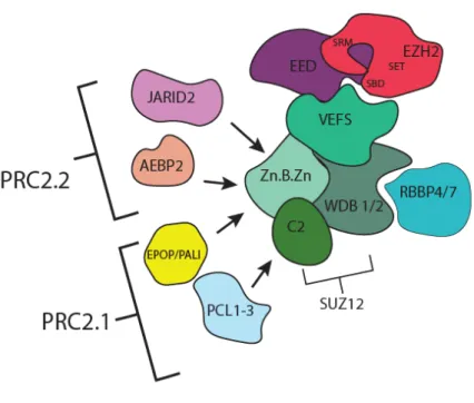
The HMT activity exerted by the SET domain of EZH2 has been shown to be exclusively responsible for establishing the three valences of H2K27me which are characterized by distinct functional disruptions across the genomic domains (as discussed in 1.2.5). This actually points out to the diversity of the aspects of PRC2, in terms of the factors influencing the HMT activity, the residence time of PRC2 subunits, the mechanisms of PRC2 recruitment that constitutes the regional preferences of PRC2 and differential deposition of the various H3K27 methylation marks. One general and plausible explanation of such diversity is the existence of PRC2 in different subsets like PRC1.[183, 186] Biochemical and structural studies have revealed that the catalytic SET domain of EZH2 is rendered “auto-inhibited” if solitarily occurs, indicating the importance of the other regulatory subunits for the functionality of the PRC2 holoenzyme.[189, 190] Analogous to PRC1, It has been shown that PRC2 occurs in two main subsets, namely PRC2.1 and PRC2.2 (Figure.6). These two subsets share three core subunits including the aforementioned catalytic EZH2, in addition to Suppressor of Zeste12 (SUZ12) and Embryonic ectoderm development (EED).[184, 191] Together, in an equimolar stoichiometry, the three subunits comprise the core PRC2 complex around which other accessory subunits assemble in sub-stoichiometric combinations, giving rise to either PRC2.1 or PRC2.2. It is suggested that these sub-stoichiometric interactions are transient and are more likely to be lineage- and developmental stage-specific.
Figure 6. The assembly of PRC2 core subunits with accessory components into PRC2.1 and PRC2.2 subsets


![Figure 10. Graphical illustration explains the conclusion of paper 1.[281]](https://thumb-us.123doks.com/thumbv2/123dok_us/11091608.2996389/54.892.256.591.490.969/figure-graphical-illustration-explains-conclusion-paper.webp)
![Figure 11. Graphical illustration explains the conclusion of paper 2.[272]](https://thumb-us.123doks.com/thumbv2/123dok_us/11091608.2996389/56.892.205.626.503.1065/figure-graphical-illustration-explains-conclusion-paper.webp)
