ORIGINAL ARTICLE
Up and down staging of TCC using
18
F-FDG
PET/CT scan
Tamer W. Kassem
Radiology Department, Cairo University, Egypt Received 15 February 2016; accepted 11 April 2016 Available online 25 April 2016
KEYWORDS
Staging; TCC;
18F-FDG PET/CT
Abstract Objective: The purpose of this study was to investigate the ability of PET/CT to restage urinary bladder transitional cell carcinoma (TCC) whether up or down after initial staging by histopathology and other imaging modalities.
Patients and methods: During three year duration 27 patients with urinary bladder TCC were prospectively evaluated. They were previously diagnosed by histopathology after biopsy and had prior imaging for initial staging. All patients underwent PET/CT examinations following a preset protocol. The images obtained were reconstructed using dedicated software and workstations. Results of PET/CT examinations were confirmed by histopathology.
Results: Prior imaging and PET/CT findings were matched in 17 patients (63%) with no changes regarding their initial TNM stages and differed in 10 patients (37%). Six patients (22.2%) showed new sites of distant metastasis and unchanged initial stages. Two patients (7.4%) were up staged and 2 patients (7.4%) were down staged for whom the plan of clinical management was justified.
Conclusion: 18F-FDG PET/CT can up and down stage TCC and can detect new sites of nodal or distant metastasis leading to optimal therapy planning.
Ó2016 The Egyptian Society of Radiology and Nuclear Medicine. Production and hosting by Elsevier. This is an open access article under the CC BY-NC-ND license ( http://creativecommons.org/licenses/by-nc-nd/4.0/).
1. Introduction
Bladder cancer represents 7% of all malignancies in men and 2% of all malignancies in women(1). More than 90% of blad-der cancers are transitional cell carcinomas(2). Bladder cancer is usually multifocal and has a high local recurrence rate(3). At least 50% of the high-grade tumors may have occult meta-static disease at initial diagnosis(3). Common sites of metas-tases include the pelvic and retroperitoneal lymph nodes,
lungs, liver and bones. Multimodal treatment, depending on preoperative stage may improve survival(4).
The TNM classification is currently the standard staging procedure for bladder cancer and is based on clinico-pathological findings(5). TNM staging system (Table 1) has become the system of choice primarily because it has the advantage of distinguishing nodal involvement from locally advanced tumors and from distant metastatic spread (6). T staging is best done by cystoscopy and deep biopsy (7). Identifying nodal disease N is critical because it has profound implication for treatment plan (6). The incidence of distal metastasis M increases with increasing T and N stages (8). Noninvasive imaging plays an important role in all stages of
Peer review under responsibility of The Egyptian Society of Radiology and Nuclear Medicine.
Egyptian Society of Radiology and Nuclear Medicine
The Egyptian Journal of Radiology and Nuclear Medicine
www.elsevier.com/locate/ejrnm
www.sciencedirect.com
http://dx.doi.org/10.1016/j.ejrnm.2016.04.011
0378-603XÓ2016 The Egyptian Society of Radiology and Nuclear Medicine. Production and hosting by Elsevier. This is an open access article under the CC BY-NC-ND license (http://creativecommons.org/licenses/by-nc-nd/4.0/).
bladder cancer. The standard preoperative method for staging is CT or MRI of the abdomen and pelvis, although these methods lack sensitivity and, therefore, may not offer an entirely accurate basis for therapeutic planning(9).
PET/CT with18F-FDG has emerged as a powerful tool for the combined metabolic and anatomic evaluation of many can-cers(10).18F-FDG uptake by bladder cancer was first demon-strated by Harney et al.(11)in rats, with an estimated uptake ratio of tumor to normal bladder of 13:1. Excreted FDG results in marked uptake throughout the urinary tract and obscures urologic malignancies. Despite this limitation, 18
F-FDG PET/CT was found to be useful in evaluating TCC primary tumor and distant disease(12)(seeTable 2).
The purpose of this study was to investigate the ability of PET/CT to restage urinary bladder TCC whether up or down after initial staging by histopathology and other imaging modalities.
2. Patients and methods 2.1. Patients
A prospective study of 27 patients aged 34–72 years old (with mean age of 55 years old) presenting to a private specialized medical center with an initial diagnosis and staging of urinary bladder TCC coming for restaging between July 2012 and August 2015 was performed. There were no set criteria for referral other than histopathologic evidence of TCC after biopsy and the presence of initial staging by post-contrast CT examination. Patients had clinical evaluation including medical history. Information regarding interventional procedures, surg-eries and histopathology was obtained from all cases.
2.2. Methods
PET/CT scan examination was obtained for each patient. All patients were scanned using a dedicated PET system (Siemens Syngo PET VG 50A Biograph 20 VA 44A, Berlin, Germany),
covering an axial field-of-view (FOV) of 15.2 cm and a resolu-tion of 4 mm axially and 3.8 mm trans axially at the center.
Patients were instructed to fast for at least 8 h before the intravenous injection of 370–410 MBq of18F-FDG. Blood cose was measured before injection of the tracer to ensure glu-cose blood levels below 120 mg/dl. Before and after injection, patients were kept sitting comfortably in a quiet room. Saline infusion (approximately 500 ml) was given before tracer injec-tion. Whole-body PET/CT images were acquired 40–50 min after radiotracer injection. CT images were acquired without breath-holding instructions. The PET emission scan was obtained immediately after acquisition of the CT scan.
Routine axial imaging consisting of CT of the chest, abdo-men and pelvis at 5 mm intervals after intravenous non-ionic contrast administration was performed in all patients. 2.3. Data analysis and interpretation
PET image datasets were reconstructed using the CT data for attenuation correction and fused images were displayed on a workstation. Images were reconstructed in coronal and sagittal multiplanar planes and read visually.
All PET/CT imaging and CT imaging were performed and evaluated by one staff radiologist with expertise in genitouri-nary imaging accompanied by one staff nuclear medicine. Standardized uptake values (SUV) were compared to back-ground vasculature to determine positivity and all foci of unphysiologic FDG uptake were considered indicative of meta-static disease during interpretation of reconstructed PET/CT images. PET/CT findings were confirmed by histopathology. 3. Results
From July 2012 to August 2015, 27 patients were entered in the study aged on average 55 years (range, 34–72 years).
All examined cases had malignant urinary bladder masses proven by histopathology to be TCC. All of them had an ini-tial staging obtained after performing post-contrast CT examination.
Prior imaging and PET/CT findings matched in 17 patients (63%) and differed in 10 patients (37%). So, the patients were classified into two groups: the first group consists of 17 patients had the same stage given before performing the PET/CT exam-ination and the second group consists of 10 patients showing different findings and was subdivided into three subgroups.
The first subgroup consists of 6 patients (22.2%) showing new sites of distant metastasis detected by current PET/CT examination; however, the final stage did not change. One patient showed newly discovered active supraclavicular lymph nodes, however it maintained its primary stage T3N3M1
Table 1 Staging groups of bladder Cancer(5).
Stage T N M
0 Ta or Tis N0 M0
I T1 N0 M0
II T2a or T2b N0 M0
III T3a, T3b or T4a N0 M0
IV T4b N0 M0
Any T N1, N2 or N3 M0 or M1
Table 2 Distribution of findings in all cases.
No % New findings Same stage Up stage Down stage
1st group 17 63 – Yes – –
2nd group 1st subgroup 6 22.2 Yes Yes – –
2nd subgroup 2 7.4 Yes Yes –
3rd subgroup 2 7.4 – – – Yes
27 100
(stage IV) (Fig. 1). Two patients showed newly discovered active cervical and mediastinal lymph nodes and their primary stage was not changed T3aN3M1 and T2bN3M1 (stage IV) (Fig. 2). One patient showed evidence of pelvic vascular infil-tration and its primary stage was the same T2aN2M1 (stage IV) (Fig. 3). Multiple new sites of metastatic bony and hepatic deposits were detected in two patients and these findings did not affect their primary stage T4bN3M1 (stage IV) (Fig. 4).
The second subgroup consists of 2 patients (7.4%) who were up staged after performing PET/CT. One patient showed newly discovered active common iliac lymph node changing its stage from T4aN0M0 (stage III) to T4aN3M0 (stage IV) (Fig. 5). The second patient showed newly discovered active mediastinal lymph nodes and few lumbar vertebrae active osseous metastatic deposits changing its stage from T3aN0M0 (stage III) to T3aN0M1 (stage IV) (Fig. 6).
Adjuvant systemic chemotherapy was added to the plan of management for these patients.
The third subgroup consists of 2 patients (7.4%) who were down staged after performing PET/CT (Fig. 7). They had external iliac lymph nodes showing no FDG uptake by PET/ CT and containing no malignant cells by histopathology. These lymph nodes were diagnosed as metastatic nodes by prior examinations. Their staging changed from T2aN1M0 (stage IV) to T2aN0M0 (stage II) in the first case and from T1N1M0 (stage IV) to T1N0M0 (stage I) in the second case. There was no need for adjuvant therapy for these patients as was planned before.
The plan of management was adjusted for patients participating in subgroups 2 and 3 (4 patients, 14.8%) either by adding needed or by omitting unnecessary systemic chemotherapy.
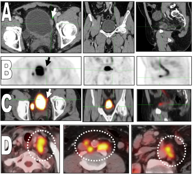
Fig. 1 62 years old male patient with urinary bladder mass (TCC). The administered activity is 370.4 MBq.18F-FDG, blood glucose
level (BGL) was 89 mg/dL. (A) Axial, coronal and sagittal post-contrast CT images, (B) axial, coronal and sagittal MIP PET images, (C) axial, coronal and sagittal fused PET/CT images showing increased FDG uptake corresponding to enhanced lesion along the left lateral surface of the urinary bladder(arrows). It measures about 1.60.7 cm in diameters with SUVmax 14.6. (D) Axial fused PET/CT images showing multiple active left supraclavicular groups measuring about 2.8 cm with SUVmax 8.3. Multiple active bilateral common iliac lymph nodes are seen largest measuring about 2.6 cm in diameter with SUVmax 6.5. Multiple active left para aortic lymph nodes are seen largest measuring about 1.6 cm in diameter with SUVmax 8.7.
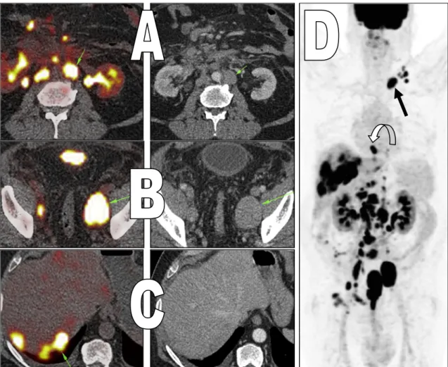
Fig. 2 64 years old male patient with urinary bladder mass (TCC). The administered activity is 379 MBq.18F-FDG, BGL was 92 mg/dL.
(A) Axial fused PET/CT and post-contrast CT image showing numerous active para-aortic lymph nodes largest measuring about 3.0 cm in diameter with SUVmax 11.8. (B) Axial fused PET/CT and post-contrast CT image showing bilateral internal iliac enlarged active lymph nodes, the largest at the left internal iliac group measuring about 4.7 cm in diameter with SUVmax 18.5. (C) Axial fused PET/CT and post-contrast CT image showing multiple hypodense hepatic focal lesions. The largest and most active is seen panning across segment VI–VII with SUVmax 12.9. (D) Whole body coronal MIP PET image showing enlarged active cervical (left levels IV and V) lymph nodes, the most active is seen at left supra-clavicular region(arrow)(2.3 cm & SUVmax 10.7). Posterior mediastinal active retro-cardiac LN is seen measuring 2.5 cm with SUVmax 11.7(curved arrow). Multiple FDG-avid lesions are seen within right pubic bone, sacrum and vertebrae. The most active lesion is seen in L1 (SUVmax 10.6).
Fig. 3 55 years old male patient with urinary bladder mass (TCC). The administered activity is 407 MBq.18F-FDG, BGL was 102 mg/
dL. (A–C) axial fused PET/CT images. Image (A) shows a metabolically active ill defined soft tissue lesion obliterating the right semino-vesical angle merging with the urinary bladder wall and the dome of the prostate (SUV max 4.4). Image (B) shows distended related pelvic vein draining into the right internal iliac vein and showing increased FDG uptake (SUV max 6.9), denoting infiltration(arrows). Image (C) shows multiple metabolically active left external iliac lymph nodes (maximum SUV 6.8 over 1.1 cm lymph node) and left internal iliac lymph nodes (maximum SUV 6.3 over 1.3 cm lymph node).
4. Discussion
Bladder carcinoma is the most frequent type of tumor of the urinary tract and is most prevalent in the fifth to seventh dec-ade of life(13). Approximately 70% of bladder cancers present as superficial tumors, which tend to recur, and 30% present as muscle-invasive disease associated with a high risk of death from distant metastases (14). Optimal therapy planning is dependent on accurate staging of the bladder tumor. For iden-tification of patients with metastatic disease, current imaging techniques including sonography, computed tomography (CT) and magnetic resonance imaging (MRI) have not proven
to be highly accurate(15).18F-FDG PET/CT has become an important noninvasive imaging modality for many malignan-cies because of its unique capability to image metabolically active lesions(16).
Patil et al. (17) found that 18F-FDG PET/CT of transi-tional cell carcinoma can be useful in evaluating the primary tumor and distant disease while Swinen et al.(18)stated that PET/CT provides good diagnostic accuracy of lymph node staging of bladder cancer.
Liu et al. (19) included 46 patients for examination of FDG-PET in staging of locoregional and distant metastases in bladder cancer. Their results were more in favor of PET/
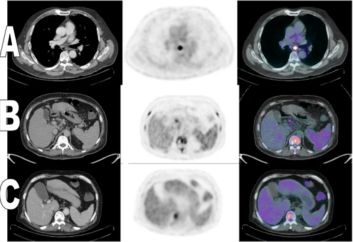
Fig. 4 47 years old male patient with urinary bladder mass (TCC). The administered activity is 398 MBq.18F-FDG, BGL was 99 mg/dL. (A) Axial post-contrast CT, PET and fused PET/CT images showing multiple ill defined hepatic hypermetabolic lesions. The most active lesion is seen in the left lobe with SUVmax 8.2. (B) Axial post-contrast CT, PET and fused PET/CT images showing hypermetabolic lytic lesions at the sacrum and pelvic bones (SUVmax 13.0 over the sacrum). (C) Sagittal post-contrast CT, fused PET/CT and PET images showing wide spread hypermetabolic lesions at the sternum and vertebrae (SUVmax 13.6 over T1).
CT, showing a sensitivity of 77%, a specificity of 97%, and an accuracy of 89% to reveal metastatic TCC.
Accurate assessment of local tumor extent and nodal dis-ease is particularly important in treatment planning. Patients with superficial tumor may be treated with transurethral resec-tion with or without intravesical chemotherapy. Those with more invasive but still localized tumor (T2 or T3) benefit from partial or radical cystectomy, or a combination of radiation
and systemic chemotherapy. Patients with unresectable tumors (T4b) or local or distant metastases should receive systemic chemotherapy(20).
This study showed surprising results regarding detection of distant metastasis as 7 patients out of 27 showed newly detected sites of distant metastasis representing 25.9% of patients. Six patients had no change regarding their stages while 1 patient was up staged from stage III to stage IV. This
Fig. 5 63 years old female patient with urinary bladder mass underwent cystoscopic and histopathology revealed (TCC). The administered activity is 395 MBq. 18F-FDG, BGL was 110 mg/dL. (A–C) Multiplanar fused PET/CT and post-contrast CT images
showing FDG-avid soft tissue thickening in the postero-inferior aspect of the urinary bladder being inseparable from another soft tissue thickening involving the vaginal vault with obliteration of the vesico-vaginal fat planes (SUVmax 11.2). (D) Axial fused PET/CT and post-contrast CT image showing hypermetabolic left common iliac lymph node is seen(arrow)measuring about 1.5 cm diameter with SUVmax 5.4.
agrees with Drieskens et al.(21) who examined the value of preoperative FDG-PET/CT in identifying locoregional lymph node metastasis and other distant metastasis in 55 patients with bladder cancer and concluded that PET/CT creates an advantage for distant metastases but, compared with CT, there was no advantage for the detection of locoregional metastasis. Swinen and colleagues (18) studied 51 patients and indi-cated that the sensitivity of CT alone and of FDG-PET/CT for the detection of regional lymph node metastasis is equal. This findings matches with the current study that showed that loco-regional lymph staging could be decided based on prior
imaging information alone, without new additional benefit from the PET/CT examination other than the advantage of precisely localizing the hypermetabolic nodes. One patient belonging to subgroup 2 in second group had a change regard-ing his N stagregard-ing after detection of active common iliac lymph node (from N0 to N3).
PET/CT affected the clinical management in 6 out of 35 patients (17% of total), by prompting additional therapy in 5 patients and a wait-and-watch strategy in 1 patient in a study done by Jadvar and colleagues(22)who reported the detection of unsuspected nodal and skeletal metastasis. When Drieskens
Fig. 6 58 years old male patient with urinary bladder mass (TCC). The administered activity is 371 MBq.18F-FDG, BGL was 95 mg/dL. (A–C) Axial post-contrast CT, PET and fused PET/CT images showing hypermetabolic subcarinal lymph node measuring about 1.5 cm in diameter (SUVmax 8.3) and multiple hypermetabolic bony deposits at the lower dorsal and lumbar vertebral bodies.
Fig. 7 60 years old male patient with urinary bladder mass (TCC). The administered activity is 381 MBq.18F-FDG, BGL was 101 mg/ dL. (A–C) Axial post-contrast CT, PET and fused PET/CT images showing left external iliac lymph node measuring about 1.2 cm in diameter. No evidence of FDG uptake was noted.
et al.(21)used PET/CT for the preoperative staging of inva-sive bladder carcinoma, they found that the addition of PET/CT to conventional imaging changed staging in 15% of the patients. The results of these two studies were very close to the current study in which the plan of management changed in 4 out of 27 patients representing 14.8% of cases.
Although FDG-PET/CT seems promising for combining metabolic and anatomic diagnosis for the detection of metasta-sis in pelvic malignancy, its use, especially in bladder malig-nancy has been hampered by technical limitations and urinary excretion(23). Several interventions such as adequate hydration, bladder irrigation and forced diuresis have been used to overcome this obstacle.
PET/CT is still limited in its spatial resolution. Another problem is the faint FDG uptake by some tumors. This behav-ior is attributed to high activity of glucose-6-phosphatase, the enzyme that converts F-FDG-6-phosphate back into F-FDG with its excretion from the tumor cells. The use of other PET tracers, such as C11-methionine and C11-choline, are cur-rent possibilities, but the short half-lives of these tracers restrict their routine use.
To avoid radical surgery in incurable patients or toxic chemotherapy in patients without metastasis, accurate staging is of utmost importance. As a metabolic and anatomic diag-nostic tool,18F-FDG PET/CT has the ability to change plan of management in patients with known TCC. This study adds to the current data that suggest that PET/CT may improve the imaging evaluation of these patients, affect the decision mak-ing and ameliorate long term outcomes.
Conflict of interest
The authors declared that there is no conflict of interest. References
(1)Jemal A, Murray T, Ward E, Samuels A, Tiwari RC, Ghafoor A, et al. Cancer statistics. CA Cancer J Clin 2005;55:10–30. (2) Ferlay J, Soerjomataram I, Ervik M, Dikshit R, Eser S, Mathers
C, et al. GLOBOCAN 2012 v1.0, Cancer incidence and mortality worldwide: IARC CancerBase No. 11 [Internet]. Lyon, France: International Agency for Research on Cancer; 2013. <http://globocan.iarc.fr> [accessed on day/month/year]. (3)Droller MJ. Bladder cancer: state-of-the-art care. CA Cancer J
Clin 1998;48:269–84.
(4)Herr HW, Cookson MS, Soloway SM. Upper tract tumors in patients with primary bladder cancer followed for 15 years. J Urol 1996;156:1286–7.
(5) American Joint Committee on Cancer. AJCC cancer staging manual. Urinary Bladder. 7th ed. New York, NY: Springer; 2010. p. 497–502.
(6)Vikram R, Sandler Carl M, Ng Chaan S. Imaging and staging of transitional cell carcinoma: part I, lower urinary tract. AJR 2009;192:1481–7.
(7)Ng CS. Radiologic diagnosis and staging of renal and bladder cancer. Semin Roentgenol 2006;41:121–38.
(8)Knap MM, Lundbeck F, Overgaard J. Prognostic factors, pattern of recurrence and survival in Danish bladder cancer treated with radical cystectomy. Acta Oncol 2003;42:160–8.
(9)Paik ML, Scolieri MJ, Brown SC, Spirnak JP, Resnick MI. Limitations of computerized tomography in staging invasive bladder cancer before radical cystectomy. J Urol 2000;163:1693–6.
(10)Purcell DD, Coakley FV, Franc BL, Hawkins RA, Boddington SE, Yeh BM. Anterior layering of excreted 18F-FDG in the bladder on PET/CT: frequency and cause. AJR 2007;189, 464; [web] W96–W99.
(11)Harney JV, Wahl RL, Liebert M, Kuhl DE, Hutchins GD, Wedemeyer G, et al. Uptake of 2-deoxy, 2-(18F) fluoro-D-glucose in bladder cancer: animal localization and initial patient positron emission tomography. J Urol 1991;145:279–83.
(12)Shreve PD, Anzai Y, Wahl RL. Pitfalls in oncologic diagnosis with FDG PET imaging: physiologic and benign variants. RadioGraphics 1999;19:61–77.
(13)Jemal A, Siegel R, Ward E, Hao Yongping, Xu J, Murray T, et al. Cancer statistics. CA Cancer J Clin 2008;58:71–96.
(14)Kaufman DS, Shipley WU, Feldman AS. Bladder cancer. Lancet 2009;374(18):239–49.
(15)Paik ML, Scolieri MJ, Brown SL, Spirnak JP, Resnick MI. Limitations of computerized tomography in staging invasive bladder cancer before radical cystectomy. J Urol 2000;163:1693–6.
(16)Antoch G, Saoudi N, Kuehl H, Dahmen G, Mueller S, Beyer T, et al. Accuracy of whole-body dual-modality fluorine-18-2-fluoro-2-deoxy-D-glucose positron emission tomography and computed tomography (FDG-PET/CT) for tumor staging in solid tumors: comparison with CT and PET. J Clin Oncol 2004;22:4357–68. (17)Patil V, Wang Z, Sollitto R, Chuang K, Konety B, Hawkins R,
et al. 18F-FDG PET/CT of transitional cell carcinoma. AJR 2009;93:W497–504.
(18)Swinen G, Maes A, Pottel H, Vanneste A, Billiet I, Lesage K, et al. FDG-PET/CT for the preoperative lymph node staging of invasive bladder cancer. Eur Urol 2010;57:641–7.
(19)Liu IJ, Lai YH, Espiritu JI, Segall GM, Srinivas S, Nino-Murcia M, et al. Evaluation of fluorodeoxyglucose positron emission tomography imaging in metastatic transitional cell carcinoma with and without prior chemotherapy. Urol Int 2006;77:69–75. (20)Konety BR, Carroll PR. Urothelial carcinoma: cancers of the
bladder, ureter, & renal pelvis. In: Tanagho EA, McAninch JW, editors. Smith’s general urology. New York, NY: McGraw Hill; 2008. p. 308–27.
(21)Drieskens O, Oyen R, Van Poppel H, Vankan Y, Flamen P, Mortelmans L. FDG-PET for preoperative staging of bladder cancer. Eur J Nucl Med Mol Imag 2005;32:1412–7.
(22)Jadvar H, Quan V, Henderson RW. [F-18]-fluorodeoxyglucose PET and PET-CT in diagnostic imaging evaluation of locally recurrent and metastatic bladder transitional cell carcinoma. Int J Clin Oncol 2008;13:42–7.
(23)Scho¨der H, Larson SM. Positron emission tomography for prostate, bladder, and renal cancer. Semin Nucl Med 2004;34:274–92.