The International Association for the Study of Lung Cancer
Staging Project
Prognostic Factors and Pathologic TNM Stage in Surgically Managed
Non-small Cell Lung Cancer
Kari Chansky, MS,* Jean-Paul Sculier, MD, PhD,† John J. Crowley, PhD,* Dori Giroux, MS,*
Jan Van Meerbeeck, MD, PhD,‡ and Peter Goldstraw, MB, FRCS,§
on behalf of the International Staging Committee and Participating Institutions
Purpose: To assess the impact of cell type, age, and gender in addition to pathologic tumor, node, metastasis (TNM) stage in surgically managed stage I-IIIA non-small cell lung cancer (NSCLC) cases from the international staging database of the International Association for the Study of Lung Cancer.
Material and Methods:From the 67,725 cases of NSCLC submit-ted to the staging database, 9137 surgically managed cases were selected for which all the following variables were available: patho-logic stage, age, gender, and specific histopatho-logic cell type. Perfor-mance status and smoking history were examined in subsets. Meth-ods used were Cox proportional hazards regression and recursive partitioning and amalgamation (RPA) analyses.
Results:Pathologic TNM stage, age, and gender were all indepen-dently prognostic for survival. The bronchioloalveolar carcinoma (BAC) subtype had superior survival over other cell types despite the potential for heterogeneity in this group. Adjusted comparisons revealed a small survival advantage for squamous cell carcinomas over non-BAC adenocarcinoma histology and also over large cell, though the effect appeared to be limited to the male patients. RPA revealed the importance of TNM stage primarily, and age was prognostic within stage groups. Cell type was not found to add prognostic value in the RPA analysis. Prognostic groups were formed based on the RPA output, and the prognostic value of these groupings was validated using the North American Surveillance, Epidemiology, and End Results Registries. Performance status and smoking history were prognostic in the subsets where data were
available. Effects of other variable were not influenced by the inclusion of smoking status in regression models.
Conclusions:Age and gender are confirmed as important prognostic factors in surgically resected NSCLC. Cell type is less important, although the small population of cases classified as BAC have a survival advantage over other histologies, and there may be a small survival advantage for squamous cell carcinomas over non-BAC adenocarcinomas. Imbalances between stage, gender, and cell type at presentation may lead to a misleading result with respect to cell type in unadjusted analyses. Pathologic TNM category is the most important prognostic factor in this analysis.
Key Words: Non-small cell lung cancer, TNM stage, Pathologic stage, Prognostic factors, Histology, Cell type, IASLC Lung Cancer Staging Project.
(J Thorac Oncol.2009;4: 792–801)
T
he International Association for the Study of Lung Cancer (IASLC) International Staging Committee has submitted proposals for revision of the tumor, node, metastasis (TNM) descriptors1– 4 and stage groupings5 for lung cancer in theforthcoming (7th) Edition of the International Union Against Cancer and American Joint Committee on Cancer TNM Classification of Malignant Tumors. These proposals were developed using a very large database that was specifically collected from individual databases for that purpose. As part of this effort, the Prognostic Factors Subcommittee of the International Staging Committee reported on the role of other prognostic factors, in addition to stage, with respect to sur-vival in 12,428 clinically staged cases of non-small cell lung cancer (NSCLC).6Age, gender, and performance status were
all found to be prognostic for survival after adjustment for stage of disease. Cell type was found to be only minimally prognostic within the non-small cell types, with the squamous cell carcinoma cell type having a slightly superior survival overall after adjustment for other factors. However, the effect of cell type only appeared important in the stage IIIA cases. Here, we examine primarily a subset of those prognos-tic factors (cell type, age, and gender) in 9137 pathologically *Statistics Department, Cancer Research And Biostatistics, Seattle,
Wash-ington; †Department of Intensive Care and Thoracic Oncology, Institut Jules Bordet, Universite´ Libre de Bruxelles (ULB), Brussels, Belgium; ‡Department of Respiratory Medicine, University Hospital, Ghent, Bel-gium; and §Royal Brompton Hospital, Imperial College, London, United Kingdom.
Disclosure: The authors declare no conflicts of interest.
Address for correspondence: Kari Chansky, MS, Cancer Research And Biostatistics, 1730 Minor Ave STE 1900, Seattle, WA 98101. E-mail: karic@crab.org
Copyright © 2009 by the International Association for the Study of Lung Cancer
staged NSCLC surgically managed cases selected from the IASLC database. The large number of cases and relatively homogenous group with respect to management (surgery as part of definitive treatment in all cases) allowed us to explore the prognostic impact of cell type in greater detail than has been possible in the analyses of single institution series or of population-based registries. The relative prognoses for the ade-nocarcinoma and squamous cell histologies have been reported with various results. In consideration of the potential for dispro-portionate representation of the different cell types within patient groups, particular care was taken to explore the relationship with respect to survival between stage, cell type, and gender.
METHODS
The methodology of the IASLC Lung Cancer Staging Project and the major proposals have been reported.2–5,7All
data were retrospective, and, by mutual agreement, were transmitted to Cancer Research And Biostatistics (CRAB) as coded data without identifiable private information, with appropriate regulatory permission from the contributing sites. The project was reviewed and determined to be exempt from further human subjects review by CRAB’s institutional re-view board.
Population
In total, 100,869 cases were submitted to the interna-tional database, of which 81,015 remained eligible for anal-ysis after exclusion of cases outside the study period (1990 – 2000), those with unknown histology, those not newly diagnosed at the point of entry and those with inadequate information on stage, treatment or follow-up. Of the eligible cases, 67,725 cases were of non-small cell histology. Of these, 15,236 were pathologically staged, surgical cases with sufficient T, N, and M descriptor information to reclassify according to the IASLC proposals for the 7th edition of TNM. From this group, 9137 stage I-IIIA cases were identi-fied as having come from databases that distinguished the bronchioloalveolar carcinoma (BAC) subtype from the other adenocarcinomas as a separate category wherever it was identi-fied and reported by the local pathologist. The time frame for these cases mostly predates the 1999 3rd edition of WHO guidelines for the classification of lung tumors,8 so that many
cases classified as BAC were potentially adenocarcinoma with a BAC component, rather than pure BAC without invasion. Al-though there was no central histopathological review of cell type and we therefore cannot be certain that the allocation of cell type was consistent across groups, especially in identifying BAC, the recognition of a separate category for BAC or adenocarcinoma with BAC features was felt to be important.
Age, gender, and cell type were available for all of these cases. Performance status was unavailable in two thirds of the cases; therefore, this factor was not included as a factor in the primary analysis but was explored in a subset. Because all of these cases were candidates for surgery, performance status typically did not exceed 1 on the Zubrod scale in those cases where performance status was provided. Smoking his-tory was also unavailable for 54% of cases; therefore it was explored separately as well. Patients who were documented as having received neoadjuvant chemotherapy were not
in-cluded in these analyses. Cases with notation of chemother-apy at some time point after surgery (in about 8.5% of the cases where the data were provided) were allowed.
Cases included in the primary analysis were from 27 separate databases representing 18 countries. The largest contributions were from the Bronchogenic Carcinoma Co-operative Group of the Spanish Society of Pneumology and Thoracic Surgery (GCCB-S, 1851 cases) and the Norway Registry (1737 cases), which collected surgical cases specif-ically. The majority of cases were from surgical series or hospital consortia submitting surgical cases to a central reg-istry. A small proportion of cases (143 cases) were from population-based registries (collecting cases from all treat-ment modalities) and 476 were from clinical trials (Table 1). Of the 9137 cases included in this analysis, 1950 had also been included in the previous analysis of clinically staged cases.
Statistical Analysis
Survival was measured from the date of surgery until death due to any cause, and median survival was calculated by the Kaplan-Meier method. Prognostic groups were as-sessed by Cox regression analysis on overall survival, using the SAS system for windows version 9.0 PHREG procedure. In regression analyses, stage and histology categories were modeled categorically using indicator variables. For ease of interpretation, age was considered as a dichotomous categor-ical variable with a cutpoint of 70. Although the cutpoint for age was 75 in the previous paper from this group, there was a smaller proportion of cases that were over age 75 in the current surgical subset. Thus, a cutpoint of 70 was chosen, which is consistent with the age cutpoint frequently cited for clinical trials in “elderly” patients. The decision to use a dichotomous age variable was reinforced by the fact that when regression models were adjusted by age as a continuous variable for comparison, the resulting hazard ratios for cell type, stage, and gender were the same to within ⫾0.03. Significance testing for binary variables (age and gender) was done using the Wald statistic. Comparisons of individual levels of stage and histology also used a Wald test for each individual hypothesis. Because of the number of variables used and models considered, the threshold for statistical significance was adjusted to 0.01.
Recursive partitioning and amalgamation (RPA) anal-yses9were performed to generate tree-based models by stage
(proposed version 7 TNM) plus the key prognostic factors: age, gender, and cell type. The tree algorithms were
per-TABLE 1. Geographical Representation of Submission Types
Total Cases
Clinical
Trial Consortium Registrya Series
Asia 1135 0 0 0 1135 Australia 1383 0 0 0 1383 Europe 4818 10 1851 1880 1077 North America 1801 466 0 0 1335 All regions 9137 476 1851 1880 4930 a
formed on a training set consisting of the entire set of 9137 cases available for analysis, and the resultant prognostic groupings were then tested for validation against appropriate surgical cases in the 1998 –2002 time frame from the U.S. National Cancer Institute’s Surveillance, Epidemiology, and End Results Registries (SEER) database. To ensure a com-parable study group for validation, only those NSCLC cases in the appropriate TNM category and with a surgery code indicating a surgical resection were selected. SEER reports best stage, which is generally pathologic stage in a surgically resected case.10 TNM stage categories were derived from
extent of disease codes (such as tumor size and other tumor descriptors) and N-stage, which are sufficient to reclassify cases to the revised staging scheme. Only cases that were reclassified as stage I-IIIA in the proposed new staging scheme were selected.
Variables entered into the RPA analysis were stage (as an ordered variable), age (as both categorical and continuous in separate iterations), gender, and cell type (as a group of indicator variables). The RPA generated tree-based models for the survival data used logrank test statistics for selecting “best splits” of the data to form the terminal groupings and bootstrap resampling to correct for the adaptive nature of the splitting algorithm. For validation of the survival tree result, the terminal nodes were then grouped according to similar hazards and the newly formed groups were evaluated using the SEER database.
RESULTS
The adenocarcinoma and squamous cell carcinoma his-tologies comprised the largest proportions of the study sam-ple (36 and 49%, respectively). Squamous cell carcinomas predominated in stages II and III and were less frequently stage I (35%) as compared with the adenocarcinomas (46%). There were imbalances with respect to gender and histology. Among female patients, 55% were adenocarcinoma and 25% squamous cell. In contrast, the male patients were 30% adenocarcinoma and 57% squamous cell (Table 2).
Survival is ordered according to pathologic TNM cat-egory (proposed version 7) as expected (Figure 1A), with median survival estimates ranging from 19 months for stage IIIA to 95 months for stage IA. For cell type across all stages combined, the BAC subtype has a median survival of 83 months, followed by adenocarcinoma, 45 months, versus 44 months for squamous cell carcinomas, 34 months for large cell, and 26 months for adenosquamous (Figure 1B).
The following variables were considered in Cox pro-portional hazards regression analyses: pathologic TNM stage (using IASLC proposals for the 7th edition of TNM), age, gender, and histologic cell type (adenocarcinoma versus squamous cell carcinoma versus large cell versus adenosqua-mous versus BAC). Unadjusted analyses (where each factor was considered independently) revealed significant differ-ences between BAC and all other cell types, between male and female patients, and between patients 70 and older versus patients less than 70 years of age (Table 3, results for unadjusted models). In unadjusted analyses across all stages and both genders, the adenocarcinomas and squamous cell carcinomas did not have a significantly different prognosis. However, in a model including all factors, (cell type, patho-logic stage, gender, and age), squamous cell has a significant survival advantage over adenocarcima and large cell, sug-gesting that, all other things being equal, the squamous cell carcinoma histology carries a slightly better prognosis (Table 3, results for adjusted models). There was no significant difference between large cell and adenocarcinoma, or be-tween adenosquamous and any other non-BAC histology.
There is a small but statistically significant interaction between histology and gender (p ⫽ 0.006 on a global test comparing full and reduced models). To illustrate the nature of the interaction, the survival statistics for histology, age, and stage are shown separately for female and male patients in Table 3. In female patients, there is no significant differ-ence between squamous cell carcinomas and adenocarcino-mas or between any other non-BAC histologies after
adjust-TABLE 2. Distribution of Cell Type Across Gender and Stage Categories for the Pathologically Staged I-IIIA (IASLC Proposals for 7th ed TNM) Database,N⫽9137
BAC Adenocarcinoma Squamous Cell Large Cell Adenosquamous Total Females Stage I 183 (16%) 648 (56%) 247 (21%) 61 (5%) 10 (1%) 1149 Stage II 59 (8%) 363 (51%) 219 (31%) 64 (9%) 11 (2%) 716 Stage III 34 (7%) 270 (57%) 117 (25%) 37 (8%) 18 (4%) 476 All females 276 (12%) 1281 (55%) 583 (25%) 162 (7%) 39 (2%) 2341 Males Stage I 154 (6%) 892 (35%) 1296 (51%) 175 (7%) 19 (1%) 2536 Stage II 59 (2%) 683 (27%) 1572 (61%) 189 (7%) 58 (2%) 2561 Stage III 28 (2%) 479 (28%) 1012 (60%) 124 (7%) 56 (3%) 1699 All males 241 (4%) 2054 (30%) 3880 (57%) 488 (7%) 133 (2%) 6796 Female⫹male Stage I 337 (9%) 1540 (42%) 1543 (42%) 236 (6%) 29 (1%) 3685 Stage II 118 (4%) 1046 (32%) 1791 (55%) 253 (8%) 69 (2%) 3277 Stage III 62 (3%) 749 (34%) 1129 (52%) 161 (7%) 74 (3%) 2175 All patients 517 (6%) 3335 (36%) 4463 (49%) 650 (7%) 172 (2%) 9137
ing for stage and other factors (though the survival estimates favor the adenocarcinomas). There is, however, a significant survival advantage for squamous cell carcinoma over adeno-carcinoma and large cell among male patients, and this seems to be the source for the finding that there is a small survival advantage for squamous cell carcinomas overall.
Within stage groups, no differences were seen between any of the non-BAC histologies that reached the significance threshold of 0.01, though the hazard ratios in the stage II and III categories favored the squamous cell cases, and BAC had a significant survival advantage (p ⱕ 0.0001) among the stage I cases only (statistics not shown).
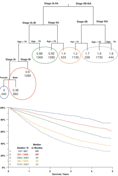
Stage, age, gender, and cell type were entered into a RPA analysis to generate a survival tree of recursive splits on the dataset. Viewing only the splits that were statistically significant after accounting for multiple tests, stage, age, and (in a limited partition of the data) gender remained as impor-tant variables (Figure 2). Unlike the results of a regression analysis, the survival tree resulting from an RPA method can
often provide an easy visualization of the relative importance of various factors in particular subsets of the data.
The most important factor overall is pTNM stage, and within stage categories, age is prognostic. Beyond that, in the under-70 stage IA group, female gender is a favorable prog-nostic factor. Using this recursive splitting algorithm, there was never a point at which cell type was found to be the most important factor in any partition of the data. Entering age as a continuous rather than categorical variable did not change the structure of the tree with respect to the relative importance and position of the factors involved in the splits, so a categorical representation (with a cutpoint at 70 years) was chosen. The RPA resulted in a survival tree with 10 terminal nodes that could be grouped according to hazard ratios to form five groups of approximately similar prognosis. Groups could be defined according to the following criteria: Stage IIIA— group 5.
Stage IIB—group 4, increase by one level (to group 5) if ageⱖ70.
0% 20% 40% 60% 80% 100% 0 2 4 6 8 10 Survival, Years BAC ADCA SQUAM LARGE ADSQ Deaths / N 230 / 517 2007 / 3335 2775 / 4463 423 / 650 125 / 172 Median in Months 83 45 44 34 26
A
B
0% 20% 40% 60% 80% 100% 0 2 4 6 8 10 Survival, Years IA IB IIA IIB IIIA Deaths / N 708 / 1715 991 / 1970 1098 / 1808 1028 / 1469 1735 / 2175 Median in Months 95 75 44 29 19FIGURE 1. Survival according toA, pathologic stage (IASLC proposals for 7th Ed.) andB, cell type. ADCA, adenocarcinoma; ADSQ, adenosqua-mous; BAC, bronchioloalveolar; LARGE, large cell; SQUAM, squa-mous cell.
Stage IIA— group 3, increase by one level if ageⱖ70. Stage IB— group 2, increase by one level if ageⱖ70. Stage IA Males— group 2, increase by one level if ageⱖ70 Stage IA Females— group 1, increase by 2 levels (to group 3)
if ageⱖ70.
For example, a stage IIA patient, age 80, would be placed in group 4. A stage IA patient, age 65, would be placed in group 2 if male, or group 1 if female.
Applying these definitions to the SEER data (n⫽9221) for validation resulted in the survival curves seen in Figure 3. All adjacent groups are significantly different from each other at the 0.0001 level, with hazard ratios between adjacent groups ranging from 1.34 to 1.75. Adding cell type as a set of indicator variables to a regression model containing the RPA group variable was a significant addition by a global test, largely because of differences between squamous cells, ade-nocarcinomas, and BAC. However, it should be noted that this addition only improved the R2 value (a measure of percent variance explained11) from 21.8 to 22.6.
In the 3027 cases where performance status data were available, performance status was independently prognostic for survival in an analysis adjusting for gender, stage, and cell type. Using the Zubrod scale, a performance status of 1 conferred a poorer prognosis than a performance status of 0 (H.R.⫽1.16,p⫽0.005), and the small number of cases (n⫽
35) with a performance status of two or higher had a worse prognosis than those with performance status 1 (H.R. ⫽ 1.61). Importantly, in this analysis of a subset of the data, the
findings for the different cell types, gender, age, and stage remained the same as those resulting from the entire dataset where performance status was not included. This suggests that the findings for these factors are independent of perfor-mance status.
Smoking history was examined in the subset where the data were available to distinguish between current (n ⫽
1258), former (n ⫽ 1155), and never-smokers (n ⫽ 54). Smoking status differed by cell type, so that 1.3% of squa-mous cell cases were never-smokers, when compared with 9.4% of adenocarcinomas and 20% of BAC. In this subset, there was no difference between former smokers versus never-smokers (H.R.⫽1.16,p⫽0.16), but current smokers had a worse prognosis than former smokers (H.R. ⫽ 1.21,
p⬍0.0001) and also worse than never-smokers (H.R.⫽1.41,
p ⫽ 0.0017). Definitions for “former smoker” may have varied, but it could be concluded that smoking conferred a negative prognosis in a univariate setting. In a multivariate analysis including stage, cell type, gender, and age, the difference between current and former smokers remained significant (p ⫽ 0.001), although the difference between former and never-smokers did not (p⫽0.93). The addition of smoking status as a factor in the model did not modify the observed effects of other factors.
DISCUSSION
Previous analyses of prognostic factors in surgically resected NSCLC have covered specific areas such as smoking
TABLE 3. Survival Statistics and Comparisons from Univariate and Multivariate Cox Proportional Hazards Regression Models for all Patients, and for Females and Males Separately
Factor
Overall Survival Median (mo)a/1
yr/5 yr All Patients Comparison
Unadjusted H.R.b All Patients
Adjusted H.R.c
All Patients Females Males
Cell type
BAC 83/92%/61%
Adenocarcinoma 45/82%/44% Adeno vs. BAC 1.56 (p⬍0.0001) 1.35 (p⬍0.0001) 1.42 (p⫽0.0009) 1.25 (p⫽0.02) Squamous cell 44/79%/43% Squam vs. Adeno 1.03 (p⫽0.291) 0.86 (p⬍0.0001) 1.02 (p⫽0.80) 0.83 (p⬍0.0001) Large cell 34/72%/41% Large vs. Squam 1.13 (p⫽0.023) 1.19 (p⫽0.0009) 1.09 (p⫽0.45) 1.19 (p⫽0.0032) Adenosquamous 26/73%/29% AdSq vs. Large 1.23 (p⫽0.046) 0.98 (p⫽0.846) 1.21 (p⫽p⫽0.38) 0.94 (p⫽0.60) Gender
Female 66/85%/52%
Male 40/79%/41% Male vs. female 1.32 (p⬍0.0001) 1.21 (p⬍0.0001) N/A N/A Age ⬍70 49/81%/46% ⱖ70 38/78%/38% Ageⱖ70 vs.⬍70 1.28 (p⬍0.0001) 1.51 (p⬍0.0001) 1.47 (p⬍0.0001) 1.52⬍.0001 TNM categoryd IA 95/93%/66% IB 75/89%/56% IB vs. IA 1.33 (p⬍0.0001) 1.30 (p⬍0.0001) 1.39 (p⫽0.0007) 1.25 (p⫽0.0002) IIA 44/82%/43% IIA vs. IB 1.39 (p⬍0.0001) 1.44 (p⬍0.0001) 1.48 (p⬍0.0001) 1.44 (p⬍0.0001) IIB 29/74%/35% IIB vs. IIA 1.28 (p⬍0.0001) 1.30 (p⬍0.0001) 1.43 (p⫽0.0003) 1.27 (p⬍0.0001) IIIA 19/65%/23% IIIA vs. IIB 1.44 (p⬍0.0001) 1.46 (p⬍0.0001) 1.44 (p⬍0.0001) 1.46 (p⬍0.0001)
Factors included were cell type, gender, age, and pathologic stage (according to proposed UICC/AJCC Version 7). All patientsN⫽9137; Females:N⫽2341; Males:N⫽6796. a
Median overall survival from Kaplan-Meier estimate. b
Hazard ratio andp-value from Cox proportional hazards regression analysis for the factors histology (by indicator variables), gender, age, and stage (by indicators), modeled separately. c
Hazard Ratio andp-value from a multivariate Cox proportional hazards regression model containing histology, gender (female as referent), age (⬍70 as referent), and stage (indicator). d
history,12,13 comorbidities,14,15 and general clinical and
de-mographic features,12,16 –22with the typical number of cases
ranging from less than 100 to approximately 5000. Other studies have had sample sizes ranging from 1000 to 19,000 and may have an impressive list of covariates to study but do not focus exclusively on surgically managed patients.23–27A
systematic review of the literature in 2002 revealed that the number of factors studied overall is narrow, and the results heterogeneous.28
Studies of gene expression11,29 –33show promise
espe-cially as a means to identify early-stage patients who (by virtue of a poor prognosis or a predictive marker) might
benefit from adjuvant therapy. However, these studies usually involve a small number of patients, often from a range of treatment modalities. Gene expression profiling studies in lung cancer are (with some exceptions31–33) limited with
respect to cell type, and the prognostic capabilities of newly discovered profiles are rarely tested in conjunction with other prognostic factors. Gene signatures will only be applicable in the clinical setting when they can be consistently proven to provide information beyond that which can be derived from readily available clinical and anatomic factors.34Until then, in
lung cancer we rely primarily on stage category, certain emerg-ing biologic markers,35and clinical patient characteristics.
stage<2
age70<1 age70<1 age70<1 age70<1
stage<3 stage<5 0.88 0.92 1.4 1.2 1.7 1.6 1.9 0.6 0 0.36 11.37 35.92 92.79 53.37 34.46 17.23 153.17 74.88 836.11 4.12 13 33.6 19.3 12.5 6.24 55.4 27.1 303 0 0 0 0 0 1300 1280 533 1130 338 1730 444 1280 440 662 Stage IIB-IIIA Stage IA-IIA
Stage IIA Stage IIB Stage IIIA Stage IA-IB Age 70 Age < 70 Age 70 Age < 70 Stage IA Stage IB Female Male Age < 70 Age < 70
FIGURE 2. Survival tree output from recursive partitioning and amalgam-ation analysis. Upper number at each node is the log hazard ratio with the leftmost group as referent. Lower number is the number of cases present in each category. Groups with similar hazards are consolidated with colored circles. Cell types were in-cluded in the analysis, but do not seem in the survival tree output as none satisfied the selection process for important split points at any point in the algorithm. 0% 20% 40% 60% 80% 100% 0 1 2 3 4 5 Survival, Years 1 2 3 4 5 Deaths / N 147 / 857 521 / 1826 1354 / 3401 581 / 1070 1319 / 2067 Median in Months NR NR 54 35 25
FIGURE 3. Validation of prognostic groups as defined by RPA, SEER data,
n⫽9221. Groups are defined in the text and shown by color in the consolidated terminal nodes of the survival tree shown in Figure 2.
The methods used in the current analyses are meant to be complementary. Recursive partitioning analyses (RPA) have been used to develop prognostic categories to inform the development of staging systems, for example in lung cancer as part of the IASLC effort5and in multiple myeloma.36This
type of application presumes that patients will, in practice, be allotted to the categories that were generated by the RPA results to provide an estimate of prognosis. However, tree-based models such as RPA are often used as part of explor-atory analyses, to gain understanding without the intention of applying the terminal groupings to clinical practice. They may be used instead, for example, to create prognostic cate-gories within which to analyze the effect of a certain treat-ment variable,37or to suggest further refinement to existing
classification schemes.38 The hierarchical structure of RPA
modeling can elucidate relationships between factors that are not easily seen with traditional regression models, allowing for the detection of relationships based on conditional infor-mation in specific subsets of the cases. In the context of this study, the survival tree provides a visualization of the most important factors and the subsets within which they are most important. The amalgamation of the terminal nodes into groups facilitates validation of the results, but in this case there is no intent to apply these groups to clinical practice.
In this analysis of surgical NSCLC cases from the IASLC lung cancer staging project, age, stage category, and gender were all prognostic for survival. Cases designated as BAC had a superior prognosis compared with other histolo-gies, and squamous cell carcinomas were slightly favored over adenocarcinomas and large cell, but only after adjusting for imbalances in gender and stage. The superior prognosis for the squamous cell carcinomas when compared with other histologies was not seen in female patients.
In a subset of the dataset with sufficient information, former smokers had a better prognosis than current smokers, and the small number of never-smokers likely hindered our ability to draw a reliable conclusion about that category. Most studies report a survival advantage for nonsmokers or lighter smokers,12,13,39 although some report that adjusting for
his-tology or other factors eliminates the significance of smoking status.15,40Adjusting for smoking status did not modify the
effects of cell type or other factors. This finding suggests that results based on gender, stage, and cell type are valid even in the absence of a smoking history. However, smoking and cell type are clearly not independent, with the majority of non-smokers shown to be in the adenocarcinoma (especially BAC) categories in this and other studies,12,40 – 42 and both
factors are also related to gender. The interactions among these factors are important in NSCLC research, and the acquisition of smoking status should be included in the data collection process for future investigations. The importance of this factor will increase as newer data collections will be more likely to be linked with laboratory data on various molecular markers. Analyses sometimes focus on smoking intensity (by means of pack-years or other measures)12,39
rather than the “ever-smoked” versus “never-smoked” cate-gories because of inadequate numbers of nonsmoking lung cancer patients for study. However, the distinction between
ever-smokers and never-smokers will be important, and studies should be designed to recruit as many never-smokers as possi-ble. Performance status was also prognostic in the subset where data were available, and this too is an important factor that should not be ignored when collecting data on surgical cases.
Although gender and stage are almost universally rec-ognized as prognostic factors in lung cancer, reports vary regarding the impact of histology, and particularly regarding comparisons between the two most common non-small cell histologies, where results have been inconsistent. Much of the disparity could potentially be explained by the omission of other important factors in such analyses.
Some examples of recent findings are given in Table 4, showing various results for survival comparisons by histol-ogy. Within cases of resectable NSCLC, failure to adjust for stage and gender is the common feature in studies where the adenocarcinoma cell type is found to be superior. For exam-ple, the recent publication from the Japanese Joint Committee of Lung Cancer Registry16reported on an impressively large
collection of over 13,000 lung cancer cases that underwent surgery at participating hospitals during a 1-year period in 2002. In addition to reporting superior survival for female patients, the adenocarcinoma cell type was also found to carry a better prognosis overall, with a 67% 5-year survival for adenocarcinoma, compared with 53% for the squamous cell type. The authors acknowledged that the analyses of prognostic factors (including histology) were not adjusted for other factors such as stage or gender. Such adjustment may have led to a different result.
The authors also noted that BAC and adenocarcinoma with BAC were not identified separately but were included in the adenocarcinoma category. This is reasonable given that the BAC subclassification has undergone several changes in definition, with new recommendations from a joint effort of the IASLC, ERS, and ATS forthcoming. In any case, a BAC designation by any criteria seems to confer a more favorable prognosis,8 and so the inclusion of BAC with the other
adenocarcinomas may boost the survival prognosis for the adenocarcinoma category overall.17Conversely, the
identifi-cation of BAC as a category distinct from other adenocarci-nomas may reveal a survival advantage for squamous cell carcinoma relative to non-BAC adenocarcinoma.18
Studies that adjusted for stage (with or without gender) found either no difference between adenocarcinoma and squamous cell, or a superior prognosis for squamous cell. For studies that reported results from unadjusted along with results from adjusted analyses, the unadjusted analyses typi-cally found in favor of adenocarcinoma, whereas the adjusted analyses found no difference (Table 4).
In this study, there are clear imbalances between stage and cell type and gender and cell type. The squamous cell cases are only 13% female, versus 38% female for the adenocarcinomas. With regard to stage, 46% of the adeno-carcinoma cases were stage I, compared with just 35% for the squamous cell carcinomas. Although none of the contributing databases drew explicitly from a CT screening program, the large registries and consortia would not have excluded such cases. It is possible that some of the stage I cases were
detected, and among adenocarcinomas the screen-detected cases seem to have a much longer tumor volume doubling time.43Furthermore, given that the IASLC database
derives from multiple sources, some BAC cases or cases of adenocarcinoma with BAC features are certain to have re-mained unidentified within the larger adenocarcinoma cate-gory, with the majority of those most likely being female patients with early stage disease. Adjusting for stage and gender mitigates any effect of imbalance that would favor the adenocarcinoma cell type, and reveals a possible survival advantage for the squamous cell type. Cell type will continue to be an important factor for data collection, especially in clinical trials, as studies of newer agents may show different effects depending upon histology.44,45
Using the database of the IASLC International Staging Project, we conclude that for surgically managed pathologi-cally staged I-IIIA NSCLC (according to the IASLC propos-als for the 7th edition of TNM), age, gender, and to a lesser degree certain cell types, in addition to pTNM stage are all prognostic. Stage remains to be the most important factor, followed by age, and in early stage cases, gender. The cases classified as BAC in this dataset would have varied from pure noninvasive BAC to invasive adenocarcinoma with BAC components. Nevertheless, this category had a prognosis distinct from the other subtypes. Regarding a comparison of the two most common NSCLC lung cancer histologies, the squamous cell carcinomas may have a better prognosis
than the non-BAC adenocarcinomas, particularly among male patients with early stage disease, but the question remains as to whether the undetected inclusion of the BAC subtype within the adenocarcinomas obscures what might otherwise be a survival advantage for squamous cells in female and male patients. Data collection for future studies should uniformly require smoking history in addition to the other factors.
ACKNOWLEDGMENTS
Supported by the AJCC grant “Improving AJCC/UICC TNM Cancer Staging.”
Eli Lilly and Company provided funding to support the International Association for the Study of Lung Cancer (IASLC) Staging Committee’s work to establish a database and to suggest revisions to the 6th Edition of the TNM classifi-cation for Lung Cancer (staging) through a restricted grant. Lilly had no input into the committee’s analysis of the data or in their suggestions for revisions to the staging system.
APPENDIX 1
IASLC International Staging Committee
P. Goldstraw (Chairperson), Royal Brompton Hospital, Imperial College, London, UK; H. Asamura, National Cancer Centre Hospital, Tokyo, Japan; D. Ball, Peter MacCallum Cancer Centre, East Melbourne, Australia; V. Bolejack,
Can-TABLE 4. A Sampling of Recent Studies Involving NSCLC Outcomes Featuring Comparison of Adenocarcinoma with Squamous Cell Carcinoma
Reference Population
Cell Type with
Superior Survival Study Features Asamura
et al.16
Japanese Joint Committee of Lung Cancer Registry
Adenocarcinoma pTNM primary focus. Not adjusted for stage or gender. BAC not identified separately.
Foegle et al.24
Bas-Rhin, France, Regional Registry Adenocarcinoma Adjusted for resectable vs. others. No further stage adjustment within resectable category. BAC identified separately.
Caldarella et al.25
Tuscany Registry No difference Adjusted for stage and gender. BAC identified but not separately analyzed. (Adeno superior in females in unadjusted analyses).
Ou et al.22 California Cancer Registry stage IA/IB
cases
No difference Adjusted for stage and gender. Kawai
et al.12
Surgical cases from a multihospital registry in Japan
No difference Stage IA only, adjusted for gender and smoking status. (Nonsquamous superior in unadjusted analyses). Ferguson
et al.20
U. of Chicago surgical series No difference Adjusted for stage (Adeno superior in unadjusted analyses). Berardi
et al.15
Surgical Series Universitaà Politecnica del Marche Stage I–IIIB
No difference Adjusted for age, gender, and smoking status. Riquet
et al.17
Surgical series from two hospitals No difference Adjusted for stage. Revealed variation within subtypes. Strand
et al.18
Resected subset of the Norway Registry Squamous cell Adjusted for stage and gender. BAC identified separately. Some cases from this analysis were also contributed to the IASLC database.
Wisnivesky et al.19
SEER database USA Stage II surgical cases
Squamous cell Adjusted for T and N stage and gender. Categorization of BAC not reported.
Pfannschmidt et al.26
Thoraxclinic Heidelberg surgical series Squamous cell Adjusted for stage and gender. Some cases from this analysis were also contributed to the IASLC database.
Alexiou et al.21
cer Research and Biostatistics, Seattle, Washington, USA; E. Brambilla, Laboratoire de Pathologie Cellulaire, Grenoble Cedex, France; P. A. Bunn, University of Colorado Health Sciences, Denver, Colorado; D. Carney, Mater Misericordiae Hospital, Dublin, Ireland; K. Chansky, Cancer Research and Biostatistics, Seattle, Washington, USA; T. Le Chevalier (resigned), Institute Gustave Roussy, Villejuif, France; J. Crowley, Cancer Research And Biostatistics, Seattle, Wash-ington, USA; R. Ginsberg (deceased), Memorial Sloan-Ket-tering Cancer Center, New York, USA; D. Giroux, Cancer Research And Biostatistics, Seattle, Washington, USA; P. Groome, Queen’s Cancer Research Institute, Kingston, On-tario, Canada; H. H. Hansen (retired), National University Hospital, Copenhagen, Denmark; P. Van Houtte, Institute Jules Bordet, Bruxelles, Belgium; J. -G. Im, Seoul National University Hospital, Seoul, South Korea; J. R. Jett, Mayo Clinic, Rochester, Minnesota, USA; H. Kato (retired), Tokyo Medical University, Tokyo Japan; C. Kennedy, University of Sydney, Sydney, Australia; H. Kondo, Shizuoka Cancer Cen-tre, Sunto-gun, Japan; M. Krasnik, Gentofte Hospital, Copen-hagen, Denmark; J. van Meerbeeck, University Hospital, Ghent, Belgium; T. Naruke, (deceased), Saiseikai Central Hospital, Tokyo, Japan; E. F. Patz, Duke University Medical Center, Durham, North Carolina, USA; P. E. Postmus, Vrije Universiteit Medical Center, Amsterdam, the Netherlands; R. Rami-Porta, Hospital Mutua de Terrassa, Terrassa, Spain; V. Rusch, Memorial Sloan-Kettering Cancer Center, New York, USA; N. Saijo, National Cancer Centre East, Kashiwashi, Japan; J. P. Sculier, Institute Jules Bordet, Bruxelles, Bel-gium; F. A. Shepherd, University of Toronto, Toronto, On-tario, Canada; Y. Shimosato (retired), National Cancer Cen-tre, Tokyo, Japan; L. Sobin, Armed Forces Institute of Pathology, Washington, DC; W. Travis, Memorial Sloan-Kettering Cancer Center, New York, USA; M. Tsuboi, Tokyo Medical University, Tokyo, Japan; R. Tsuchiya (retired), National Cancer Centre, Tokyo, Japan; E. Vallieres, Swedish Cancer Institute, Seattle, Washington, USA; J. Vansteenkiste, Leuven Lung Cancer Group, Belgium; Yoh Watanabe (de-ceased), Kanazawa Medical University, Uchinada, Japan; and H. Yokomise (retired), Kagawa University, Kagawa, Japan.
Participating Institutions
O. Visser, Amsterdam Cancer Registry, Amsterdam, The Netherlands; R. Tsuchiya and T. Naruke (deceased), Japanese Joint Committee of Lung Cancer Registry; J. P. Van Meerbeeck, Flemish Lung Cancer Registry-VRGT, Brussels, Belgium; H.〉u¨lzebruck, Thorax-klinik am Universitatsklini-kum, Heidelberg, Germany; R. Allison and L. Tripcony, Queensland Radium Institute, Herston, Australia; X. Wang, D. Watson and J. Herndon, Cancer and Leukemia Group B (CALGB), USA; R. J. Stevens, Medical Research Council Clinical Trials Unit, London, England; A. Depierre, E. Quoix and Q. Tran, Intergroupe Francophone de Cancerologie Tho-racique (IFCT), France; J. R. Jett and S. Mandrekar, North Central Cancer Treatment Group (NCCTG), USA; J. H. Schiller and R. J. Gray, Eastern Cooperative Oncology Group (ECOG), USA; J. L. Duque-Medina and A. Lopez-Encuentra, Bronchogenic Carcinoma Co-operative Group of the Spanish Society of Pneumology and Thoracic Surgery (GCCB-S),
Spain; J. J. Crowley, Southwest Oncology Group (SWOG); J. J. Crowley and K. M. W. Pisters, Bimodality Lung Oncol-ogy Team (BLOT), USA; T. E. Strand, Cancer Registry of Norway; S. Swann and H. Choy, Radiation Therapy Oncol-ogy Group (RTOG), USA; R. Damhuis, Rotterdam Cancer Registry, The Netherlands; R. Komaki and P. K. Allen, MD Anderson Cancer Center-Radiation Therapy (MDACC-RT), Houston, Texas; J. P. Sculier and M. Paesmans, European Lung Cancer Working Party (ELCWP); Y. L. Wu, Guang-dong Provincial People’s Hospital, Peoples Republic of China; M. Pesek and H. Krosnarova, Faculty Hospital Plzen, Czech Republic; T. Le Chevalier and A. Dunant, International Ad-juvant Lung Cancer Trial (IALT), France; B. McCaughan and C. Kennedy, University of Sydney, Sydney, Australia; F. Shepherd and M. Whitehead, National Cancer Institute of Canada (NCIC); J. Jassem and W. Ryzman, Medical Univer-sity of Gdansk, Poland; G. V. Scagliotti and P. Borasio, Universita’ Degli Studi di Torino, S Luigi Hospital, Orbas-sano, Italy; K. M. Fong and L. Passmore, Prince Charles Hospital, Brisbane, Australia; V. W. Rusch and B. J. Park, Memorial Sloan-Kettering Cancer Center, New York, USA; H. J. Baek, Korea Cancer Centre Hospital, Seoul, South Korea; R. P. Perng, Taiwan Lung Cancer Society, Taiwan; R. C. Yung, A. Gramatikova, John Hopkins University, USA; J. Vansteenkiste, Leuven Lung Cancer Group (LLCG), Bel-gium; C. Brambilla and M. Colonna, Grenoble University Hospital-Isere Cancer Registry, France; J. Hunt and A. Park, Western Hospital, Melbourne Australia; J. P. Sculier and T. Berghmans, Institute of Jules Bordet, Brussels, Belgium; A. K. Cangir, Ankara University School of Medicine, An-kara, Turkey; D. Subotic, Clinical Centre of Serbia, Belgrade, Serbia; R. Rosell and V. Aberola, Spanish Lung Cancer Group (SLCG), Spain; A. A. Vaporciyan and A. M. Correa, MD Anderson Cancer Center-Thoracic and Cardiovascular Surgery (MDACC-TCVS), Houston, Texas, USA; J. P. Pig-non, T. Le Chevalier and R. Komaki, Institut Gustave Roussy (IGR), Paris, France; T. Orlowski, Institute of Lung Diseases, Warsaw, Poland; D. Ball and J. Matthews, Peter MacCallum Cancer Institute, East Melbourne, Australia; M. Tsao, Prin-cess Margaret Hospital, Toronto, Ontario, Canada; S. Dar-wish, Policlinic of Perugia, Italy; H. I. Pass and T. Stevens, Karmanos Cancer Institute, Wayne State University, USA; G. Wright, St. Vincent’s Hospital, Victoria, Australia; C. Legrand and J. P. van Meerbeeck, European Organization for Research and Treatment of Cancer (EORTC), Brussels, Belgium.
REFERENCES
1. Goldstraw P, Crowley JJ; On behalf of the IASLC International Staging Project. The international association for the study of lung cancer international staging project on lung cancer.J Thorac Oncol 2006;1: 281–286.
2. Rami-Porta R, Ball D, Crowley JJ, et al. International Staging Commit-tee; Cancer Research and Biostatistics; Observers to the CommitCommit-tee; Participating Institutions. The IASLC lung cancer staging project: pro-posals for the revision of the T descriptors in the forthcoming (seventh) edition of the TNM classification for lung cancer. J Thorac Oncol
2007;2:593– 602.
3. Rusch VW, Crowley JJ, Giroux DJ, et al. International Staging Com-mittee; Cancer Research and Biostatistics; Observers to the ComCom-mittee; Participating Institutions. The IASLC Lung Cancer Staging Project: proposals for the revision of the N descriptors in the forthcoming
seventh edition of the TNM classification for lung cancer. J Thorac Oncol2007;2:603– 612.
4. Postmus PE, Brambilla E, Chansky K, et al. International Association for the Study of Lung Cancer International Staging Committee; Cancer Research and Biostatistics; Observers to the Committee; Participating Institutions. The IASLC lung cancer staging project: proposals for revision of the M descriptors in the forthcoming (seventh) edition of the TNM classification of lung cancer.J Thorac Oncol2007;2:686 – 693. 5. Goldstraw P, Crowley JJ, Chansky K, et al. International Association for
the Study of Lung Cancer International Staging Committee; Participat-ing Institutions. The IASLC Lung Cancer StagParticipat-ing Project: proposals for the revision of the TNM stage groupings in the forthcoming (seventh) edition of the TNM Classification of malignant tumours.J Thorac Oncol
2007;8:706 –714.
6. Sculier JP, Chansky K, Crowley JJ, et al. International Staging Com-mittee and Participating Institutions. The impact of additional prognostic factors on survival and their relationship with the anatomical extent of disease expressed by the 6th edition of the TNM classification of malignant tumors and the proposals for the 7th edition.J Thorac Oncol
2008;3:457– 466.
7. Groome PA, Bolejack V, Crowley JJ, et al. IASLC International Staging Committee; Cancer Research and Biostatistics; Observers to the Com-mittee; Participating Institutions. The IASLC lung cancer staging project: validation of the proposals for revision of the T, N, and M descriptors and consequent stage groupings in the forthcoming (seventh) edition of the TNM classification of malignant tumours.J Thorac Oncol
2007;2:694 –705.
8. Travis WD, Colby TV, Corrin B, Shimosato Y, Brambilla E. Histolog-ical Typing of Lung and Pleural Tumors, 3rd Ed. Berlin: Springer, 1999. 9. Crowley JJ, Leblanc M, Jacobson J, Salmon S. Some Exploratory Tools for Survival Analysis. In Lin DY, Fleming TR, (Eds.), New York: Springer, 1997.
10. Cancer Statistics Branch Surveillance Program DCCPS. The SEER Program Code Manual, 3rd Ed. Bethesda, MD: National Cancer Insti-tute, National Institutes of Health, Public Health Service, U.S. Depart-ment of Health and Human Services, 1998.
11. Bianchi F, Nuciforo P, Vecchi M, et al. Survival prediction of stage I adenocarcinomas by expression of 10 genes. J Clin Invest2007;117: 3436 –3444.
12. Kawai H, Tada A, Kawahara M, et al. The Japan National Hospital Study Group for Lung Cancer. Smoking history before surgery and prognosis in patients with Stage IA non-small cell lung cancer—a multicenter study.Lung Cancer2005;49:63–70.
13. Nordquist LT, Simon GR, Cantor A, Alberts M, Bepler G. Improved survival in never-smokers vs. current smokers with primary adenocar-cinoma of the lung.Chest2004;126:347–351.
14. Moro-Silot D, Aubert A, Diab S, et al. Comorbidities and Charlson score in resected stage I nonsmall cell lung cancer. Eur Respir J2005;26: 480 – 486.
15. Berardi R, Brunelli A, Tamburrano T, et al. Perioperative anemia and blood transfusions as prognostic factors in patients undergoing resection for non-small cell lung cancer.Lung Cancer2005;49:371–376. 16. Asamura H, Goya T, Koshiishi Y, et al. Japanese Joint Committee of
Lung Cancer Registry. A Japanese lung cancer registry study. Prognosis of 13,010 resected lung cancers.J Thorac Oncol2008;3:46 –52. 17. Riquet M, Foucault C, Berna P, Assouad J, Dujon A, Danel C.
Prog-nostic value of histology in resected lung cancer with emphasis on the relevance of the adenocarcinoma subtyping.Ann Thorac Surg2006;81: 1988 –1995.
18. Strand TE, Rostad H, Moller B, Norstein J. Survival after resection for primary lung cancer: a population-based study of 3211 resected patients.
Thorax2006;61:710 –715.
19. Wisnivesky JP, Henschke C, McGinn T, Iannuzzi MC. Prognosis of stage II non-small cell lung cancer according to tumor and nodal status at diagnosis.Lung Cancer2005;49:181–186.
20. Ferguson MK, Wang J, Hoffman PC, et al. Sex-associated differences in survival in patients undergoing resection for lung cancer.Ann Thorac Surg2000;69:245–250.
21. Alexiou C, Onyeaka CV, Beggs D, et al. Do women live longer following lung resection for carcinoma? Eur J Cardiothorac Surg
2002;21:319 –325.
22. Ou SHI, Zell JA, Ziogas A, Anton-Culver H. Prognostic factors for survival of Stage I non-small cell lung cancer patients: a population-based analysis of 19,702 Stage I patients in the California Cancer Registry from 1989 –2003.Cancer2007;110:1532–1541.
23. Sanchez de Cos J, Miravet L, Abal J, et al. Lung cancer survival in Spain and prognostic factors: a prospective, multiregional study.Lung Cancer
2008;59:246 –254.
24. Foegle J, Hedelin G, Lebitasy M, Purohit A, Velten M, Quoix E. Specific features of non-small cell lung cancer in women: a retrospective study of 1738 cases diagnosed in Bas-Rhin between 1982 and 1997.
J Thorac Oncol2007;2:466 – 474.
25. Caldarella A, Crocetti E, Comin CE, Janni A, Lopes Pegna A, Paci E. Gender differences in non-small cell lung cancer: a population study.
Eur J Surg Oncol2007;33:763–768.
26. Pfannschmidt J, Muley T, Bulzebruck H, Hoffman H, Dienemann H. Prognostic assessment after surgical resection for non-small cell lung cancer: experiences in 2,083 patients.Lung Cancer2007;55:371–377. 27. Madrekar SJ, Schild SE, Hillman SL, et al. A prognostic model for
advanced stage non-small cell lung cancer. Pooled analysis of North Central Cancer Treatment Group trials.Cancer2006;107:781–792. 28. Brundage MD, Davies D, Mackillop WJ. Prognostic factors in non-small
cell lung cancer: a decade of progress.Chest2002;122:1037–1057. 29. Raponi M, Zhang Y, Yu J, et al. Gene expression signatures for
predicting prognosis of squamous cell and adenocarcinomas of the lung.
Cancer Res2006;66:7466 –7472.
30. Raz DJ, Ray MR, Kim JY, et al. A multigene assay is prognostic of survival in patients with early stage lung adenocarcinoma.Clin Cancer Res2008;14:5565–5570.
31. Sun Z, Wigle DA, Yang P. Non-overlapping and non-cell-type-specific gene expression signatures predict lung cancer survival.J Clin Oncol
2008;26:877– 883.
32. Lau SK, Boutros PC, Pintilie M, et al. Three-gene prognostic classifier for early-stage non-small cell lung cancer.J Clin Oncol2007;25:5562–5569. 33. Skrzypski M, Jassem E, Taron M, et al. Three-gene expression signature
predicts survival in early-stage squamous cell carcinoma of the lung.
Clin Cancer Res2008;14:4794 – 4799.
34. Shedden K, Taylor JM, Enkemann S, et al. Gene expression-based survival prediction in lung adenocarcinoma: a multi-site, blinded vali-dation study.Nat Med2008;14:822– 827.
35. Herbst RS, Heymach JV, Lippman SM. Lung cancer.N Engl J Med
2008;359:1367–1380.
36. Greipp PR, San Miguel J, Durie BG, et al. International staging system for multiple myeloma.J Clin Oncol2005;23:3412–3420.
37. Albain KS, Crowley JJ, Leblanc M, Livingston RB. Survival determi-nants in extensive-stage non-small-cell lung cancer: the Southwest Oncology Group experience.J Clin Oncol1991;9:1618 –1626. 38. Albain KS, Crowley JJ, Leblanc M, Livingston RB. Determinants of
improved outcome in small-cell lung cancer: an analysis of the 2,580-patient Southwest Oncology Group Database. J Clin Oncol 1990;8: 1563–1574.
39. Yoshino I, Kawano D, Oba T, Yamazaki K, Kometani T, Maehara Y. Smoking status as a prognostic factor in patients with stage I pulmonary adenocarcinoma.Ann Thorac Surg2006;81:1189 –1193.
40. Sakao Y, Miyamoto H, Oh S, et al. The impact of cigarette smoking on prognosis in small adenocarcinomas of the lung: the association between histologic subtype and smoking status.J Thorac Oncol2008;3:958 –962. 41. Wynder EL, Muscat JE. The changing epidemiology of smoking and lung cancer histology.Environ Health Perspect1995;103(Suppl. 8):143–148. 42. Yano T, Miura N, Takenaka T, et al. Never-smoking non-small cell lung
cancer as a separate entity: clinicopathologic features and survival.
Cancer2008;113:1012–1018.
43. Detterbeck FC, Gibson CJ. Turning gray: the natural history of lung cancer over time.J Thorac Oncol2008;3:781–792.
44. Scagliotti GV, Parikh P, von Pawel J, et al. Phase III study comparing cisplatin plus gemcitabine with cisplatin plus pemetrexed in che-motheapy-naive patients with advanced-stage non-small-cell lung can-cer.J Clin Oncol2008;26:3543–3551.
45. Einhorn LH. First-line chemotherapy for non-small-cell lung cancer: is there a superior regimen based on histology?J Clin Oncol 2008;26: 3485–3486.