Regenerating the damaged central
nervous system
Philip J. Horner & Fred H. Gage
The Laboratory of Genetics, 10010 North Torrey Pines Road, The Salk Institute, La Jolla, California 92037, USA
...
It is self-evident that the adult mammalian brain and spinal cord do not regenerate after injury, but recent discoveries have forced a reconsideration of this accepted principle. Advances in our understanding of how the brain develops have provided a rough blueprint for how we may bring about regeneration in the damaged brain. Studies in developmental neurobiology, intracellular signalling and neuroimmunology are bringing the regeneration ®eld closer to success. Notwithstanding these advances, clear and indisputable evidence for adult functional regeneration remains to be shown.
I
njury to the adult central nervous system (CNS) isdevastat-ing because of the inability of central neurons to regenerate correct axonal and dendritic connections. The consequences of injury are not just a break in communication between healthy neurons, but a cascade of events that can lead to neuronal degeneration and cell death (Box 1). In contrast to ®sh, amphibia, and the mammalian peripheral nerves and developing central nerves, adult central mammalian neurons do not regrow functional axons after damage. The inability of the adult neurons to regrow after injury cannot be entirely attributed to intrinsic differ-ences between adult CNS and all other neurons. As reported by
RamoÂn y Cajal1 in 1928, Tello showed in 1911 that adult CNS
neurons could regrow if they were provided access to the permissive environment of a conditioned sciatic nerve. Seventy years passed
before Aguayo and colleagues2 replicated these studies with new
methods that de®nitively con®rmed that adult CNS neurons have regenerative capabilities. This ®nding revealed that the failure of CNS neurons to regenerate was not an intrinsic de®cit of the neuron, but rather a characteristic feature of the damaged environ-ment that either did not support or prevented regeneration. In the past 20 years, progress has been made in identifying the elements that are responsible for the differences between the adult CNS and peripheral nervous system environments, and in the past few years the molecular and cellular bases of regenerative compared with non-regenerative responses are beginning to be revealed.
Regeneration strategies developed from these new discoveries will be applicable to many CNS disorders. Spinal cord and, to a lesser degree, brain injury could be the most approachable, owing to the well de®ned loss of cells and axons and the relative lack of chronic pathological sequelae. Genetic disorders that result in aberrant axonal path®nding (such as MASA (mental retardation, aphasia, shuf¯ing gait and adducted thumbs) or Kallmann syndrome) or neuronal cell loss (such as macular degeneration or retinitis pig-mentosa) may also be amenable to regeneration. Degenerative diseases where a de®ned cell phenotype is lost, such as Parkinson's disease, Alzheimer's disease or amyotrophic lateral sclerosis, are also good targets, but may be more challenging because of the potential for continued cell loss or axonal degeneration. Finally, regeneration strategies may also be applied to less well de®ned disorders where diffuse cell and axonal loss can occur, such as vascular disease, tumour and infection of the CNS.
Strategies for regenerating the adult CNS
Regeneration in the adult CNS requires a multistep process. First, the injured neuron must survive, and then the damaged axon must extend its cut processes to its original neuronal targets. Once contact is made, the axon needs to be remyelinated and functional synapses
need to form on the surface of the targeted neurons. Several strategies have been undertaken that target different aspects of this process. We will summarize and evaluate critically the latest data emerging from the following regeneration strategies: cellular
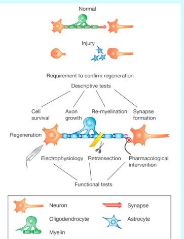
Regeneration
Retransection Requirement to confirm regeneration
Descriptive tests Electrophysiology Cell survival Re-myelination Axon growth Synapse formation Functional tests Pharmacological intervention Normal Injury Neuron Oligodendrocyte Myelin Synapse Astrocyte
Figure 1Steps to functional regeneration. Several criteria need to be met before functional regeneration can be validated. A combination of descriptive and functional analyses needs to be used to critically evaluate an experiment. Descriptive tests can be used to determine the survival and integrity of the injured system, whether axonal regeneration is present, and if appropriate synaptic connections and remyelination have occurred. The goal is to correlate the descriptive data with direct physiological and behavioural evidence for regeneration. Electrophysiological and pharmacological intervention can be used to assess the function and speci®city of the regenerated pathway. Ultimately, elimination of the regenerated pathway (for example, retransection) is important to determine its role in any reported functional recovery.
replacement, neurotrophic factor delivery, axon guidance and removal of growth inhibition, manipulation of intracellular signal-ling, bridging and arti®cial substrates, and modulation of the immune response.
As described below, an increasing number of studies have shown that an adult cut axon can be induced to regrow by either increasing the permissive cues or decreasing the non-permissive cues of the existing environment. Furthermore, a growing list of reports show that one strategy or another can induce some level of functional recovery following damage. However, it is not suf®cient to demon-strate axon elongation and behavioural improvement after injury to conclude that authentic functional regeneration is responsible for the outcome. There are many mechanisms that may account for observed functional recovery that do not require regeneration. These non-regenerative mechanisms are common in most experi-mental models of traumatic injury and need to be excluded before invoking functional regeneration as the cause of repair and recovery. The reason for sorting out the authentic mechanisms of functional recovery is that, without understanding the underlying basis of regeneration, little progress can be made beyond the phenomen-ological observation of recovery from injury.
Cellular replacement
In most cases of CNS trauma and disease, both neurons and glia are lost. Cell replacement is a vital step in CNS lesions, in which spared systems cannot supplant the function of lost cells. An excellent example is the loss of motor neurons in the case of injury to the
cervical spine. No matter how well the lesion is bridged or re-innervated, the target motor neurons that project to the muscles are irrevocably lost. Recognizing the need for cell replacement, several laboratories have taken advantage of fetal tissue grafts to replace cells after a variety of CNS insults3. These studies have even led to
clinical trials for humans. However, the inherent mechanical, physical and ethical hurdles of using fetal tissue limit its large-scale use. Ideally, a cell replacement source should be more homo-geneous, readily obtainable and syngeneic.
Stem cells have great promise as a source for introducing new neurons or glial cells to the damaged CNS. Neural stem cells have now been isolated from diverse regions of the developing and adult rodent brain and cultured using either epidermal or ®broblast
growth factor4. Stem cells are advantageous for research and
potential therapy because they are multipotent, and can be propa-gatedin vitro, genetically tagged with markers or therapeutic genes,
and grafted into the developing and mature intact CNS5±7. Once
transplanted into the brain, neural stem cells can adapt to the region of engraftment by differentiating into the appropriate neuronal and glial subpopulations8. In an extreme example, a single study has
reported that cells derived from the adult brain can become cells of the blood lineage when transplanted into irradiated mice9.
Replica-tion of the study has not been reported and is necessary if we are to consider this potential further. In addition, several studies have reported that stem-cell grafts can lead to neurogenesis in animal models of degeneration. For example, cortical neurons undergoing damage-induced apoptosis can be replaced morphologically by Inhibitory guidance or growth inhibition
[Ca2+↑cAMP/cGMP] [Ca2+↓cAMP/cGMP] Growth Retraction PTPase PKC/G Guidance or fasciculation Injured neuron Growth cone Denervated target Surviving neuron Target cell
P P P P Receptor Growth promoter Growth inhibitor Neuron Oligodendrocyte Myelin Synapse Growth cone Astrocyte Microglial cell Receptor (phosphorylated) = = = = (unphosphorylated) = = Cytoskeleton-associated protein
Figure 2Intracellular mediators of axon growth. Regeneration in the CNS requires that axons migrate long distances through the adult CNS to reconnect with an appropriate target. The axon growth cone contains the machinery for growth and must navigate the intact and injured CNS to reconnect with a target. Along its path, a growth cone can be diverted or converted to a non-migratory retraction bulb when it encounters inhibitory factors. Inhibition can be induced by molecules expressed on the surface of reactive cells and exported to the extracellular matrix, by inhibitory molecules found on myelin and by lack of diffusion or tropic molecules. Axonal growth and fasciculation can be induced by molecules expressed on other regenerating axons or by surviving axons, and by supportive molecules expressed by reactive cells. Many of these effectors appear to feed into the growth cone through a common mechanism involving the regulation of intracellular messengers. Changes in the levels of intracellular messengers affect the phosphorylation state of cytoskeleton-asso-ciated proteins, which leads to axon growth or collapse. Modi®cation of these intracellular mediators may be a powerful tool for converting inhibition to attraction within the injured CNS. PKC, protein kinase C.
transplanted embryonic neural stem cells that morphologically resemble pyramidal cells10. Replication and subsequent tests are
necessary to determine whether grafted cells are functional.
Evaluating functional replacement.Despite encouraging data that stem cells can morphologically replace cells of the CNS, few studies have shown a functional impact for this approach. One exception is a study showing the replacement of oligodendrocytes by trans-planted embryonic stem (ES) cells in a spinal-cord-injured rat11.
The result was improved locomotion; however, it is not clear whether the ES cells that survived contributed to the structural reorganization following injury, or whether the cells secreted some factor that in¯uenced the remaining damaged tissue. Although it is still unclear how these transplants may work, they illustrate an important point, speci®cally that neural replacement cells may not necessarily need to be derived from the brain. For example, ES cells and bone marrow stromal cells have been transplanted to the adult brain, where they are reported to differentiate into neural cells12±14.
This is promising, but the criteria to determine that a surviving neural cell is differentiated and functional need to become more rigorous as this ®eld advances.
Stimulation of self repair. The presence of neural stem cells resident within the CNS and the ability to regulate their numbers and fate may provide an alternative to transplantation. The exis-tence of neural stem cells has recently been shown in non-human primates and humans15,16. In addition, the adult brain4and spinal
cord17have been shown to contain stem cells that continually divide
throughout adult life. Recent studies have shown that cell genesis and the synaptic plasticity of these cells can be in¯uenced by stress, an enriched environment and physical exercise18±20. With respect to
injury, progenitor cells are capable of proliferation and
differentia-tion into mature myelinating oligodendrocytes21±23 in models of
acute demyelination. In addition, cortical neurons are replaced by dividing progenitor cells in a model of selective pyramidal cell apoptosis24. Strikingly, these newborn neurons extend new axons
several millimetres through the intact CNS. These ®ndings show that under the correct lesion conditions, CNS stem cells are capable of participating in cell replacement. Despite their inherent plasticity, however, it is clear that endogenous stem cells do not produce complete recovery in cases of severe trauma.
Whether a strategy of transplantation or endogenous replace-ment is used, the molecules and genes that govern cell proliferation, migration and differentiation need to be determined. In addition,
new cells that are grafted or generated in situ will need to be
engineered to be resistant to, or pharmacologically protected from, the toxic environment of the injured CNS. Finally, even when dead cells can be replaced with new ones, the new cells will need to be functionally integrated into the remaining circuitry, perhaps by surviving cells or by externally driven programs. Neurotrophic factor delivery
Many studies have shown both the cell survival and axon growth-promoting effects of neurotrophic molecules after injury to the
adult CNS in vivo25. Neurotrophins are not typically used as a
singular effector, however, but rather are combined with growth-promoting cells or matrices26. The assumption is that neurotrophins
will induce axonal growth only when the appropriate growth-permissive substrate is present. Schwann cells provide a growth-permissive substrate for propriospinal, sensory and supraspinal axons,
although axons do not then re-enter the CNS27. For example,
Menei et al.28used engineered Schwann cells that express
brain-derived neurotrophic factor (BDNF) to regrow supraspinal axons across a transected adult spinal cord. Schwann cells were seeded into a polymer channel that was placed in between the spinal cord stumps, and a Schwann cell trail was created by injecting engineered cells into the proximal and distal stump. In this experiment, BDNF did provide increased selection for trkB-expressing axons, which demonstrates the utility of neurotrophin selection; however, axons
once again did not leave the permissive substrate of the guidance channel or Schwann cell trails.
Are neurotrophins suf®cient?These studies illustrate one of the principal obstacles to the use of neurotrophins: the need to drive regenerating axons out of a permissive substrate or bridge and into the injured CNS. Some studies have shown that experimentally induced increases in neurotrophin levels within the tissue may be enough to facilitate this process. Grillet al.29used NT-3-producing
®broblasts to stimulate the regeneration of the cortical spinal tract in the hemisected adult rat spinal cord. In a similar study, Liuet al.30
used BDNF-producing ®broblasts to increase the growth of rubrospinal axons in the hemisected spinal cord. In both of these studies, tract tracing showed axons that regenerated around the lesion site and several millimetres into the target region, with a correlated improvement in behaviour. These ®ndings suggest that neurotrophins alone can stimulate axonal migration through the injured CNS, and that regeneration beyond the injury site can be associated with functional improvements. However, these studies do not prove a direct relationship between axonal regeneration and behavioural outcome. Alternative explanations include axonal sprouting of non-injured axons, activation of redundant pathways and alterations in the receptor number/phenotype or excitability of surviving neurons or glia. The existence of these mechanisms is well demonstrated in three studies in which behavioural changes were documented to occur as a result of neurotrophin administration, but no evidence for long-tract regeneration was found31±33. Evaluation of functional regeneration.The ®ndings of functional changes without substantial regeneration argue for a more detailed analysis of reported axonal regeneration (Fig. 1). One recent
example comes from Rameret al.34, who showed that
phenotypi-cally appropriate regeneration of sensory axons into the CNS could be accomplished by pumping neurotrophins intrathecally. This study is particularly thorough because, in addition to anatomical tracing, the investigators used both speci®c behavioural tests corresponding to the regenerated ®bre phenotypes and electro-physiology for con®rmation of connectivity. Most importantly, they subsequently re-injured the regenerated ®bres to demonstrate their requirement for functional recovery. Extension of these types of analyses to injured central axons is needed.
Despite the extensive use of neurotrophins, there are many remaining challenges. Neurotrophic factors comprise a large and complex family of molecules, and many subclasses of neurons and glia express unique complements of neurotrophic factor receptors. Many questions remain regarding practical application of these molecules; for example, diffusion of exogenously delivered neuro-trophins is variable. In addition, neuroneuro-trophins have many proper-ties aside from their roles in neuronal survival and axonal growth. For example, neurotrophins are anterogradely transported and released from presynaptic to postsynaptic targets. Neurotrophins also modulate membrane excitability, induce neuronal hypertro-phy, affect cell differentiation, and result in broad systemic effects. These functions for neurotrophins will need to be considered when interpreting future studies.
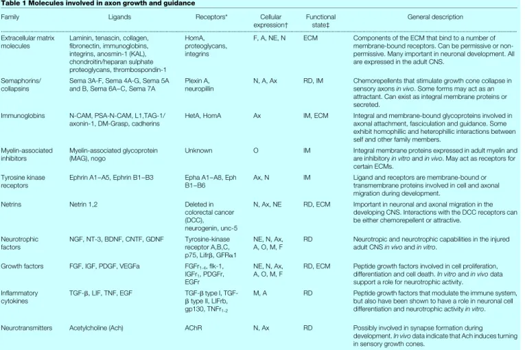
Axon guidance and removal of growth inhibition
Several growth-promoting molecules have a developmental role in axon guidance, fasciculation, synapse formation and, in the intact or injured adult, regeneration and activity-dependent plasticity (Table 1). Although the existence of axon guidance molecules can be inferred from transplantation experiments35, few direct data exist
to show that speci®c growth-promoting molecules in¯uence regen-eration in the adult CNS. Notable exceptions include L1 and polysialic acid neural cell adhesion molecule (PSA-NCAM), which have been shown to be associated with regenerating axons in the hippocampus or spinal cord-lesioned rat36,37.
Identi®cation of axon guidance factors on a molecular level has been a relatively recent event and, as a result, the use of these factors
has been limited in studies of repair. With the knowledge that adult central neurons can grow when given an alternative to the CNS environment, considerable effort has been placed on identifying and reversing the effects of growth inhibitory molecules. Proof that the CNS contains actual growth inhibitors was ®rst provided by Caroni, Schwab and Savio38,39in 1988. They discovered that the inhibitory
nature of cultured oligodendrocytes could be blocked by the monoclonal antibody, IN-1. Recently, the gene for this inhibitor
has been sequenced and namednogo40. Since Caroni and Schwab's
original discovery, several other factors have been identi®ed that can inhibit axon growth, including a variety of glycoproteins and proteoglycans41,42. In addition, the concept of axonal guidance by
inhibitory molecules was introduced from studies in the developing nervous system43±45.
Inhibition by the gliotic scar. Many of the inhibitory factors identi®ed for the immature and adult brain may be re-expressed after CNS injury in the adult. The chemorepellant semaphorin III/ collapsin I is re-expressed in scar tissue after injury to several areas
of the adult CNS46. The axon growth inhibitors chondroitin
sulphate and keratin are re-expressed after injury to the mature brain47,48and spinal cord49. The proteoglycan NG2, thought to be
expressed on immature glial cells, is increased after cortical injury and can inhibit axon growth when produced by astrocytes50.
Inter-estingly, an important characterization of the astrocytic response to injury has revealed heterogeneity in the expression of inhibitors by glial cells51. This ®nding underscores the complexity of the
environ-ment through which an injured axon must regrow and may reveal opportunities for intervention. Another important ®nding is the physical evidence that glial scarring can inhibit diffusion between cells at the injury site. Calculating diffusion coef®cients with
tetramethyl ammonium-sensitive electrodes, Roitbak and Sykova52
have shown that diffusion is decreased in regions of astrocytic hypertrophy and increased chondroitin sulphate. This ®nding indicates that scar may not only contain molecular inhibitors but may also act as a simple barrier to the diffusion of growth-promoting molecules.
The contribution of scar formation to axonal inhibition has been tested by using chondroitinase to increase the permissiveness of injured spinal cord sections to embryonic dorsal root ganglion axons53. Similarly, deletion of scar-forming astrocytes by gene
targeting results in increased axonal sprouting accompanied by a dysfunctional blood-brain barrier and increased immune activation in vivo54. Work by Stichelet al.55has suggested that the impermeable
nature of scar is due to its basal membrane. Reduction of collagen type IV in the lesioned fornix resulted in axonal migration in the scar region despite continued expression of proteoglycans and glial activation. However, this approach did not improve growth of the injured cortico-spinal tract37.
Inhibition by adult myelin. Considerable attention has been directed towards the disruption of myelin inhibitors. The addition of monoclonal antibodies to the neurite inhibitor IN-1 alone resulted in axonal regeneration of the cortical spinal tract and functional return of forelimb use in the adult rat56. Disruption of
myelin by immune activation57 or induction of an autoimmune
reaction to myelin58also enhanced regeneration in the adult spinal
cord. These data illustrate that blocking or eliminating endogenous white matter inhibitors may permit the natural regeneration poten-tial of injured nerves. However, the role of myelin inhibitors has been challenged by a study in which micro-injected adult peripheral sensory neurons were capable of extending axons along myelin tracts in the intact adult rat spinal cord59. These data indicate that,
although myelin inhibitors exist, the geometry of white matter may be such that myelin is not always inhibitory and can even, in this case, be permissive. The concept of myelin geometry was further
addressed in an in vitromodel of regeneration where peripheral
axons were inhibited only when the alignment of white matter was perpendicular to the direction of axonal growth60. It is vital to keep
in mind that these two experiments examined peripheral sensory neurons, and their behaviour may differ from that of neuronal cells that reside within the CNS.
Collectively these data indicate that growth inhibition may be caused by molecules that are present in the uninjured state, but also by production of the glial scar, which represents a molecular and physical barrier to regeneration. However, it is important to con-sider the novel role of growth inhibitory molecules in guiding axons along a white matter tract or around a cyst that no longer contains an appropriate target or scaffold. In the future, we will undoubtedly see increased manipulation of axon guidance molecules, both inhibitory and permissive, as a method for circumventing scar and stimulating axon growth.
Manipulation of intracellular signalling
Potential mediators of cell and axon fate.There are many diffu-sible and substrate-bound factors vital to cell survival and axonal growth. The intracellular mediators of these effects are beginning to be understood. One potential method for preventing cell death and stimulating regeneration is to alter the intracellular signals that a cell uses to transduce responses to injurious or growth-inhibiting substrates. For example, in apoptotic cell death, several transcrip-tion factors (such as fas, p53, c-Jun, bax and bcl-2) are involved and may represent common pathways to intervene and prevent cell loss61. Apoptotic cell death has been inhibited by blocking the ionic
changes that occur near the cell, increasing protease inhibitors that block the downstream effectors of cell death signals62or
upregulat-ing anti-apoptotic genes63. With regards to axon growth, several
intracellular factors, including growth-associated proteins, Ca2+ concentrations, transcription factors, and second messengers such as the cyclic nucleotides and inositol triphosphate, appear to have key roles (Fig. 2). Growth-associated proteins such as tubulin, actin and GAP-43 are thought to be harbingers of critical growth periods after injury and during development64±67. Manipulation of their
expression may be important for stimulating a cell to re-enter a growth mode. The anti-apoptotic transcription factor bcl-2 also plays a role in axonal plasticity68and regeneration following injury69.
Although high intracellular Ca2+has long been thought to be a
mediator of cell death, recent data suggest that frequent Ca2+
transients within a growth cone are correlated with retarded growth70. Several reports suggest that the intracellular
concentra-tions of cyclic AMP and cyclic GMP can modulate the response of a growth cone to inhibitory and stimulatory factors found in its environment71,72. In general, it appears that raising intracellular
cyclic nucleotide concentrations is suf®cient to convert an inhibi-tory response to an attractive one. For example, axonal repulsion of Xenopus spinal axons induced by semaphorin III/collapsin-1 is reversed in the presence of an analogue of cGMP73. The next step
is for these intriguingin vitro®ndings to be translated toin vivo contexts.
Application of signalling molecules in vivo. Modi®cation of intracellular responses has the bene®t of generating a broad general-ized response from the cell. This approach may be appealing for cell preservation and regeneration, as it may eliminate the need for multiple inhibitors of the many factors shown to induce apoptosis and growth cone collapse. However, there are several concerns
about in vivoapplication. Systemic approaches do not take into
account the variety of cell types that exist in the CNS and their individual responses to injury. In addition, Ca2+channel blockers and cyclic nucleotide analogues have adverse systemic side effects and can be lethal. Finally, timing is probably a vital issue. A recent study suggests that, once a sprouting axon has encountered inhibi-tory substrates, it loses its ability to respond to neurotrophins and their intracellular mediators74.
Bridging and arti®cial substrates
lesions of the CNS where a portion of the CNS is lost, a cyst has formed or regions of extensive scarring develop. A bridge is used to guide axons across these barriers and re-introduce axons into the remaining intact parenchyma. This familiar concept has led to some of the most promising evidence for regeneration using peripheral nerves or fetal tissue in the visual system and spinal cord. Several
new approaches are being developed that may complement the task of collecting peripheral nerves or embryonic tissue. However, the important advances will be guided by our molecular understanding of how the Schwann cell or the immature nervous system provides a permissive substrate for axonal growth. Genetic engineering of skin
®broblasts has shown that overexpression of neurotrophins75can
Box 1
Consequences of neuronal injury and strategies for repair
Following a speci®c traumatic or chemotoxic event, or as a result of ongoing degenerative processes, long-term structural and functional de®cits occur in the adult CNS. In severe cases, these insults are not repaired or compensated for by surviving systems. On a cellular level, these de®cits include demyelination, degeneration, abortive or aberrant sprouting, and cell death.
Demyelination.A demyelinated axon may maintain both its afferent and efferent connections but, due to a loss of myelination, poor or failed conduction results.
Axonal retraction.Injury to an axon itself or to the original cellular target of the axon can result in degeneration. Presynaptic, retrograde and trans-synaptic degeneration can occur. Synaptic conduction across a pathway is lost and a reactive cellular response, including
astrocytes and microglia, forms.
Sprouting.Axonal sprouting has been described for surviving neurons. It is typically abortive when a sprouting axon encounters an inhibitory matrix or scar, loss of neurotrophin support, or the presence of continuing in¯ammation or toxicity. Aberrant sprouting can occur when an axon reconnects to an inappropriate target. Synaptic conduction is restored but this pathway does not result in functional restoration.
Cell death.When a neuron is completely deprived of its source of growth factors and exposed to high levels of toxic molecules or in¯ammatory attack, it can undergo cell death. These patterns represent the anatomical correlates of brain dysfunction but also the speci®c processes that must be targeted for repair.
Demyelination
Conduction block
Axon retraction Reactive gliosis
Presynaptic Retrograde Trans-synaptic Sprouting Abortive Aberrant Cell death Injury
Neurotrophic factor delivery Manipulation of intracellular signalling Cellular replacement
Strategies for repair Neurotrophic factor delivery Cellular replacement
Modulation of immune response
Neurotrophic factor delivery Removal of growth inhibition Axon guidance
Manipulation of intracellular signalling Bridging
Modulation of immune response
Neurotrophic factor delivery Removal of growth inhibition Axon guidance
Manipulation of intracellular signalling Target cell Intact = Action potential = Excitatory synaptic potential = Conduction block Neuron Oligodendrocyte Myelin Synapse Retraction bulb Astrocyte Microglial cell
make these cells more permissive for axonal regeneration. In addition, cultured Schwann cells have been used to replace whole peripheral nerves37,76. Type I collagen has been used as a substrate for
grafting cells77, or alone as a bridge mixed with neurotrophins32.
Several recent reports have shown that cultured ensheathing glial cells taken from the olfactory epithelium may have the unique ability to guide injured axons back into the intact CNS78±80. In each
case, ensheathing glia grafted into the injured spinal cord show extensive migration and facilitate the regeneration of axons. These qualities are somehow tied to their intrinsic role of guiding and ensheathing growing axons across the border between the CNS and the peripheral nervous system in the adult olfactory bulb. As the mechanism by which ensheathing glial cells promote axonal re-growth remains to be reported, a call for a more physiological and molecular understanding of this process is warranted.
Arti®cial substrates. In the future, arti®cial substrates may be useful for repair of lesions where bridging is necessary. The ideal arti®cial substrate will have a molecular make-up that is easily manipulated and immune tolerant, and will contain a porous scaffold for nerve regeneration and cell repopulation that will be easily absorbed by the CNS. In the transected spinal cord, regenera-tion has been guided by arti®cial tubes that are seeded with growth-promoting Schwann cells27,81. These are composed of acrylonitrile
and vinylchloride and are semi-rigid, porous tubes that can be implanted and survive inde®nitely. Remaining concerns include mechanical hindrance and lack of absorption. Rapidly absorbable substrates are being tested, such as poly-a-hydroxyacids in the transected spinal cord82. Freeze-dried alginate gels and gel foam
soaked in neurotrophins are reabsorbable materials that have been
used to promote cell survival and regeneration83. One of the primary
limitations of arti®cial substrates is that invasive surgery is often necessary to install them. This downside is particularly relevant to the CNS. Although there are many hurdles to overcome, reabsorb-able substrates that are integrated with cells, guidance molecules or electrical ®elds represent an exciting new approach for repair. Modulation of the immune response
Does the immune system exacerbate injury?The immune system's role in injury and the process of regeneration involves peripheral and central, and cellular and humoral immune responses84. For a
considerable time there has been speculation about dualism of the immune response to injury in the brain. On the positive side, several investigators have suggested that the immune system may be protective and even assist regeneration. On the negative side, the immune system may cause bystander damage and progressive necrosis during the process of eliminating dead or dying tissue. Recently, investigators have begun to dissect out detrimental aspects of the immune system that may be responsible for unnecessary tissue loss and restrictions on axonal regeneration. For example, early inhibition of tumour necrosis factors or transforming growth factor-b2 signi®cantly decreases scarring and tissue loss, and can lead to improvement in functional outcome after CNS injury85,86.
Application of interferon-gmodulates several extracellular matrix molecules found in the CNS scar and inhibits astrocyte proliferation87. These observations demonstrate that certain aspects
of the immune response may substantially restrict axonal plasticity following trauma.
Regeneration and the central immune response.Blinzinger and Table 1 Molecules involved in axon growth and guidance
Family Ligands Receptors* Cellular
expression² Functionalstate³ General description
...
Extracellular matrix
molecules Laminin, tenascin, collagen,®bronectin, immunoglobins, integrins, anosmin-1 (KAL), chondroitin/heparan sulphate proteoglycans, thrombospondin-1
HomA, proteoglycans, integrins
F, A, NE, N ECM Components of the ECM that bind to a number of membrane-bound receptors. Can be permissive or non-permissive. Many important in neuronal development. All are expressed in the adult CNS.
Semaphorins/
collapsins Sema 3A-F, Sema 4A-G, Sema 5Aand B, Sema 6A±C, Sema 7A Plexin A,neuropillin N, A, Ax RD, IM Chemorepellents that stimulate growth cone collapse insensory axonsin vivo. Some forms may act as an attractant. Can exist as integral membrane proteins or secreted.
Immunoglobins N-CAM, PSA-N-CAM, L1,TAG-1/
axonin-1, DM-Grasp, cadherins HetA, HomA Ax IM, ECM Integral and membrane-bound glycoproteins involved inaxonal attachment, fasciculation and guidance. Some exhibit homophillic and heterophillic interactions between self and other family members.
Myelin-associated
inhibitors Myelin-associated glycoprotein(MAG), nogo Unknown O IM Integral membrane proteins expressed in adult myelin andare inhibitoryin vitroandin vivo. May act as receptors for certain ECMs.
Tyrosine kinase
receptors Ephrin A1±A5, Ephrin B1±B3 Epha A1±A8, EphB1±B6 Ax, N IM Ligand and receptors are membrane-bound ortransmembrane proteins involved in cell and axonal migration during development.
Netrins Netrin 1,2 Deleted in
colorectal cancer (DCC), neurogenin, unc-5
N, Ax, NE RD, ECM Important in neuronal and axonal migration in the developing CNS. Interactions with the DCC receptors can be either chemorepellent or attractive.
Neurotrophic
factors NGF, NT-3, BDNF, CNTF, GDNF Tyrosine-kinasereceptor A,B,C, p75, Lifrb, GFRa1
NE, N, Ax,
A, O, M, F RD Neurotropic and neurotrophic capabilities in the injuredadult CNSin vivoandin vitro. Growth factors FGF, IGF, PDGF, VEGFa FGFr1-4, ¯k-1,
IGFr1, PDGFr, EGFr
NE, N, Ax,
A, O, M, F RD, ECM Peptide growth factors involved in cell proliferation,differentiation and cell death.In vitroandin vivodata support a role for neurotrophic activity.
In¯ammatory
cytokines TGF-b, LIF, TNF, EGF TGF-btype II, LIFrb,btype I, TGF-gp130, TNFr1-2
M, A RD Peptide growth factors that modulate the immune system, but also have been shown to have a role in neuronal cell differentiation and neurotrophic activityin vitro. Neurotransmitters Acetylcholine (Ach) AChR N, Ax RD Possibly involved in synapse formation during
development.In vivodata indicate that Ach induces turning in sensory growth cones.
...
* HetA, heterophillic adhesion; HomA, homophillic adhesion.
² A, astrocyte; Ax, axon; F, ®broblast; M, microglia; NE, neuroepithelial; N, neuron; O, oligodendrocyte. ³ ECM, extracellular matrix; IM, integral membrane; RD, released/diffusible.
Kreutzburg88 originally suggested a proregenerative role for the
central immune system when they described the microglial response after injury to the facial nerve. Microglial cells are found to contact the membrane of neurons that will not undergo apoptosis and will eventually regenerate their axons. Because this lesion does not involve the peripheral immune response, these anatomical data suggested that microglia may somehow orchestrate the recovery of these cells by assuming a protective role. Supportive cytokines are released after injury to the facial nerve lesion or spinal cord, which suggests that candidate cytokines are involved in immune-mediated repair89. Finally, depletion of peripheral macrophages results in
tissue sparing and increased neuronal sprouting following spinal cord injury90. These data further indicate a disparity between central
and peripheral immune responses to injury. In the future, this ®eld will need to continue to identify the pathways that lead to the bene®cial and harmful immune responses.
Conclusions
The inherent complexity of the adult CNS as well as its amazing plasticity and adaptiveness make interpretingin vivoregeneration experiments challenging. As reports of functional regeneration become more common, it is important to critically evaluate the mechanisms of the observed functional changes. Case by case, it will be important to consider the inherent plasticity and variability of sparing in each injury model. In addition, several questions should be asked when evaluating a regeneration experiment. To what degree does sprouting or compensation by spared systems contri-bute to recovery? Do anatomical changes occur in conjunction with behavioural and physiological recovery? When regenerated path-ways are experimentally interrupted, is functional recovery once again lost? Technological advances will lead to more and better tools, allowing us to address these questions and identify the most promising therapeutic approaches. Identi®cation of the molecules that induce or inhibit regeneration will propel us closer to achieving functional regeneration. It is conceivable that most of these mol-ecules will be discovered in the next 5 years and the ®rst rational, functional therapy for regeneration, probably for spinal cord injury, may be in the clinic just 10 years from now.
Despite the progress in the last century of research on regenera-tion, however, Cajal's ¯owery decree, as translated by Raoul May, still resonates: ``once the development was ended, the founts of growth and regeneration of the axons and dendrites dried up irrevocably. In the adult centres the nerve paths are something ®xed, ended and immutable. Everything may die, nothing may be regenerated. It is for the science of the future to change, if possible, this harsh decree''.
The decree is lifted; the solution remains elusive. M
1. Ramon y Cajal, S.Degeneration and Regeneration of the Nervous System(Hafner, New York, 1928). 2. Richardson, P. M., McGuinness, U. M. & Aguayo, A. J. Axons from CNS neurones regenerate into PNS
grafts.Nature284,264±265 (1980).
3. Kordower, J. H. & Tuszynski, M. H.CNS Regeneration: Basic Science and Clinical Advances(eds Kordower, J. H. & Tuszynski, M. H.) 159±182 (Academic, San Diego, 1999).
4. Gage, F. H. Mammalian neural stem cells.Science287,1433±1438 (2000).
5. Flax, J. D.et al. Engraftable human neural stem cells respond to developmental cues, replace neurons, and express foreign genes.Nature Biotechnol.16,1033±1438 (1998).
6. Gage, F. H.et al. Survival and differentiation of adult neuronal progenitor cells transplanted to the adult brain.Proc. Natl Acad. Sci. USA92,11879±11883 (1995).
7. Liu, Y.et al. Intraspinal delivery of neurotrophin-3 using neural stem cells genetically modi®ed by recombinant retrovirus.Exp. Neurol.158,9±26 (1999).
8. Gaiano, N. & Fishell, G. Transplantation as a tool to study progenitors within the vertebrate nervous system.J. Neurobiol.36,152±161 (1998).
9. Bjornson, C. R., Rietze, R. L., Reynolds, B. A., Magli, M. C. & Vescovi, A. L. Turning brain into blood: a hematopoietic fate adopted by adult neural stem cells in vivo.Science283,534±537 (1999).
10. Snyder, E. Y., Yoon, C., Flax, J. D. & Macklis, J. D. Multipotent neural precursors can differentiate toward replacement of neurons undergoing targeted apoptotic degeneration in adult mouse neocortex.Proc. Natl Acad. Sci. USA94,11663±11668 (1997).
11. McDonald, J. W.et al. Transplanted embryonic stem cells survive, differentiate and promote recovery in injured rat spinal cord.Nature Med.5,1410±1412 (1999).
12. Brustle, O.et al. Embryonic stem cell-derived glial precursors: a source of myelinating transplants. Science285,754±756 (1999).
13. Ono, K.et al. Migration of exogenous immature hematopoietic cells into adult mouse brain parenchyma under GFP-expressing bone marrow chimera.Biochem. Biophys. Res. Commun.262,
610±614 (1999).
14. Bartlett, P. F. Pluripotential hemopoietic stem cells in adult mouse brain.Proc. Natl Acad. Sci. USA79,
2722±2725 (1982).
15. Kornack, D. R. & Rakic, P. Continuation of neurogenesis in the hippocampus of the adult macaque monkey.Proc. Natl Acad. Sci. USA96,5768±5773 (1999).
16. Eriksson, P. S.et al. Neurogenesis in the adult human hippocampus.Nature Med.4,1313±1317 (1998). 17. Horner, P. J.et al. Proliferation and differentiation of progenitor cells throughout the intact adult rat
spinal cord.J. Neurosci.20,2218±2228 (2000).
18. Gould, E. & Tanapat, P. Stress and hippocampal neurogenesis.Biol. Psychiatry46,1472±1479 (1999). 19. Kempermann, G., Kuhn, H. G. & Gage, F. H. More hippocampal neurons in adult mice living in an
enriched environment.Nature386,493±495 (1997).
20. van Praag, H., Kempermann, G. & Gage, F. H. Running increases cell proliferation and neurogenesis in the adult mouse dentate gyrus.Nature Neurosci.2,266±270 (1999).
21. McTigue, D. M., Horner, P. J., Stokes, B. T. & Gage, F. H. Neurotrophin-3 and brain derived neurotrophic factor induce oligodendrocyte proliferation and myelination of regenerating axons in the contused rat spinal cord.J. Neurosci.18,5354±5365 (1998).
22. Franklin, R. J., Gilson, J. M. & Blakemore, W. F. Local recruitment of remyelinating cells in the repair of demyelination in the central nervous system.J. Neurosci. Res.50,337±344 (1997).
23. Gensert, J. M. & Goldman, J. E. Endogenous progenitors remyelinate demyelinated axons in the adult CNS.Neuron19,197±203 (1997).
24. Magavi, S. S., Leavitt, B. R. & Mackliss, J. D. Induction of neurogenesis in the neocortex of adult mice. Nature405,951±955 (2000).
25. Stichel, C. C. & Muller, H. W. Experimental strategies to promote axonal regeneration after traumatic central nervous system injury.Prog. Neurobiol.56,119±148 (1998).
26. Cheng, H., Cao, Y. & Olson, L. Spinal cord repair in adult paraplegic rats: partial restoration of hind limb function.Science273,510±513 (1996).
27. Xu, X. M., Guenard, V., Kleitman, N., Aebischer, P. & Bunge, M. B. A combination of BDNF and NT-3 promotes supraspinal axonal regeneration into Schwann cell grafts in adult rat thoracic spinal cord. Exp. Neurol.134,261±272 (1995).
28. Menei, P., Montero-Menei, C., Whittemore, S. R., Bunge, R. P. & Bunge, M. B. Schwann cells genetically modi®ed to secrete human BDNF promote enhanced axonal regrowth across transected adult rat spinal cord.Eur. J. Neurosci.10,607±621 (1998).
29. Grill, R., Murai, K., Blesch, A., Gage, F. H. & Tuszynski, M. H. Cellular delivery of neurotrophin-3 promotes corticospinal axonal growth and partial functional recovery after spinal cord injury.J. Neurosci.17,5560±5572 (1997).
30. Liu, Y.et al. Transplants of ®broblasts genetically modi®ed to express BDNF promote regeneration of adult rat rubrospinal axons and recovery of forelimb function.J. Neurosci.19,4370±4387 (1999). 31. Jakeman, L. B., Wei, P., Guan, Z. & Stokes, B. T. Brain-derived neurotrophic factor stimulates
hindlimb stepping and sprouting of cholinergic ®bers after spinal cord injury.Exp. Neurol.154,170± 184 (1998).
32. Houweling, D. A., Lankhorst, A. J., Gispen, W. H., Bar, P. R. & Joosten, E. A. Collagen containing neurotrophin-3 (NT-3) attracts regrowing injured corticospinal axons in the adult rat spinal cord and promotes partial functional recovery.Exp. Neurol.153,49±59 (1998).
33. Ribotta, M. G.et al. Activation of locomotion in adult chronic spinal rats is achieved by transplantation of embryonic raphe cells reinnervating a precise lumbar level.J. Neurosci.20,5144± 5152 (2000).
34. Ramer, M. S., Priestley, J. V. & McMahon, S. B. Functional regeneration of sensory axons into the adult spinal cord.Nature403,312±316 (2000).
35. Bahr, M. Target-speci®c guidance cues for regenerating axons are reexpressed in the lesioned adult mammalian central nervous system.Adv. Neurol.73,83±90 (1997).
36. Aubert, I., Ridet, J. L., Schachner, M., Rougon, G. & Gage, F. H. Expression of L1 and PSA during sprouting and regeneration in the adult hippocampal formation.J. Comp. Neurol.399,1±19 (1998). 37. Weidner, N., Blesch, A., Grill, R. J. & Tuszynski, M. H. Nerve growth factor-hypersecreting Schwann cell grafts augment and guide spinal cord axonal growth and remyelinate central nervous system axons in a phenotypically appropriate manner that correlates with expression of L1.J. Comp. Neurol.413,
495±506 (1999).
38. Caroni, P. & Schwab, M. E. Two membrane protein fractions from rat central myelin with inhibitory properties for neurite growth and ®broblast spreading.J. Cell Biol.106,1281±1288 (1988). 39. Caroni, P., Savio, T. & Schwab, M. E. Central nervous system regeneration: oligodendrocytes and
myelin as non-permissive substrates for neurite growth.Prog. Brain Res.78,363±370 (1988). 40. Chen, M. S.et al. Nogo-A is a myelin-associated neurite outgrowth inhibitor and an antigen for
monoclonal antibody IN-1.Nature403,434±439 (2000).
41. Fawcett, J. W. & Asher, R. A. The glial scar and central nervous system repair.Brain Res. Bull.49,377± 391 (1999).
42. Fitch, M. T. & Silver, J. Glial cell extracellular matrix: boundaries for axon growth in development and regeneration.Cell Tissue Res.290,379±384 (1997).
43. Bernhardt, R. R. Cellular and molecular basis of axonal regeneration in the ®sh central nervous system.Exp. Neurol.157,223±240 (1999).
44. Goodman, C. S. Mechanisms and molecules that control growth cone guidance.Annu. Rev. Neurosci.
19,341±377 (1996).
45. Tessier-Lavigne, M. Eph receptor tyrosine kinases, axon repulsion, and the development of topographic maps.Cell82,345±348 (1995).
46. Pasterkamp, R. J.et al. Expression of the gene encoding the chemorepellent semaphorin III is induced in the ®broblast component of neural scar tissue formed following injuries of adult but not neonatal CNS.Mol. Cell Neurosci.13,143±166 (1999).
47. Jaworski, D. M., Kelly, G. M. & Hock®eld, S. Intracranial injury acutely induces the expression of the secreted isoform of the CNS-speci®c hyaluronan-binding protein BEHAB/brevican.Exp. Neurol.157,
327±337 (1999).
48. Haas, C. A., Rauch, U., Thon, N., Merten, T. & Deller, T. Entorhinal cortex lesion in adult rats induces the expression of the neuronal chondroitin sulfate proteoglycan neurocan in reactive astrocytes.J. Neurosci.19,9953±9963 (1999).
49. Lemons, M. L., Howland, D. R. & Anderson, D. K. Chondroitin sulfate proteoglycan immuno-reactivity increases following spinal cord injury and transplantation.Exp. Neurol.160,51±65 (1999).
50. Fidler, P. S.et al. Comparing astrocytic cell lines that are inhibitory or permissive for axon growth: the major axon-inhibitory proteoglycan is NG2.J. Neurosci.19,8778±8788 (1999).
51. Fitch, M. T. & Silver, J. Activated macrophages and the blood-brain barrier: in¯ammation after CNS injury leads to increases in putative inhibitory molecules.Exp. Neurol.148,587±603 (1997).
52. Roitbak, T. & Sykova, E. Diffusion barriers evoked in the rat cortex by reactive astrogliosis.Glia28,
40±48 (1999).
53. Zuo, J., Neubauer, D., Dyess, K., Ferguson, T. A. & Muir, D. Degradation of chondroitin sulfate proteoglycan enhances the neurite-promoting potential of spinal cord tissue.Exp. Neurol.154,654± 662 (1998).
54. Bush, T. G.et al. Leukocyte in®ltration, neuronal degeneration, and neurite outgrowth after ablation of scar-forming, reactive astrocytes in adult transgenic mice.Neuron23,297±308 (1999). 55. Stichel, C. C.et al. Inhibition of collagen IV deposition promotes regeneration of injured CNS axons.
Eur. J. Neurosci.11,632±646 (1999).
56. Thallmair, M.et al. Neurite growth inhibitors restrict plasticity and functional recovery following corticospinal tract lesions.Nature Neurosci.1,124±131 (1998).
57. Keirstead, H. S., Morgan, S. V., Wilby, M. J. & Fawcett, J. W. Enhanced axonal regeneration following combined demyelination plus schwann cell transplantation therapy in the injured adult spinal cord. Exp. Neurol.159,225±236 (1999).
58. Huang, D. W., McKerracher, L., Braun, P. E. & David, S. A therapeutic vaccine approach to stimulate axon regeneration in the adult mammalian spinal cord.Neuron24,639±647 (1999).
59. Davies, S. J.et al. Regeneration of adult axons in white matter tracts of the central nervous system. Nature390,680±683 (1997).
60. Pettigrew, D. B. & Crutcher, K. A. White matter of the CNS supports or inhibits neurite outgrowth in vitro depending on geometry.J. Neurosci.19,8358±8366 (1999).
61. Bredesen, D. E. Neural apoptosis.Ann. Neurol.38,839±851 (1995).
62. Harvey, N. L. & Kumar, S. The role of caspases in apoptosis.Adv. Biochem. Eng Biotechnol.62,107±128 (1998).
63. Blomer, U., Kafri, T., Randolph-Moore, L., Verma, I. M. & Gage, F. H. Bcl-xL protects adult septal cholinergic neurons from axotomized cell death.Proc. Natl Acad. Sci. USA95,2603±2608 (1998). 64. Bisby, M. A. & Tetzlaff, W. Changes in cytoskeletal protein synthesis following axon injury and during
axon regeneration.Mol. Neurobiol.6,107±123 (1992).
65. Skene, J. H. Axonal growth-associated proteins.Annu. Rev. Neurosci.12,127±156 (1989). 66. Benowitz, L. I. & Routtenberg, A. GAP-43: an intrinsic determinant of neuronal development and
plasticity.Trends Neurosci.20,84±91 (1997).
67. Hopker, V. H., Shewan, D., Tessier-Lavigne, M., Poo, M. & Holt, C. Growth-cone attraction to netrin-1 is converted to repulsion by laminin-netrin-1.Nature401,69±73 (1999).
68. Davies, A. M. The Bcl-2 family of proteins, and the regulation of neuronal survival.Trends Neurosci.
18,355±358 (1995).
69. Chen, D. F., Schneider, G. E., Martinou, J. C. & Tonegawa, S. Bcl-2 promotes regeneration of severed axons in mammalian CNS.Nature385,434±439 (1997).
70. Gomez, T. M. & Spitzer, N. C. In vivo regulation of axon extension and path®nding by growth-cone calcium transients.Nature397,350±355 (1999).
71. Qiu, J., Cai, D. & Filbin, M. T. Glial inhibition of nerve regeneration in the mature mammalian CNS. Glia29,166±174 (2000).
72. Song, H. J., Ming, G. L. & Poo, M. M. cAMP-induced switching in turning direction of nerve growth cones.Nature388,275±279 (1997).
73. Song, H.et al. Conversion of neuronal growth cone responses from repulsion to attraction by cyclic neuclotides.Science281,1515±1518 (1998).
74. Cai, D., Shen, Y., De Bellard, M., Tang, S. & Filbin, M. T. Prior exposure to neurotrophins blocks inhibition of axonal regeneration by MAG and myelin via a cAMP-dependent mechanism.Neuron22,
89±101 (1999).
75. Tuszynski, M. H.et al. Nerve growth factor delivery by gene transfer induces differential outgrowth of sensory, motor, and noradrenergic neurites after adult spinal cord injury.Exp. Neurol.137,157±173 (1996).
76. Sitchel, C. C., Hermanns, S., Lausberg, F. & Muller, H. W. Effects of schwann cell suspension grafts on axon regeneration in subacute and chronic CNS traumatic injuries.Glia28,156±165 (1999). 77. Kawaja, M. D. & Gage, F. H. Reactive astrocytes are substrates for the growth of adult CNS axons in the
presence of elevated levels of nerve growth factor.Neuron7,1019±1030 (1991).
78. Li, Y., Field, P. M. & Raisman, G. Repair of adult rat corticospinal tract by transplants of olfactory ensheathing cells.Science277,2000±2002 (1997).
79. Ramon-Cueto, A., Plant, G. W., Avila, J. & Bunge, M. B. Long-distance axonal regeneration in the transected adult rat spinal cord is promoted by olfactory ensheathing glia transplants.J. Neurosci.18,
3803±3815 (1998).
80. Ramon-Cueto, A., Cordero, M. I., Santos-Benito, F. F. & Avila, J. Functional recovery of paraplegic rats and motor axon regeneration in their spinal cords by olfactory ensheathing glia.Neuron25,425±435 (2000).
81. Xu, X. M., Zhang, S. X., Li, H., Aebischer, P. & Bunge, M. B. Regrowth of axons into the distal spinal cord through a Schwann-cell-seeded mino-channel implanted into hemisected adult rat spinal cord. Eur. J. Neurosci.11,1723±1740 (1999).
82. Gautier, S. E.et al. Poly(alpha-hydroxyacids) for application in the spinal cord: resorbability and biocompatibility with adult rat Schwann cells and spinal cord.J. Biomed. Mater. Res.42,642±654 (1998).
83. Houle, J. D. & Ye, J. H. Survival of chronically-injured neurons can be prolonged by treatment with neurotropic factors.Neuroscience94,929±936 (1999).
84. Matyszak, M. K. In¯ammation in the CNS: balance between immunological privilege and immune responses.Prog. Neurobiol.56,19±35 (1998).
85. Brewer, K. L., Bethea, J. R. & Yezierski, R. P. Neuroprotective effects of interleukin-10 following excitotoxic spinal cord injury.Exp. Neurol.159,484±493 (1999).
86. Logan, A., Green, J., Hunter, A., Jackson, R. & Berry, M. Inhibition of glial scarring in the injured rat brain by a recombinant human monoclonal antibody to transforming growth factor-b2.Eur. J. Neurosci.11,2367±2374 (1999).
87. DiProspero, N. A., Meiners, S. & Geller, H. M. In¯ammatory cytokines interact to modulate extracellular matrix and astrocytic support of neurite outgrowth.Exp. Neurol.148,628±639 (1997). 88. Blinzinger, K. & Kreutzberg, G. Displacement of synaptic terminals from regenerating motoneurons
by microglial cells.Z. Zellforsch. Mikrosk. Anat.85,145±157 (1968).
89. Streit, W. J.et al. Cytokine mRNA pro®les in contused spinal cord and axotomized facial nucleus suggest a bene®cial role for in¯ammation and gliosis.Exp. Neurol.152,74±87 (1998). 90. Popovich, P. G.et al. Depletion of hematogenous macrophages promotes partial hindlimb recovery
and neuroanatomical repair after experimental spinal cord injury.Exp. Neurol.158,351±365 (1999).
Acknowledgements
We thank E. Brandon, S. Colamarino, M.-C. Senut, L. Shihabuddin, H. van Praag, B. Benish and L. Horky for providing input during the preparation of this review. We appreciate the editorial assistance of M. L. Gage and the assistance of E. Grabowski in the preparation of illustrations. We are grateful for the continued support of The Christopher Reeve Paralysis Foundation, The Lookout Fund, The Parkinson's Disease Foundation and the National Institutes of Health. The content of this publication does not necessarily re¯ect the views or policies of the Department of Health and Human Services, nor does mention of trade names, commercial products, or organizations imply endorsement by the US Government.