doi:10.20776/S03035476-96E-2-P33
〔Chiba Medical Society Award Review〕
Novel approach for management of endometrial cancer:
drug repositioning of metformin
Akira Mitsuhashi
Department of Reproductive Medicine, Graduate School of Medicine, Chiba University, Chiba 260-8670. (Received January 21, 2020, Accepted February 5, 2020, Published April 10, 2020.)
Address correspondence to Dr. Akira Mitsuhashi.
Department of Reproductive Medicine, Graduate School of Medicine, Chiba University, 1-8-1, Inohana, Chuou-ku, Chiba 260-8670, Japan.
Phone: +81-43-226-2121. Fax: +81-43-226-2122. E-mail: antira@faculty.chiba-u.jp
Abstract
Metformin, a mainstay drug used clinically for over 60 years, improves insulin sensitivity and is widely prescribed for type 2 diabetes mellitus. Since population-based studies have suggested that metformin decreases the incidence of cancer and cancer-related mortality in patients with diabetes, metformin has attracted attention in cancer research as well. Endometrial cancer (EC) is the most common gynecological cancer and exhibits the strongest association with obesity and insulin resistance, and as such, is hypothesized as a suitable target cancer for metformin evaluation.
In the in vitro experiment, metformin suppressed EC cell proliferation via AMPK-dependent and inAMPK-dependent pathways, as in other cancer cell lines. Metformin also showed an additive effect on cell proliferation when administered in combination with anticancer drugs. Since the dose that was necessary to suppress cell proliferation in vitro was notably higher than the serum concentration in metformin-administered patients, we next examined whether a clinical dose of metformin was effective in patients. Preoperative metformin treatment decreased serum-stimulated DNA synthesis and significantly reduced Ki-67 and topoisomerase IIα levels in endometrial tissues, indicating reduced growth-supporting potential in the serum of patients with EC. Collectively, metformin may act both directly and indirectly, with both pathways contributing to its anti-neoplastic activity. In a clinical setting, we conducted a phase II study of medroxyprogesterone (MPA) in combination with metformin as a fertility-sparing treatment for patients with atypical endometrial hyperplasia or EC. Combining metformin with MPA reduced relapse after remission and improved patient metabolic status. Following the trial, we have initiated a prospective randomized, open, blinded-endpoint, dose-response trial (phase IIb) of MPA plus metformin (jRCT 2031190065) to verify an appropriate metformin dose in a fertility-sparing treatment, which is ongoing.
Ⅰ.Introduction
Endometrial cancer (EC) is the most common gynecological cancer, and the fourth most common cancer among women in the US[1]. Its prevalence in Japan has increased almost fivefold over the past two decades. Among all cancers, EC shows the strongest association with obesity, with a 5 kg/m2increase in body
mass index (BMI) strongly associated with EC at a risk ratio of 1.59 with 95 % confidence interval (CI) of
1.50-1.68(P<0.0001)[2]. Moreover, the risk for EC in women with BMI ≥ 25 is two to four times greater than in women with BMI<25[3]. Additionally, the prevalence of hyperinsulinemia and insulin resistance
(IR) is high among patients with EC regardless of their obesity status[4]. EC risk also increases with diabetes mellites (DM)[5,6]. This association can be largely accounted for by obesity.
Mechanisms that link excess weight and cancer risk have not been fully understood. Nonetheless, insulin and the insulin-like growth factor (IGF) axis are the most studied candidates. Insulin and IGF-1 signaling via the insulin and IGF-1 receptors, respectively, promote cellular proliferation and inhibit apoptosis via the phosphoinositide 3 kinase/AKT/mammalian target of rapamycin (mTOR) pathway and mitogen-activated protein kinase (MAPK) pathway in many tissue types
[7]. Metformin is a biguanide widely prescribed to treat type 2 DM that inhibits complex I of the mitochondrial electron transport chain[8]and stimulates adenosine monophosphate-dependent kinase (AMPK)[9], which reduces gluconeogenesis while enhancing glucose uptake in skeletal muscles to reduce insulin levels. Recently, metformin has attracted attention in cancer research since population-based studies suggest that it decreases the incidence of cancer and cancer-related mortality in patients with diabetes. Multiple meta-analyses have reported that metformin is associated with a decreased risk of breast, colon, liver, pancreas, prostate, endometrial, and lung cancer[10,11].
We hypothesized that metformin could be useful for EC prevention and treatment because of its direct and indirect actions improving insulin resistance and
abnormal glucose tolerance. Therefore, we conducted basic and clinical research with the aim of applying metformin to the treatment of endometrial cancer.
Ⅱ. Insulin resistance, diabetes, and endometrial cancer
A common link between obesity and EC is classically but not fully explained by unopposed estrogen[12]. Estrogen is produced via aromatization in peripheral adipocytes and does not encounter antagonization by progestin, and is associated with increased risk of EC[13,14]. However, unopposed estrogen alone cannot fully explain the risk of EC, and insulin resistance is also believed to be involved. A high prevalence of IR and abnormal glucose metabolism in patients with EC has been reported at 36-67%[4,15,16]. The marked difference in IR prevalence in the previous reports is thought to be race-dependent, where IR occurs more frequently among Hispanic women[4], for example. There is an apparent association between DM and increased risk of EC[5,6], and the prevalence of pre existing DM in patients with EC is around 15.5%-25%[17-19]. However, little is known about the exact prevalence of abnormal glucose metabolism, since not all studies examined glucose tolerance.
We evaluated 279 patients with EC among Japanese women[16], among whom 55(20.1%) were already diagnosed with DM. A 75-g oral glucose tolerance test was performed on the remaining 225 patients. Impaired fasting glucose, impaired glucose tolerance, and DM were newly identified in 7(2.5%), 69(24.7%), and
33(11.8%) patients, respectively. Eventually, more than half of EC patients (58.4%) were categorized to be in the pre-DM or DM stage, and IR was identified in 144(51.6%). In each age group, BMI ≥ 25 kg/m2
was 71% in patients<40 years old, 58% in patients
41-59 years old, 56% in patients 51-59 years old, and
17% in patients>60 years old in type I EC patients alone. Patients<40 years of age were more likely to be obese and have IR. Summarily, abnormal glucose metabolism, IR, and obesity were highly prevalent in
patients with EC. These results indicate that physicians should consider a patient’s metabolic status during the postoperative management of EC patients.
Ⅲ. In vitro effect of metformin on endometrial cancer cells
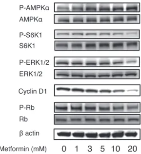
Metformin is an AMPK-dependent growth inhibitor in breast cancer[20], and similar antiproliferative effects have been demonstrated in several cancer cell lines. We treated established endometrial cancer cells with metformin, and similarly observed that cell growth substituted by thymidine uptake was suppressed in a concentration-dependent manner[21](Fig. 1A). Flow cytometry indicated that metformin caused a significant increase in the number of cells in the G1 phase of the cell cycle (Fig. 1B), where the cell cycle was arrested in G0/G1 accompanied by a strong decrease in cyclin D1 and phospho-RB and increased
expression of p27 Kip1 proteins. In addition, metformin induced phosphorylation of AMPKα and decreased
phosphorylation of S6K1 and ERK1/2(Fig. 2). However, inhibition of the AMPK pathway via siRNA-mediated AMPKα knockdown did not prevent the
antiproliferative effect of metformin, suggesting that its effects on the cell cycle are independent of the AMPK pathway (unpublished).
We also tested metformin in combination with cisplatin and adriamycin and found it to have additive effects such as increased apoptosis and suppression of thymidine uptake[22]. However, the antitumor effect of metformin in combination with cisplatin was attenuated under hypoxia compared to normoxia (Fig. 3). Mito Tracker staining indicated that metformin reduced mitochondrial fragmentation, indicating that metformin caused morphological and functional changes to the mitochondria. The additive effects of metformin on cisplatin-induced inhibition of cell proliferation were attenuated under hypoxic conditions, while metformin compromised mitochondrial structure and function.
Fig. 1 Metformin inhibits the proliferation of cells in a dose-dependent manner.(A) Ishikawa cells and HEC IB cells were treated with metformin for 48 h. Cell proliferation was measured using the thymidine incorporation assays. Results are presented as means ± standard errors of the mean. Asterisks indicate significant differences compared with metformin-free controls (P< 0.05, Mann-Whitney U-test). From Mitsuhashi et al., modified with permission[21]. (B) Flow cytometry analysis of Ishikawa cells after 24 h treatment with 5 or 10 mM metformin. The cells in the G1 phase of the cell cycle is indicated. After the addition of metformin, G1 cells increased by 18.6% in those treated with 5 mM metformin and 23% in those treated with 10 mM metformin compared to 16.8% in the control.
Fig. 2 Western blotting for MAPK·AMPK·mTOR signaling pathways and cell cycle proteins showing the dose-dependent change. Proteins extracted from Ishikawa cells that had been exposed to metformin (1-20 mM) for 24 h were subjected to western blotting for the analysis of cyclin D1 expression and the evaluation of AMPK, S6K1, ERK1/2, and Rb phosphorylation.
Ⅳ. In vivo effect of metformin on endometrial cancer cells
A number of unanswered questions remain prior to a clinical application. First is that the metformin concentration to suppress cell growth in vitro studies was much higher than the reported serum concentration of metformin in patients with orally administered
metformin for DM (1mM vs. 13µM by Cmax from interview form). Can such a low concentration of metformin reduce EM cell growth in vivo? Accordingly, we carried out window-of-opportunity studies using scheduled preoperative administration of clinical doses[21]by administering 1500-2250 mg/day to
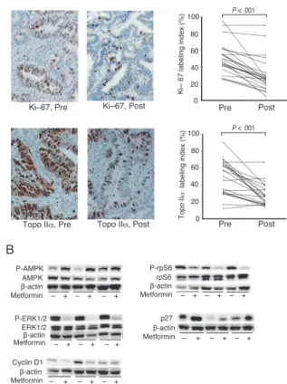
31 endometrial cancer patients for 4-6 weeks prior to surgical treatment. Preoperative metformin treatment significantly reduced Ki-67 labeling indices with a mean proportional decrease of 21.2% (95% CI 16.1-26.3; P
<.0001) and topoisomerase IIαlabeling indices with a
mean proportional decrease of 20.4% (95% CI, 13.0
-Fig. 3 Effect of combined metformin and cisplatin treatment on thymidine incorporation in Ishikawa cells cultured at different O2levels. (A) In cells cultured under
normoxic conditions (21% O2), higher concentrations
of metformin resulted in a greater reduction in thymidine incorporation relative to cells treated with cisplatin only. (B) The additive effects of metformin and cisplatin were attenuated under hypoxic conditions (1% O2). Data are
shown as mean ± standard error of six samples from three independent experiments. *P<0.005 and **P<0.001. From Uehara et al., modified with permission[22].MET: metformin, CDDP: cisplatin.
Fig. 4 Preoperative metformin administration reduces proliferative activity and alters cell proliferation signaling. (A) Preoperative metformin administration decreased immunostaining of Ki-67 and topoisomerase IIα in endometrial cancer tissues. Representative changes in immunostaining are shown for paired specimens. The change in labeling indices, expressed as a percentage of positively stained nuclei among a total of 500 nuclei, are shown for each pair and evaluated using the Wilcoxon signed-rank test. (B) Cell signaling molecules in endometrial cancer tissues were detected by western blotting, quantitated by densitometry, and normalized to β-actin. Pre: before the commencement of metformin treatment; Post: after metformin treatment. From Mitsuhashi et al., modified with permission[21].
reduces cancer proliferative activity by changing the endocrine environment.
We next investigated the effect of metformin on the expression of protein phosphatase 2A (PP2A) in EC tissues using immunohistochemistry[23]. Preoperative metformin treatment resulted in significantly reduced PP2A expression. Additionally, metformin administration resulted in significantly reduced PP2A regulatory subunit 4(PPP2R4) mRNA expression
(mean proportional decrease of 31.3% with 95% CI of
13-50; P=.039) in EC tissues. PPP2R4 knockdown reduced proliferation and induced apoptosis by activating caspases 3/7 in HEC265 and HEC1B cells. However, metformin was not capable of directly altering PPP2R4 mRNA expression in EC cancer cell lines. These findings indicate that metformin downregulated PPP2R4 expression indirectly in patients with EC, which might lead to apoptosis of endometrial cancer cells.
Ⅴ. Application of metformin in fertility-sparing treatment for patients with atypical endometrial hyperplasia and endometrial cancer
Progestin therapy is one of the most popular treatment options for preserving fertility in patients with atypical endometrial hyperplasia (AEH) and EC. The guidelines of the National Comprehensive Cancer Network (Version 3.2019) and the European Society of Gynecological Oncology Task Force for fertility-sparing treatment recommend progestin therapy for patients with AEH and EC who wish to preserve fertility[24]. Although fertility-sparing treatments have high rates of remission, they are also associated with high rates of relapse[25,26]. Based on our results of in vitro and in vivo analyses, metformin use may be a promising therapy for controlling recurrence after progestin treatment. In our phase II study of medroxyprogesterone (MPA)
plus metformin as a fertility-sparing treatment for AEH and EC patients, we found that metformin inhibits disease relapse after remission (UMIN 000002210) [27]. In this trial, patients received a daily oral dose
27.8; P<.0001)(Fig. 4A). Phospho-ribosomal protein S6 and phospho-extracellular signal-regulated kinase 1/2
were significantly decreased, and phospho-adenosine monophosphate activated protein kinase and p27 were significantly increased (Fig. 4B).
We then evaluated metformin concentration in serum and endometrial tissues of patients taking a clinical dose and found quite low concentrations, at 6.8-18.1 µM in serum and 1.2-5.1 µmol/kg in endometrium 2 h after administration (likely representative of Cmax). Since the minimum concentration required to suppress growth is approximately 1 mM in vitro, these concentrations represent<1/400 of the estimated required dose. DNA synthesis-stimulating activity in patient serum decreased significantly after metformin administration from pre-treatment levels (Fig. 5). Preoperative metformin use also caused a significant decrease in circulating factors (including insulin, glucose, insulin-like growth factor 1, and leptin), with oral administration decreasing insulin and
IGF-1 by approximately 40% and 15%, respectively. These findings support the possibility that metformin indirectly
Fig. 5 Humoral factors were responsible for the stimulatory action of metformin on thymidine uptake.
Thymidine uptake (DNA synthesis) activity of Ishikawa cells is measured in the presence of serum collected from patients before and during metformin treatment. Serum samples during metformin treatment were collected before and 2 h after a 75 g oral glucose tolerance test. Connecting lines represent paired samples obtained from the same patients. OGTT, oral glucose tolerance test; N.S., not significant. From Mitsuhashi et al., modified with permission[21].
Following this trial, we reported long-term outcomes for patients with AEH and EC on MPA plus metformin
[28]. Ninety-seven percent of patients treated with MPA and metformin (61/63) achieved CR within 18 months. Metformin significantly reduced relapse in comparison to a historical control of 24 patients treated only with MPA. Relapse-free survival at 3 years in patients with EC treated with MPA plus metformin and MPA alone was 79.7% and 43.0%, respectively (P=.025 Fig.
7). Notably, metformin may be more efficacious for patients with BMI≥25 kg/m2, as these patients showed
significantly better prognoses than patients with BMI<
25 kg/m(2 odd ratio 0.27; 95% CI, 0.08-0.88; P=0.03).
We recently initiated a prospective randomized, open, blinded-endpoint design, dose-response trial
(phase IIb) of MPA plus metformin in fifteen institutions of Japan (jRCT 2031190065)(Fig. 8)
in order to define the appropriate metformin dose in a fertility-sparing treatment involving combined metformin and MPA among patients with AEH and EC and to further investigate long-term efficacy and safety. Patients were randomized to receive MPA only, MPA + 750 mg/day metformin, or MPA + 1500 mg/ day metformin taken simultaneously. If patients achieve remission during MPA treatment, metformin therapy will be continued until conception or disease recurrence in MPA +metformin group. The primary endpoint of the study is a 3-year relapse-free survival rate, which would indicate the achievement of remission without recurrence 3 years from the entry date of study for all subjects. Secondary endpoints include the relapse-free of 400 mg MPA for 24-36 weeks and metformin at
an initial dose of 750 mg/day (increased weekly by
750 mg up to 2250 mg/day in the absence of adverse effects) administered concurrently from the initiation of treatment until pregnancy was confirmed. Of the 36
patients, 29(81%) achieved a complete response (CR), with three (10%) relapsing during a median follow-up period of 38 months (Fig. 6). Metformin additionally prevented weight gain induced by MPA and improved insulin resistance.
Fig. 6 Kaplan-Meier curves for relapse-free survival in patients who achieved remission after treatment with metformin plus medroxyprogesterone acetate. AEH, atypical endometrial hyperplasia; EC, endometrial cancer. From Mitsuhashi et al., modified with permission[27].
Fig. 8 Study design of a prospective randomized, open, blinded-endpoint design, dose-response trial (phase IIb) of MPA plus metformin. See the text for
detailed explanation.
Fig. 7 Relapse-free survival of patients with endometrial cancer treated with metformin plus medroxyprogesterone acetate (MPA) compared with historical controls treated with MPA alone. From Mitsuhashi et al., modified with permission[28].
prospective cohort study. Cancer Epidemiol Biomarkers Prev 16, 276-80.
6) Zhang ZH, Su PY, Hao JH, Sun YH. (2013) The role of preexisting diabetes mellitus on incidence and mortality of endometrial cancer: a meta-analysis of prospective cohort studies. Int J Gynecol Cancer 23, 294-303. 7) Calle EE, Kaaks R. (2004) Overweight, obesity
and cancer: epidemiological evidence and proposed mechanisms. Nat Rev Cancer 4, 579-91.
8) Owen MR, Doran E, Halestrap AP. (2000) Evidence that metformin exerts its antidiabetic effects through inhibition of complex 1 of the mitochondrial respiratory chain. Biochem J 348, 607-14.
9) Zhou G, Myers R, Li Y, et al. (2001) Role of AMP-activated protein kinase in mechanism of metformin action. J Clin Invest 108, 1167-74.
10) Evans JM, Donnelly LA, Emslie-Smith AM, Alessi DR, Morris AD. (2005) Metformin and reduced risk of cancer in diabetic patients. BMJ 330, 1304-5.
11) Heckman-Stoddard BM, DeCensi A, Sahasrabuddhe VV, Ford LG. (2017) Repurposing metformin for the prevention of cancer and cancer recurrence. Diabetologia 60, 1639-47.
12) Key TJA, Pike MC. (1988) The dose-effect relationship between ‘unopposed’ oestrogens and endometrial mitotic rate: Its central role in explaining and predicting endometrial cancer risk. Br J Cancer 57, 205-12. 13) Crosbie EJ, Zwahlen M, Kitchener HC, Egger M,
Renehan AG. (2010) Body mass index, hormone replacement therapy, and endometrial cancer risk: a meta-analysis. Cancer Epidemiol Biomarkers Prev 19, 3119-30.
14) Potischman N, Hoover RN, Brinton LA, et al. (1996) Case-Control Study of Endogenous Steroid Hormones and Endometrial Cancer. J Natl Cancer Inst 88, 1127-35. 15) Berstein LM, Kvatchevskaya JO, Poroshina TE, et
al. (2004) Insulin resistance, its consequences for the clinical course of the disease, and possibilities of correction in endometrial cancer. J Cancer Res Clin Oncol 130, 687-93.
16) Mitsuhashi A, Uehara T, Hanawa S, Shozu M. (2017) Prospective evaluation of abnormal glucose metabolism and insulin resistance in patients with atypical endometrial hyperplasia and endometrial cancer. Support Care Cancer 25, 1495-501.
17) Zanders MM, Boll D, van Steenbergen LN, van de Poll-Franse LV, Haak HR. (2013) Effect of diabetes on endometrial cancer recurrence and survival. Maturitas 74, 37-43.
18) Nevadunsky NS, Van Arsdale A, Strickler HD, et al. (2014) Metformin use and endometrial cancer survival.
Gynecol Oncol 132, 236-40.
19) Ko EM, Walter P, Clark L, et al. (2014) The complex triad of obesity, diabetes and race in Type I and II endometrial cancers: prevalence and prognostic significance. Gynecol Oncol 133, 28-32.
20) Zakikhani M, Dowling R, Fantus IG, et al. (2006)
survival rate, the overall response rate to MPA therapy, the conception rate following treatment, the outcome of pregnancy, the toxicity evaluation, and the changes in IR and BMI. This study is presently ongoing.
Ⅵ.Conclusion
The direct and indirect effects of metformin on EC contribute to its anti-neoplastic activity. Due to its observed metabolic involvement, EC may be one of the best candidate cancers for metformin use. Studies, including ours, have indicated the potential uses of metformin against EC in combination with anticancer chemotherapeutic drugs and progestins. In the future, in addition to using fertility-sparing therapy, metformin may be applied in postoperative maintenance therapy for EC or as a preventative measure against endometrial carcinogenesis.
Acknowledgments
I would like to express my gratitude to Prof. Makio Shozu for critically reading the manuscript.
Conflict of interest
The author declares that he has no conflicts of interest, either financial or non-financial, with the contents of this article.
References
1) Siegel RL, Miller KD, Jemal A. (2019) Cancer statistics, 2019. CA Cancer J Clin 69, 7-34.
2) Bhaskaran K, Douglas I, Forbes H, dos-Santos-Silva I, Leon DA, Smeeth L. (2014) Body-mass index and risk of 22 specific cancers: a population-based cohort study of 5·24 million UK adults. Lancet 384, 755-65.
3) Bianchini F, Kaaks R, Vainio H. (2002) Overweight, obesity, and cancer risk. Lancet Oncol 3, 565-74. 4) Burzawa JK, Schmeler KM, Soliman PT, et al. (2011)
Prospective evaluation of insulin resistance among endometrial cancer patients. Am J Obstet Gynecol 204, 355 e351-357.
5) Friberg E, Mantzoros CS, Wolk A. (2007) Diabetes and risk of endometrial cancer: a population-based
and live birth rates with fertility-sparing therapy for endometrial cancer and atypical complex endometrial hyperplasia: a systematic review and metaanalysis. Am J Obstet Gynecol 207, 266 e261-212.
26) Gunderson CC, Fader AN, Carson KA, Bristow RE. (2012) Oncologic and reproductive outcomes with progestin therapy in women with endometrial hyperplasia and grade 1 adenocarcinoma: a systematic review. Gynecol Oncol 125, 477-82.
27) Mitsuhashi A, Sato Y, Kiyokawa T, Koshizaka M, Hanaoka H, Shozu M. (2016) Phase II study of medroxyprogesterone acetate plus metformin as a fertility-sparing treatment for atypical endometrial hyperplasia and endometrial cancer. Ann Oncol 27, 262-6.
28) Mitsuhashi A, Habu Y, Kobayashi T, et al. (2019) Long-term outcomes of progestin plus metformin as a fertility-sparing treatment for atypical endometrial hyperplasia and endometrial cancer patients. J Gynecol Oncol 30, e90.
Metformin is an AMP kinase-dependent growth inhibitor for breast cancer cells. Cancer Res 66, 10269-73. 21) Mitsuhashi A, Kiyokawa T, Sato Y, Shozu M. (2014)
Effects of metformin on endometrial cancer cell growth in vivo: a preoperative prospective trial. Cancer 120, 2986-95.
22) Uehara T, Mitsuhashi A, Tsuruoka N, Shozu M. (2015) Metformin potentiates the anticancer effects of cisplatin under normoxic conditions in vitro. Oncol Rep 33, 744-50.
23) Hanawa S, Mitsuhashi A, Shozu M. (2018) Antitumor effects of metformin via indirect inhibition of protein phosphatase 2A in patients with endometrial cancer. PLoS One 13, e0192759.
24) Rodolakis A, Biliatis I, Morice P, et al. (2015) European Society of Gynecological Oncology Task Force for Fertility Preservation: Clinical Recommendations for Fertility-Sparing Management in Young Endometrial Cancer Patients. Int J Gynecol Cancer 25, 1258-65. 25) Gallos ID, Yap J, Rajkhowa M, Luesley DM,