Original Article
Trichosanthin inhibits human ovarian cancer cells
growth due to apoptosis and autophagy
Chuanhua Cao1*, Huixiong Qi1*, Fang Chen2, Jingbo He1
1Department of Oncology, Xiangyang Central Hospital, Affiliated Hospital of Hubei University of Arts and Science, Xiangyang 441021, Hubei, P.R. China; 2Department of Gerontology, Affiliated Taihe Hospital, Hubei University of Medicine, Shiyan 442000, Hubei, P.R. China. *Equal contributors.
Received January 21, 2016; Accepted May 12, 2016; Epub March 15, 2017; Published March 30, 2017
Abstract: Trichosanthin (TCS) exhibits an anti-cancer effect on various human cancer cells. However, the mecha-nism of anti-cancer effect at the molecular level remains to be elucidated. In this study, the underlying anti-cancer mechanism of TCS in human ovarian cancer cells (OVCAR-3) was investigated using various molecular biology
tech-niques, such as flow cytometry, western blotting. We major focus on the potential roles of apoptosis and autophagy
in TCS inhibition of human ovarian cancer cells. The results demonstrated TCS inhibits human ovarian cancer cells
growth due to apoptosis and autophagy. TCS triggers autophagy firstly, which was confirmed by an increased ATG5
expression and promoted LC3 cleavage, and subsequent apoptotic cell death. The apoptotic cell death induced by TCS was attenuated by both pharmacological and genetic inhibition of autophagy. The autophagy inhibitor 3-MA,
which functions at the early stage of autophagy, significantly reduced TCS-induced cell death and caspase-3 activity in human ovarian cancer cells. We also demonstrated that inhibition of apoptosis had no effect on TCS-induced au -tophagy in OVCAR-3 cells. In conclusion, we found that a common pathway between au-tophagy and apoptosis exists in TCS-induced cell death in human ovarian cancer cells, which might shed light on drug therapy.
Keywords: Trichosanthin (TCS), ovarian cancer, autophagy, apoptosis
Introduction
Ovarian cancer is one of the major causes of death for women globally. The worldwide inci-dence of this cancer is 238700 diagnoses in 2012, and this leads to 151900 deaths [1]. The number is increasing year by year. Ovarian can-cer patients are usually diagnosed at an ad- vanced stage which substantially increases the risk of recurrence and early death [2]. Treatment of advanced ovarian cancer remains a major challenge because of the poor efficacy of cur-rent therapies and chemotherapy. New effec-tive drugs for ovarian cancer are urgently needed.
Trichosanthin (TCS), or Tian Hua Fen, a 27 kDa protein, is a bioactive component obtained from the root tuber of trichosanthes kirilowii. TCS possesses only a 247-amino -acid polypep-tide chain which belongs to the type I ribosome-inactivating protein (RIP). Like all other RIPs, TCS can inactivate the ribosomes of eukaryotic
cells by removing adenine-4324 in 28S rRNA and results in protein synthesis inhibition and ultimately cell death [3, 4]. Because of its effect on cells, TCS has been used for centuries in tra-ditional Chinese medicine as an abortifacient drug in early and middle-gestation for over 1500 years. Only in recent decades, a wide spectrum of biological and pharmacological activities of TCS has been reported, especially its anti-tumor and anti-HIV activities. TCS has been found to be active against a variety of tumors, including cervical cancer, choriocarci-noma, leukemia/lymphoma, stomach cancer, colon cancer, hepatoma, breast cancer, and prostate cancer.
that besides their role in the maintenance of the organismal and cellular homeostasis serve as a main target of tumor therapeutics [5]. Autophagy is a conservative self-degradation pathway occurred both in healthy tissues and cancer cells. It is reported that autophagy has dual roles in cancer, acting as both a tumor suppressor by preventing the accumulation of damaged proteins and organelles and as a mechanism of cell survival that can promote the growth of established tumors [6, 7]. The potential in cancer cells either suppress or induce the growth of cancer cells depending on the cellular microenvironment. In our research, autophagy inhibitor can partially inhibits apop-tosis induced by TCS but the apopapop-tosis inhibi- tor doesn’t influence autophagy markers. Our study established an important role of autoph-agy in TCS-induced cell apoptosis in human ovarian cancer cells.
Materials and methods
Materials and reagents
TCS was obtained from Sigma (St. Louis, MO, USA) and resolved in DMSO for use. The pri- mary antibody of ATG5, caspase-3 substrate, LC3-I and LC3-II antibody were purchased from Santa Cruz Biotechnology, Inc. (Santa Cruz, CA, USA). The autophagy inhibitor 3 methylade- nine (3-MA), caspase inhibitor z-VAD-FMK and Mono-Dansyl Cadaverine (MDC) were pur-chased from Sigmal. All other chemicals used in this study were of analytical reagent grade. Cell line and cell culture
Human epithelial ovarian cancer cell line OVCAR-3 was established in 1982 by T.C. Hamilton, et al. from the malignant ascites of a patient with progressive adenocarcinoma of the ovary [8]. It is an appropriate model system in which to study drug resistance in ovarian cancer. OVCAR-3 cell was obtained from ATCC. The cells were cultured in RPMI 1640 (Life Technology, USA) supplemented with 20% fetal bovine serum, 0.01 mg/mL insulin and 100 mg/L streptomycin, 100 IU/mL penicillin. The cell line was grown in a 5% CO2 incubator at 37°C
CellTiter-Glo® luminescent cell viability assay
Cell viability assay was performed using CellTiter-Glo luminescent assay (Promega)
according to manufacturer’s instructions. OVCAR-3 cells were cultured in 96-cell plates (plate type) at a density of 1×10⁵ cells/mL for
stimulation. Cells were divided into three groups for three different TCS treatment time. Each group was treated with DMSO or TCS with the final concentration 0.33, 0.67, 1, 3.3, 6.7, 10 μM. CellTiter-Glo measurements were taken at 12, 24 and 48 h to track cell proliferation. All experiments were repeated at least three times and the average values were used as the final results.
Flow cytometry analysis of apoptosis
We test the apoptosis using Annexin V and Propidium Iodide labeling analyzed on flow cytometry. Annexin V is a marker of early tosis. Propidium Iodide is a marker of late apop-tosis and necrotic cells. Cells were cultured in 24-well plate, after treated with DMSO, increas-ing concentrations of TCS or other inhibitors for indicated time, OVCAR3 cells (both viable and death cells) were harvested by trypsinization, washed with cold PBS and centrifuged at 100× g to obtain a pellet. The pellet was suspended in binding buffer and stained with fluorescein isothiocyanate (FITC)-labeled Annexin V and Propidium Iodide(TACS Annexin V-FITC kit, R&D Systems), then analyzed by a flow cytometry on FACScan (Becton Dickinson, USA) using CellQuest software.
Western blotting analysis
MDC staining analysis of autophagy
Mono-Dansyl Cadaverine (MDC) has been reported to label autophagic vacuoles in cells [9], which can be tracked with flow cytometry. In our study, MDC staining was used to analyze autophagy rate. MDC was purchased from Sigma. Autophagic vacuoles were labeled with 0.05 mM MDC in PBS at 37°C for 10 min, and then washed three times with PBS. Flow cytom-etry was then used to determine the fluores-cence intensity at the excitation wavelength of 488 nm.
Caspase 3 activity assay
Caspase-3 activity was assayed in cellular extracts using the Caspase-3 Colorimetric Assay kit (R&D Systems). Cells were cultured and treated with DMSO, TCS or inhibitors, then harvested and lysed in lysis buffer supplied in the kit. A total of 20 μL cell lysate with 100 μg of protein was needed for testing. The enzymat-ic reaction for caspase activity is carried out in a 96 well plate. Then the lysate was added with the caspase-3 substrate, DEVD-pNA in the reaction buffer and incubated at 37°C for 2 h. The plate was measured at an absorbance of 405 nm with an ELISA reader.
Statistical analysis
All data and results presented are representa-tive of, or calculated from, at least three inde-pendent experiments. The data collected for each group are expressed as means ±
stan-dard deviation (SD). P < 0.05 was regarded as statistically significant.
Results
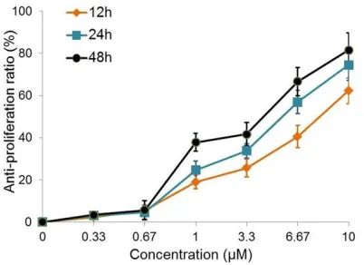
TCS inhibited cell viability in ovarian cancer cells
CellTiter-Glo® luminescent cell viability assay
was used to evaluate the cytotoxic effect of TCS towards OVCAR3 cells. As Figure 1 showed that the anti-proliferation rate increased with the increasing treatment time and concentra-tion, suggesting that TCS induced cell death in a time and concentration dependent manner. The anti-proliferation ratio reached above 80% upon 10 μM TCS treatment for 48 h. In the fol-lowing studies, we used 48 h as the treatment time and a higher range of TCS concentration (3.3, 6.7, 10 μM).
TCS induced cell apoptosis in ovarian cancer cells
The effect of TCS on the apoptotic cell death of OVCAR3 cells were examined by flow cytometry using Annexin V-FITC and propidium iodide labeling. Treatment with TCS increased the per-centage of early apoptotic and late apoptotic/ necrotic cells compared to the vehicle group and it was also in a dose-dependent (3.33 to 10 μM) manner (Figure 2A). Annexin V and PI double staining cells (late apoptosis and necrot-ic cells) reached above 30% at the concentra-tion of 10 μM TCS treatment (Figure 2B). As we know, apoptosis pathway is mediated upon the activation of the caspase cascade. Caspase-3 is characterized as both a marker and an ultimate executioner of cell apoptosis [10]. Western blotting was used to determine the caspase-3 activity. As showed in Figure 2D, caspase-3 activity was increased upon stimula-tion of 10 μM TCS, as the normalized caspase-3 proteolytic cleavage level was enhanced com-pared with control. This is consistent with the result tested by the Caspase-3 Colorimetric Assay kit (Figure 3B). It is indicated that TCS induced ovarian cancer cell apoptosis is medi-ated via caspase signal.
TCS induced cell autophagy in ovarian cancer cells
[image:3.612.90.291.71.217.2]We also evaluated the effects of TCS on autoph-agy. During the autophagy initiation phase, Figure 1.Anti-proliferation effect of TCS on ovarian
cancer. OVCAR3 cells were cultured and treated with
0, 0.33, 0.67, 1, 3.3, 6.67 or 10 μM TCS for 12, 24
or 48 hours. The cell viability was determined using
ATG5 plays a key role in the formation of autophagosomes, and LC system is required for autophagosome transport and maturation [11, 12]. ATG5 and LC3-II had been used for autophagy markers. As Figure 2D indicated, LC3-II and ATG5, were both upregulated in TCS treated cells. To confirm TCS-induced autopha-gy in OVCAR-3 cells, MDC staining assay was used to label the autophagic vacuoles. As shown in Figure 2C, the number of MDC stain-ing positive cells increased in each concentra-tion of TCS treated group compared with DMSO group, which is consistent with the western blot data. These results indicated that TCS can induce ovarian cancer cell autophagy.
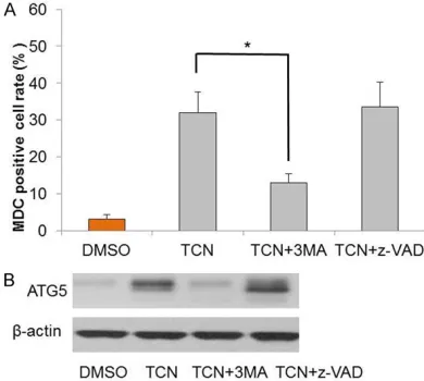
Inhibition of autophagy attenuated TCS-induce cell death
As TCS can both induce ovarian cancer cell autophagy and apoptosis, we tested whether
autophagy is linked with apoptotic cell death. OVCAR3 cells were pretreated with an autopha-gy inhibitor prior to TCS for apoptosis testing on flow cytometry. 3 methyladenine (3-MA) is a commonly used early stage inhibitor of autoph-agy which inhibits the activity of PI3K and blocks the formation of pre autophagosomes, autophagosomes and autophagic vacuoles [13]. As showed in Figure 3A, the number of Annexin V and PI staining double positive cells induced by TCS at 10 μM was significantly attenuated by pretreatment with 1 mM 3-MA for 1 h (P < 0.05). Another apoptosis marker caspase-3 activity was also attenuated by 3-MA pretreatment (Figure 3B), which is consis-tent with the result of western blotting (Figure 3C). All these data showed that autophagy inhibitor 3-MA partially abolished cell apoptosis induced by TCS. In addition, TCS-induced MDC autophagic vacuoles and ATG5 expression were Figure 2.TCS treatments induced apoptosis and autophagy in OVCAR3 cells. Cell treatment
condi-tion are DMSO only, 3.33, 6.67 or 10 μM TCS for
48 h. (A) Dot plot of untreated and treated cells
analyzed on flow cytometry. (B) Bar graph showing
the percentage of late apoptotic or necrotic cells (Annexin V+/PI+) analyzed in (A). (C) Flow cytometry determination of MDC staining positive cells
treat-ed with DMSO or TCS. (D) Western blotting analy
-ses of OVCAR3 total cell lysates with ATG5, LC3 and cleaved caspase-3 antibody. β-actin was used as
both confirmed decreasing upon 3-MA treat-ment (Figure 4A, 4B).
Inhibition of apoptosis had no impact on TCS-induced autophagy
As TCS induced apoptosis can be significantly reduced by autophagy inhibitor, autophagy may
be one cause of cell apoptotic death in OVCAR3 cells. An apoptosis pathway inhibitor z-VAD-FMK (a pan-caspase inhibitor) was used to investigate the effects of apoptosis on autoph-agy. Compared with TCS only group, z-VAD-FMK pre-treatment decreased both the percentage of apoptotic cells and caspase-3 activity as pre-dicted (Figure 3). But MDC positive cell rate and ATG5 expression were not impacted by the inhibitor z-VAD-FMK, as shown in Figure 4A and
4B. These data suggested that apoptosis inhib-itor z-VAD-FMK had no impact on autophagy in ovarian cancer cells.
Discussion
[image:5.612.94.518.72.285.2]In this study, we demonstrated that TCS-induced cell death in human ovarian cancer cells was mediated by both apoptosis and autophagy. In flow cytometry test of apoptosis, Annexin-V-FITC and PI positive cells were both significantly increased by TCS treatment. TCS enhanced caspase-3 activity which is a key player of cell apoptosis. TCS induced ATG5 expression, autophagic vacuoles and promoted LC3 cleavage. We had also confirmed that inhi-bition of autophagy by 3-MA attenuated TCS-induced cell apoptosis, otherwise apoptosis inhibitor Z-VAD-FMK had no impact on auto- phagy.
Figure 3.The effects of 3-MA or z-VAD-FMK on TCS-induced OVCAR3 cell apoptosis. Cell treatment conditions are
DMSO only, 10 μM TCS only, 10 μM TCS+1 mM 3-MA, 10 μM TCS+20 μM z-VAD-FMK. A. Bar graph showing the per
-centage of late apoptotic or necrotic cells (Annexin V+PI+) analyzed on flow cytometry. B. Caspase 3 activity tested by Caspase-3 Colorimetric Assay kit (R&D). C. Western blotting determination of caspase-3 activity. β-actin was used
as the internal control. *P < 0.05.
Figure 4.The effects of 3-MA or z-VAD-FMK on TCS-induced OVCAR3 cell autophagy. A. Flow cytometry determination of MDC staining positive cells. B.
[image:5.612.90.285.374.549.2]It is reported that TCS had been used as an anticancer agent in many cancer cell lines, including breast cancer, cervical cancer, cho- riocarcinoma, and leukemia/lymphoma, etc. Although the molecular mechanism differs from cancer to cancer, TCS induces a typical apoptosis process in most of the cancer cell lines. Zhang et al. found out TCS stimulated the production of reactive oxygen species (ROS) in JAR cells and this may leads to cell apoptosis [14]. It was reported that TCS inhibits protein kinase C (PKC) activity in HeLa and K562 cells, and the activation of PKC by PKC agonists inhibits TCS-induced apoptosis [15, 16]. Li et al. reported that when TCS induced the apopto-sis of HL-60 cells, caspase-9-mediated mito-chondrial pathway and the caspase-4-mediat-ed endoplasmic reticulum pathway are both involved [17]. The molecular mechanism of TCS on ovarian cancer is still unclear. We used a typical ovarian cancer cell line OVCAR3 cell to test the anticancer effects of TCS on ovarian cancer. Caspase cascade is a traditional pa- thway mediates cell apoptosis. The detailed mechanism of TCS on ovarian cancer needs further investigation.
A key observation of our study is that autopha-gy played an important role in TCS-induced apoptotic cell death. Autophagy has recently gained attention because of its paradoxical roles in cancer cell survival and death [18]. Many studies have indicated that autophagy can function as a protective mechanism in cells that are exposed to antitumor agents and that blocking autophagy can trigger the activation of apoptosis [19, 20]. Besides, autophagy can suppress tumorgenesis under different mecha-nism [21, 22]. In our study, autophagy was invoked to promote OVCAR3 cell death upon TCS treatment, while inhibiting autophagy at- tenuated apoptosis. It’s indicated that there is a common pathway between apoptosis and autophagy in TCS-induced ovarian cancer, au- tophagy may be one cause leading to apoptotic cell death. This result is consistent with a recent study on resveratrol by Lang et al. [23]. Apoptosis inhibitor z-VAD-FMK had no effect on autophagy also confirmed that autophagy may functioned up streaming of apoptosis in ovari-an covari-ancer cells. Scientists have focused on the crosstalk between apoptosis and autophagy. Several proteins have been reported regulating both apoptosis and autophagy [24, 25].
In conclusion, our research revealed a new insight into the complex role of autophagy on OVCAR3 cell apoptotic death which may be a target for the development of novel ovarian cancer therapies. Combination of TCS with an autophagy trigger would possibly enhance the anticancer effect. More evidence is needed to confirm this in the future.
Disclosure of conflict of interest
None.
Address correspondence to: Jingbo He, Department
of Oncology, Xiangyang Central Hospital, Affiliated
Hospital of Hubei University of Arts and Science, Xiangyang 441021, Hubei, P.R. China. E-mail: jing-bo_he1@126.com
References
[1] Torre LA, Bray F, Siegel RL, Ferlay J,
Lortet-Tieulent J, Jemal A. Global cancer statistics,
2012. CA Cancer J Clin 2015; 65: 87-108. [2] Jayson GC, Kohn EC, Kitchener HC, Ledermann
JA. Ovarian cancer. Lancet 2014; 384: 1376-1388.
[3] Sha O, Niu J, Ng TB, Cho EY, Fu XY, Jiang WQ.
Anti-tumor action of trichosanthin, a type 1 ri-bosome-inactivating protein, employed in tra-ditional Chinese medicine: a mini review. Can- cer Chemother Pharmacol 2013; 71: 1387-1393.
[4] Fang EF, Ng TB, Shaw PC, Wong RN. Recent
progress in medicinal investigations on tricho-santhin and other ribosome inactivating
pro-teins from the Plant Genus Trichosanthes. Curr
Med Chem 2011; 18: 4410-4417.
[5] El-Khattouti A, Selimovic D, Haikel Y, Hassan M. Crosstalk between apoptosis and autopha-gy: molecular mechanisms and therapeutic strategies in cancer. J Cell Death 2013; 6: 37-55.
[6] Yang ZJ, Chee CE, Huang S, Sinicrope FA. The role of autophagy in cancer: therapeutic impli-cations. Mol Cancer Ther 2011; 10: 1533-1541.
[7] Hippert MM, O’Toole PS, Thorburn A. Autophagy in cancer: good, bad, or both? Cancer Res 2006; 66: 9349-9351.
[8] Hamilton TC, Young RC, McKoy WM, Grotzinger KR, Green JA, Chu EW, Whang-Peng J, Rogan AM, Green WR, Ozols RF. Characterization of a
human ovarian carcinoma cell line (NIH:OVC-AR-3) with androgen and estrogen receptors. Cancer Res 1983; 43: 5379-5389.
vacuoles with a degradative compartment, us-ing monodansylcadaverine (MDC) and DQ-BSA. Methods Enzymol 2009; 452: 85-95. [10] Philchenkov A. Caspases: potential targets for
regulating cell death. J Cell Mol Med 2004; 8: 432-444.
[11] Yang ZJ, Chee CE, Huang S, Sinicrope FA. The Role of Autophagy in Cancer: Therapeutic Implications. Mol Cancer Ther 2011; 10: 1533-1541.
[12] Matsushita M, Suzuki NN, Obara K, Fujioka Y, Ohsumi Y, Inagaki F. Structure of Atg5.Atg16, a complex essential for autophagy. J Biol Chem 2007; 282: 6763-6772.
[13] Petiot A, Ogier-D enis E, Blommaart EF, Meijer AJ, Codogno P. Distinct classes of phosphati-dylinositol 3’-kinases are involved in signaling pathways that control macroautophagy in HT-29 cells. J Biol Chem 2000; 275: 992-998. [14] Zhang C, Gong Y, Ma H, An C, Chen D, Chen ZL.
Reactive oxygen species involved in trichosan-thin-induced apoptosis of human choriocarci-noma cells. Biochem J 2001; 355: 653-661. [15] Wang P, Chen LL, Yan H, Li JC. Trichosanthin
suppresses HeLa cell proliferation through in-hibition of the PKC/MAPK signaling pathway. Cell Biol Toxicol 2009; 25: 479-488.
[16] Li J, Xia XC, Nie H, Smith MA, Zhu XW. PKC
in-hibition is involved in trichosanthin-induced apoptosis in human chronic myeloid leukemia cell line K562. Biochim Biophys Acta 2007; 1770: 63-70.
[17] Li J, Xia XC, Ke YB, Nie HL, Smith MA, Zhu XW.
Trichosanthin induced apoptosis in HL-60 cells via mitochondrial and endoplasmic reticulum stress signaling pathways. Biochim Biophys Acta 2007; 1770: 1169-1180.
[18] Amaravadi RK, Thompson CB. The roles of therapy-induced autophagy and necrosis in cancer treatment. Clin Cancer Res 2007; 13: 7271-7279.
[19] Liu DL, Yang Y, Liu Q, Wang JJ. Inhibition of au -tophagy by 3-MA potentiates cisplatin-induced apoptosis in esophageal squamous cell carci-noma cells. Med Oncol 2011; 28: 105-111. [20] Ren Y, Huang F, Liu Y, Yang Y, Jiang Q, Xu
CM. Autophagy inhibition through PI3K/Akt in-creases apoptosis by sodium selenite in NB 4 cells. BMB Rep 2009; 42: 599-604.
[21] Hoyer-Hansen M, Bastholm L, Mathiasen IS, Elling F, Jaattela M. Vitamin D analog EB1089 triggers dramatic lysosomal changes and Beclin 1-mediated autophagic cell death. Cell Death Differ 2005; 12: 1297-309.
[22] Mathew R, Karp CM, Beaudoin B, Vuong N,
Chen G, Chen HY, Bray K, Reddy A, Bhanot G, Gelinas C, Dipaola RS, Karantza-Wadsworth V, White E. Autophagy suppresses tumorigenesis
through elimination of p62. Cell 2009; 137: 1062-1075.
[23] Lang FF, Qin ZY, Li F, Zhang HL, Fang ZH, Hao EK. Apoptotic cell death induced by resveratrol is partially mediated by the autophagy path- way in human ovarian cancer cells. PLoS One 2015; 10: e0129196.
[24] Li XH, Yan J, Wang LH, Xiao FJ, Yang YF, Guo XZ.
Beclin1 inhibition promotes autophagy and de-creases gemcitabine-induced apoptosis in Miapaca 2 pancreatic cancer cells. Cancer Cell Int 2013; 13: 26.
[25] Shi M, Zhang T, Sun L, Luo Y, Liu DH, Xie ST,
Song XY, Wang GF, Chen XL, Zhou BC, Zhang
YZ. Calpain, Atg5 and Bak play important roles in the crosstalk between apoptosis and