Original Article
Serum TIMP-2, NGAL and angiopoietin-2 as
biomarkers of coronary artery stenosis
Yeoeun Han*, Hyung Joon Joo*, Ha-Rim Seo, Seung-Cheol Choi, Jae Hyoung Park, Cheol Woong Yu, Soon Jun
Hong, Do-Sun Lim
Department of Cardiology, Cardiovascular Center, Korea University Anam Hospital, Seoul, Korea. *Equal contribu-tors.
Received March 9, 2016; Accepted June 3, 2016; Epub July 15, 2016; Published July 30, 2016
Abstract: Background and Aim: Coronary artery atherosclerosis develops through the interplay of lipid metabolism, vascular endothelial activation and immune cell activation. In this study, newly discovered biomarkers for vascular inflammation and activation were tested for diagnostic strength in angiographically significant coronary artery ste-nosis. Methods: Serum levels of NGAL, TIMP2, IL-8, GRO alpha, angiopoietin-2, bFGF and hsCRP were measured in 70 patients undergoing coronary angiography. Severity of disease was evaluated from angiographic findings based on the modified Gensini score. Results: Serum TIMP-2, NGAL and angiopoietin-2 levels were significantly elevated in patients with coronary artery stenosis (P = 0.002, P = 0.01 and P = 0.01, respectively) and correlated significantly with the number of stenotic coronary arteries. Receiver operating characteristic analysis of TIMP-2, NGAL and an-giopoietin-2 showed significantly increased areas under the curve (AUC = 0.795, 0.728, and 0.722, respectively). Multivariate analysis revealed that the elevated serum angiopoietin-2, old age and current smoking significantly and independently predicted the presence of coronary artery stenosis (P = 0.022, P = 0.006 and P = 0.004, respec-tively). Conclusion: Serum TIMP-2, NGAL and angiopoietin-2 levels may be useful in predicting the presence and severity of coronary artery stenosis.
Keywords: TIMP-2, NGAL, angiopoietin-2, coronary artery stenosis
Introduction
Coronary angiography may be regarded as the gold standard for diagnosis of coronary artery stenosis (CAS). Although an invasive endovas-cular technique, angiography only rarely results in serious complications such as vascular dis-section or thrombosis [1, 2]. Among the non-invasive methods for assessment of CAS, stre- ss tests, including the treadmill test, stress echocardiography, and myocardial perfusion scanning provide the mainstay. Overall accura-cy of the CAS diagnosis by using those non-invasive methods remains unsatisfactory, how-ever, as more than 30% of patients without suspected cardiac disease may be found by coronary angiography to have significant coro-nary artery stenosis [3]. The recent introdu- ction of computed tomographic angiography (CTA), a non-invasive alternative to coronary an- giography [4] may potentially extend the
accu-racy of CAS diagnosis. The overall sensitivity of CTA for coronary artery stenosis is reportedly as high as 85% to 95% and specificity, 90% to 97% [5, 6]. However, increases in heart rate and cardiac arrhythmia, use of a nephrotoxic contrast dye, and extensive radiation exposure related to CTA urge caution in its clinical appli-cation [7]. Technology with less hazardous, in- vasive and complex is needed to provide an accurate and acceptable population screening method for CAS.
Vascular inflammation and angiogenesis ap- pear to link the genetic and ecological compo-nents of CAD to the development and progre- ssion of vascular lesions. Biomarkers associ- ated with vascular inflammation and angio- genesis, and coronary atherosclerosis include neutrophil gelatinase-associated lipocalin (NG-
[image:2.612.91.524.84.294.2]AL), reported to be highly expressed in human arterial plaques [10]. Tissue inhibitor of me- talloproteinase-2 (TIMP-2) may be associated with plaque stability [11]. Angiopietin-2 and basic fibroblast growth factor (bFGF) influence the stability of atherosclerotic plaque through endothelial cell and vascular smooth muscle
Table 1. Baseline patient characteristics
Variables (n = 70)Total (n = 13)Normal 1-vessel disease (n = 22) 2-vessel disease (n = 17) 3-vessel disease (n = 18) valueP
-Men/Women 57/13 11/2 19/3 12/5 15/3 0.65
Age (years) 58.5 52.3±10.0 57.0±9.4 56.8±9.8 60.4±9.8 0.07 Body mass index (kg/m2) 24.0 (3.9) 24.0±3.2 23.4±2.9 24.9±4.1 25.5±3.1 0.82
Hypertension 39 (55.7%) 5 (38.5%) 11 (50.0%) 11 (64.7%) 12 (66.7%) 0.83 Diabetes mellitus 14 (20%) 1 (7.7%) 4 (18.2%) 4 (23.5%) 5 (27.8%) 0.16 Dyslipidemia 13 (18.8%) 5 (38.5%) 3 (13.6%) 3 (17.6%) 2 (11.8%) 0.14
Smoking status 0.74
Current smoker 30 (42.9%) 4 (30.8%) 11 (50.0%) 8 (47.1%) 7 (38.9%) Past smoker 16 (22.9%) 2 (15.4%) 6 (27.3%) 4 (23.5%) 4 (22.2%) Never smoker 24 (34.3%) 7 (53.8%) 5 (22.7%) 5 (29.4%) 7 (38.9%)
LDL cholesterol (mg/dl) 91.2±30.1 102.4±31.4 85.9±32.8 86.4±28.3 89.0±31.7 0.61 Triglycerides (mg/dl) 125.0 (88) 186.0 (146) 126.9±68.1 116.0±43.5 119.0±40.9 0.61 Creatinine (mg/dl) 1.0 (0.3) 1.1±0.2 0.9±0.2 1.0±0.2 1.0±0.2 0.62 eGFR (ml/min/1.73 m2) 77.5±14.6 68.7±9.1 81.9±19.2 71.6±10.6 76.6±11.9 0.28 Data are presented as the mean ± SD for continuous, normally distributed variables; median (interquartile range) for continu-ous, non-normally distributed variables; and number (percent) for categorical variables. eGFR, Estimated glomerular filtration rate.
[image:2.612.95.520.349.557.2]cell activation [12, 13]. Growth-regulated pro-tein alpha (GROα) and interleukin-8 (IL8) are associated with immune cell adhesion to ath-erosclerotic plaque [14].
The purpose of this study was to test a panel of biomarkers related to immune cell recruit-ment and plaque inflammation (NGAL, TIMP2, IL-8, and GROα), as well as vascular activa- tion (Angiopoietin-2 and bFGF) for diagnostic significance in coronary artery disease.
Materials and methods
Study subjects
Patients with clinical suspicion of coronary artery disease who were admitted to our clinic for the elective coronary angiography were prospectively selected for this study. The ex- clusion criteria were acute coronary syndrome, acute infection, autoimmune disease and in- flammatory disease, significant hepatic or re- nal diseases and anti-inflammatory drug use. In 70 patients, coronary artery stenosis was confirmed by coronary angiography. These pa- tients were enrolled for study through the de- partment of cardiology at Korea University Anam Hospital (Seoul, South Korea) beginning in July 2012 and ending in March 2013. The ethics committees of Korea University Anam Hospital approved this study. All patients gave their informed consent to use part of their blood for scientific purposes.
Coronary angiography interpretation
Significant coronary artery stenosis was defined as luminal stenosis of at least 50% in more than one major coronary artery. Measurement of coronary artery disease burden was based on the modified Gensini score [15, 16].
Measurements of serum NGAL, angiopoietin-2, TIMP2, bFGF, IL8, GRO alpha
Blood samples were obtained in EDTA tubes just before coronary angiography. Each serum sample was divided into three aliquots and stored at -80°C until analysis. Repeated free- ze-thawing was avoided. Serum NGAL, angio-poietin-2, TIMP2, bFGF, IL8 and GROα were measured by enzyme-linked immunosorbent assays (ELISA) using commercially available kits (Raybiotech, Inc., GA, USA) according to the manufacturer’s instructions. All samples were assayed in duplicate by a researcher who was blinded to sample identities.
Other laboratory parameters including high-sensitive C-reactive protein (hsCRP), creatinine, and lipids were evaluated at the chemistry laboratory of Korea University Anam Hospital. Statistics
Median values with interquartile range are reported for continuous variables, and counts with percent for categorical variables. The Kol- mogorov-Smirnov test was used to test con- tinuous variables for normality. Comparisons between two groups were performed using Student’s t-tests or a Mann-Whitney test for continuous variables, and chi2 tests or Fish-
[image:4.612.91.288.109.212.2]er’s exact test for categorical variables. The Jonckheere-Terpstra test was used to com- pare data from more than two groups. Corre- lations between variables were tested using Pearson’s method. Factors predicting signifi-cant coronary artery stenosis were tested us- ing multiple stepwise logistic regression ana- lysis. Variables tested for predictive power in- cluded age, sex, hypertension, diabetes, dyslip-idemia, smoking, body mass index, estimated glomerular filtration rate and serum creatinine, NGAL, TIMP-2, angiopoietin-2, bFGF and hs- CRP concentrations. Receiver operating char-acteristic (ROC) curves were constructed for the diagnosis of significant coronary artery stenosis. Two academic authors analyzed the database independently and reconciled any differences. All tests were two-sided and a P-value less than 0.05 was considered to in- dicate significance. All calculations were per-formed using the Statistical Package for the Social Sciences (SPSS) software (Version 18.0, SPSS Inc., Chicago, IL, USA).
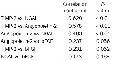
Table 2. Correlations between serum TIMP-2, NGAL, angiopoietin-2 and bFGF concentra-tions
Correlation
Results
Baseline demographic characteristics, strati-fied by number of coronary arteries with angio-graphically significant stenosis, are presented in Table 1. The mean age of participants was 58.5±10.2 years; 81% were male, 55.7% had hypertension, 20% had type 2 diabetes, and 18.8% had hypercholesterolemia.
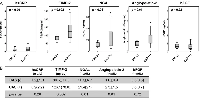
We measured the serum hsCRP, TIMP-2, NGAL, angiopoietin-2, bFGF, IL-8 and GROα. The IL-8 and GROα were not analyzed further because IL-8 levels in 20 samples and GROα levels in 59 samples were below the detection limits. TIMP-2, NGAL and angiopoietin-2 levels were signifi-cantly elevated in patients with coronary artery stenosis (P = 0.002, P = 0.01 AND P = 0.01, respectively; Figure 1).
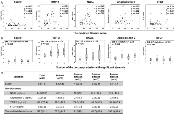
The serum TIMP-2, NGAL and angiopoietin- 2 levels were also significantly correlated with the number of stenotic coronary arteries and the angiographical severity based on the modi-fied Gensini score (Figure 2). When adjusted for age and sex, these markers still correlated significantly with severity of stenosis by angio- graphy. The strongest associations were obse- rved between NGAL and the modified Gensini score (r = 0.345, P = 0.004 (crude); *r = 0.343, P = 0.007 (adjusted)).
The serum TIMP-2, NGAL, angiopoietin-2, and bFGF levels correlated significantly with each other (Table 2). The correlation coefficient for TIMP-2 and NGAL was highest (r = 0.620, P < 0.01).
Correlations between biomarkers and clinical variables are shown in Table 3. The TIMP-2 level was statistically correlated with sex (r = 0.284, P = 0.019) and hypertension (r = 0.269, P = 0.027). NGAL was correlated with body mass index (r = 0.292, P = 0.017), and bFGF with age (r = -0.311, P = 0.011). Other clinical variables did not correlate significantly with TIMP-2, NGAL, angiopoietin-2, bFGF or hsCRP. In multiple step-wise logistic regression analy-ses, angiopoietin-2, age and current smoking were predictive for angiographically significant coronary artery stenosis (P = 0.22, P = 0.006 AND P = 0.004, respectively; Table 4).
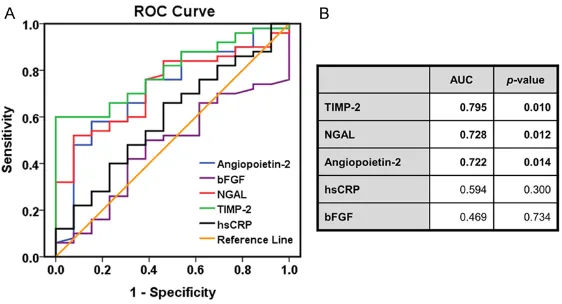
Based on the receiver operating characteris- tic curves, we analyzed the accuracy of these putative biomarkers in predicting angiogra- phically significant coronary artery stenosis. ROC curves of TIMP-2, NGAL, angiopoietin-2, bFGF, and hsCRP are shown in Figure 3A. The areas under the curve (AUC) were significant- ly greater for TIMP-2, NGAL and angiopoietin- 2 (Figure 3B). Using a cut-off value of 77.87 ng/ml for TIMP-2, a sensitivity of 80.0% and specificity 53.8% were obtained while a cut- off value of 91.46 ng/ml produced 67.3% sen-sitivity and 69.2% specificity. A cut-off value of 9.15 ng/ml for NGAL gave 77.4% sensiti- vity and 61.5% specificity, while a cut-off at 15.15 ng/ml showed 56.6% sensitivity and 76.9% specificity. A cut-off value of 1.57 ng/ml angiopoietin-2 showed 75.5% sensitivity and 61.5% specificity, and a cut-off value of 1.91 ng/ml yielded 56.6% sensitivity and 76.6% specificity.
Table 3. Correlations between proposed biomarkers and clinical variables
TIMP-2 NGAL Angiopoietin-2 bFGF hsCRP Age Correlation coefficient 0.060 -0.040 -0.108 -0.311 0.091
P-value 0.624 0.747 0.386 0.011 0.462
Sex Correlation coefficient 0.284 0.163 0.237 0.152 -0.135
P-value 0.019 0.188 0.54 0.222 0.274
BMI Correlation coefficient 0.238 0.292 0.192 0.108 0.056
P-value 0.050 0.017 0.120 0.387 0.650
Hypertension Correlation coefficient 0.269 0.134 0.91 0.006 0.169
P-value 0.027 0.278 0.465 0.960 0.168
Table 4. Factors predicting a diagnosis of angio-graphically significant coronary artery stenosis
OR (95% CI) P-value Angiopoietin-2 5.408 (1.279-22.865) 0.022 Age 1.261 (1.068-1.489) 0.006 Current smoking 66.311 (3.793-1159.318) 0.004 Body mass index 1.579 (0.993-2.511) 0.54
Discussion
The principal aim of this study was to evaluate putative biomarkers of inflammation, angiogen-esis, and plaque remodeling for the power to predict coronary artery stenosis as confirmed by coronary angiography. The main findings of the study were as follows: 1) serum levels of TIMP-2, NGAL and angiopoietin-2 were signifi-cantly higher in patients with coronary artery stenosis; 2) serum levels of TIMP-2, NGAL and angiopoietin-2 were correlated with coronary artery disease severity; 3) TIMP-2, NGAL and angiopoietin-2 in patient serum showed moder-ate diagnostic accuracy in screening for coro-nary artery disease; 4) angiopoietin-2 was a significant and independent predictor of coro-nary artery stenosis.
The role of cholesterol metabolism in athero-sclerotic coronary artery disease is complex. A high serum cholesterol level, or hypercho- lesterolemia, is associated with coronary ath-erosclerosis and its progression, and the low-density lipoprotein-cholesterol (LDL-C) fraction shows an especially strong correlation. Statins are shown to lower LDL-C levels, and to mo- dify other components of arterial disease. Randomized controlled trials demonstrate that statin drugs lower LDL-C levels and reduce the risk of cardiovascular events in coronary artery disease [17].
High-sensitivity C-reactive protein (hsCRP), a nonspecific marker of inflammation, is one of the most extensively studied biomarkers for coronary artery disease, notably because
ele-As risk indicators, LDL-C and hsCRP also show limited value in diagnosis [20-22]. In the pres-ent study, the serum hsCRP level was not asso-ciated with the presence of coronary artery ste-nosis or its severity. One possible explanation for the poor correlation of those conventional biomarkers in the diagnosis of coronary artery disease is that the wide use of statins for car-diovascular risk prevention in the enrolled patients may affect and lower their statistical correlation.
[image:6.612.93.374.73.225.2]Research into the pathogenesis of coronary atherosclerosis reveals serum proteins that may potentially be used to diagnose and moni-tor coronary artery disease [23]. Through a lit-erature review we identified TIMP2, NGAL, angiopoietin-2, bFGF, IL8 and GROα as serum proteins with plausible involvement in develop-ment and progression coronary atherosclero-sis. Of these, we detected TIMP2, NGAL, angio-poietin-2 and bFGF in all of the patients in this study. In patients with angiographically con-firmed coronary artery stenosis, serum levels of TIMP2, NGAL and angiopoietin-2 were signifi-cantly higher than in patients without angio-graphically confirmed coronary artery stenosis. TIMP-2, an inhibitor of endogenous matrix metalloproteinases (MMPs), is associated with both vascular inflammation and platelet activa-tion during atherosclerosis. In patients with stable coronary artery disease, serum levels of TIMP-2 and MMP-9 may increase [24]. In patients with early coronary atherosclerosis, on the other hand, plasma TIMP-2 levels may decrease [25].
Figure 3. ROC analysis of biomarker performance in the diagnosis of the an-giographically significant coronary artery stenosis. A. ROC curves for serum hsCRP, TIMP2, NGAL, angiopoietin-2 and bFGF. B. Table shows biomarker sensitivities and specificities derived from ROC analysis.
NGAL, which is expressed in neutrophils and associated with degradation of MMP-9, influ-ences remodeling of atherosclerotic plaque. Serum NGAL is reported to increase in coronary artery disease and to correlate with disease severity [26].
Angiopoietin-2 is selectively expressed in co- ronary endothelial cells and is related to in- tegrity of the coronary artery endothelium, as shown in a porcine model system [27]. Plasma angiopoietin-2 may increase in patients with acute coronary syndrome [28]; however, recent study shows that serum angiopoietin-2 level may also increase significantly in patients with stable coronary heart disease [29].
In the present study we compared serum levels of several proposed biomarkers and hsCRP between patients with and without angiograph-ically significant coronary arterial stenosis. While serum concentrations of TIMP-2, NGAL and angiopietin-2 were significantly higher in the patients with significant stenosis, serum hsCRP was not (Figure 1). Moreover, serum lev-els of TIMP-2, NGAL and angiopietin-2 correlat-ed closely with the angiographically assesscorrelat-ed severity of the stenosis (Figure 2).
In the present study, we tested various scoring methods to improve diagnostic accuracy results for TIMP-2, NGAL and angiopietin-2 but without success (data not shown). This may plausibly be attributed to the low specificity of the roles those biomarker proteins play in the develop-ment of atherosclerosis. Moreover, the close correlations among those markers (Table 2) suggested that those biomarkers act by the same or closely related pathogenic mecha-nisms. Based on a recent study, the plasma biomarkers monocyte chemoattractant pro-tein-1, galectin-3 and N-terminal fragment of brain natriuretic peptide may independently and significantly predict cardiovascular events in patients with high-risk coronary artery dis-ease [30]. Possibly the combination of one or more of those markers with markers investi-gated in the present study would improve the accuracy of our diagnostic panel. Meanwhile, identification of biomarkers that specifical- ly represent the molecular and cellular media-tors of atherosclerosis remains an important research target.
Acknowledgements
The authors thank Long-Hui Cui for skilled tech-nical assistance.
Disclosure of conflict of interest
None.
Address correspondence to: Do-Sun Lim, Depart- ment of Cardiology, Cardiovascular Center, Korea University Anam Hospital, 126-1, 5ka, Anam-dong, Sungbuk-ku, Seoul 136-705, Korea. Tel: +82-2-920-5445; Fax: +82-2-927-1478; E-mail: dslmd@kumc. or.kr
References
[1] Levin DC. Invasive evaluation (coronary arteri-ography) of the coronary artery disease pa-tient: clinical, economic and social issues. Circulation 1982; 66: III71-79.
[2] Kennedy JW. Complications associated with cardiac catheterization and angiography. Ca- thet Cardiovasc Diagn 1982; 8: 5-11.
[3] Patel MR, Peterson ED, Dai D, Brennan JM, Redberg RF, Anderson HV, Brindis RB, Douglas PS. Low diagnostic yield of elective coronary angiography. N Engl J Med 2010; 362: 886-895.
[4] Rumberger JA. Noninvasive coronary angiogra-phy using computed tomograangiogra-phy: ready to kick it up another notch? Circulation 2002; 106: 2036-2038.
[5] Leschka S, Alkadhi H, Plass A, Desbiolles L, Grunenfelder J, Marincek B, Wildermuth S. Accuracy of MSCT coronary angiography with 64-slice technology: first experience. Eur Heart J 2005; 26: 1482-1487.
[6] Raff GL, Gallagher MJ, O’Neill WW, Goldstein JA. Diagnostic accuracy of noninvasive coro-nary angiography using 64-slice spiral com-puted tomography. J Am Coll Cardiol 2005; 46: 552-557.
[7] Einstein AJ, Henzlova MJ, Rajagopalan S. Esti- mating risk of cancer associated with radi- ation exposure from 64-slice computed to- mography coronary angiography. JAMA 2007; 298: 317-323.
[8] Siasos G, Tousoulis D, Athanasiou D, Oiko- nomou E, Tourikis P, Gouliopoulos N, Limperi M, Kampoli AM, Toutouzas K, Papavassiliou AG, Stefanadis C. Novel risk factors related to stable angina. Curr Pharm Des 2013; 19: 1550-1561.
kis E, Toutouzas C, Stougianos P, Tentolouris C, Stefanadis C. Clinical utility of biomarkers in premature atherosclerosis. Curr Med Chem 2012; 19: 2521-2533.
[10] te Boekhorst BC, Bovens SM, Hellings WE, van der Kraak PH, van de Kolk KW, Vink A, Moll FL, van Oosterhout MF, de Vries JP, Doevendans PA, Goumans MJ, de Kleijn DP, van Echteld CJ, Pasterkamp G, Sluijter JP. Molecular MRI of murine atherosclerotic plaque targeting NGAL: a protein associated with unstable human plaque characteristics. Cardiovasc Res 2011; 89: 680-688.
[11] Dabek J, Glogowska-Ligus J, Szadorska B. Transcription activity of MMP-2 and MMP-9 metalloproteinase genes and their tissue in-hibitor (TIMP-2) in acute coronary syndrome patients. J Postgrad Med 2013; 59: 115-120. [12] Post S, Peeters W, Busser E, Lamers D, Sluijter
JP, Goumans MJ, de Weger RA, Moll FL, Doevendans PA, Pasterkamp G, Vink A. Balance between angiopoietin-1 and angiopoi-etin-2 is in favor of angiopoiangiopoi-etin-2 in athero-sclerotic plaques with high microvessel densi-ty. J Vasc Res 2008; 45: 244-250.
[13] Sigala F, Savvari P, Liontos M, Sigalas P, Pa- teras IS, Papalampros A, Basdra EK, Kolettas E, Kotsinas A, Papavassiliou AG, Gorgoulis VG. Increased expression of bFGF is associated with carotid atherosclerotic plaques instability engaging the NF-kappaB pathway. J Cell Mol Med 2010; 14: 2273-80.
[14] Papadopoulou C, Corrigall V, Taylor PR, Poston RN. The role of the chemokines MCP-1, GRO-alpha, IL-8 and their receptors in the adhesion of monocytic cells to human atherosclerotic plaques. Cytokine 2008; 43: 181-186.
[15] Gensini GG. A more meaningful scoring system for determining the severity of coronary heart disease. Am J Cardiol 1983; 51: 606.
[16] Ringqvist I, Fisher LD, Mock M, Davis KB, Wedel H, Chaitman BR, Passamani E, Russell RO Jr, Alderman EL, Kouchoukas NT, Kaiser GC, Ryan TJ, Killip T, Fray D. Prognostic value of angiographic indices of coronary artery dis-ease from the Coronary Artery Surgery Study (CASS). J Clin Invest 1983; 71: 1854-1866. [17] Baigent C, Keech A, Kearney PM, Blackwell L,
Buck G, Pollicino C, Kirby A, Sourjina T, Peto R, Collins R, Simes R; Cholesterol Treatment Trialists’ (CTT) Collaborators. Efficacy and safety of cholesterol-lowering treatment: pro-spective meta-analysis of data from 90,056 participants in 14 randomised trials of statins. Lancet 2005; 366: 1267-1278.
[18] Jialal I, Devaraj S. Inflammation and athero-sclerosis: the value of the high-sensitivity
C-reactive protein assay as a risk marker. Am J Clin Pathol 2001; 116 Suppl: S108-115. [19] Bayturan O, Kapadia S, Nicholls SJ, Tuzcu EM,
Shao M, Uno K, Shreevatsa A, Lavoie AJ, Wolski K, Schoenhagen P, Nissen SE. Clinical predic-tors of plaque progression despite very low lev-els of low-density lipoprotein cholesterol. J Am Coll Cardiol 2010; 55: 2736-2742.
[20] Rasouli M, Kiasari AM, Bagheri B. Total and dif-ferential leukocytes counts, but not hsCRP, ESR, and five fractioned serum proteins have significant potency to predict stable coronary artery disease. Clin Chim Acta 2007; 377: 127-132.
[21] Cheng VY, Wolak A, Gutstein A, Gransar H, Wong ND, Dey D, Thomson LE, Hayes SW, Friedman JD, Slomka PJ, Berman DS. Low-density lipoprotein and noncalcified coronary plaque composition in patients with newly di-agnosed coronary artery disease on computed tomographic angiography. Am J Cardiol 2010; 105: 761-766.
[22] Veselka J, Prochazkova S, Duchonova R, Bolo- mova I, Urbanova T, Tesar D, Honek T. Rela- tionship of C-reactive protein to presence and severity of coronary atherosclerosis in pa- tients with stable angina pectoris or a patho-logical exercise test. Coron Artery Dis 2002; 13: 151-154.
[23] Garg A. What is the role of alternative bio- markers for coronary heart disease? Clin Endocrinol (Oxf) 2011; 75: 289-293.
[24] Tayebjee MH, Lip GY, Tan KT, Patel JV, Hughes EA, MacFadyen RJ. Plasma matrix metallopro-teinase-9, tissue inhibitor of metalloprotein-ase-2, and CD40 ligand levels in patients with stable coronary artery disease. Am J Cardiol 2005; 96: 339-345.
[25] Nanni S, Melandri G, Hanemaaijer R, Cervi V, Tomasi L, Altimari A, Van Lent N, Tricoci P, Bacchi L, Branzi A. Matrix metalloproteinases in premature coronary atherosclerosis: influ-ence of inhibitors, inflammation, and genetic polymorphisms. Transl Res 2007; 149: 137-144.
[26] Zografos T, Haliassos A, Korovesis S, Giazit- zoglou E, Voridis E, Katritsis D. Association of neutrophil gelatinase-associated lipocalin with the severity of coronary artery disease. Am J Cardiol 2009; 104: 917-920.
[27] Kim I, Moon SO, Han CY, Pak YK, Moon SK, Kim JJ, Koh GY. The angiopoietin-tie2 system in coronary artery endothelium prevents oxi-dized low-density lipoprotein-induced apopto-sis. Cardiovasc Res 2001; 49: 872-881. [28] Lee KW, Lip GY, Blann AD. Plasma
tie-2, and vascular endothelial growth factor levels in acute coronary syndromes. Circulation 2004; 110: 2355-2360.
[29] Wang X, Yong H, Mi L, Bai Y, Guo L, Gao W, Cui M, Zhang Y. Changes and significance of se-rum angiopoietin-2 levels in patients with coro-nary heart disease. Biomarkers 2012; 17: 745-749.
[30] Tunon J, Blanco-Colio L, Cristobal C, Tarin N, Higueras J, Huelmos A, Alonso J, Egido J,