Original Article
Mechanisms of Raddeanin A-induced autophagy and
apoptosis in human colorectal cancer cells
Chunqin Meng1,2, Xue Zhou3, Yuhao Teng1, Cunen Wu2, Jian Wu1, Fang Tian1, Yao Zhou2, Xi Zou1, Ruiping Wang1
1Department of Oncology, Affiliated Hospital of Nanjing University of Chinese Medicine, Jiangsu Province Hospital
of Traditional Chinese Medicine, Nanjing 210029, Jiangsu Province, China; 2Nanjing University of Chinese
Medicine, Department of First Clinical Medical College, Nanjing 210023, Jiangsu Province, China; 3Department of
Acupuncture, Nanjing Hospital of Traditional Chinese Medicine, Nanjing 210029, Jiangsu Province, China
Received June 17, 2017; Accepted November 7, 2017; Epub November 15, 2017; Published November 30, 2017
Abstract: Several studies have confirmed that the Chinese medicine Raddeanin A (RA), which is extracted from the plant Anemone raddeana Regel, can inhibit the proliferation of a variety of tumor cell lines. Previously, our team demonstrated that RA could induce apoptosis and autophagy in human gastric cancer cells. Objective: This experiment was intended to explore whether RA can induce autophagy and apoptosis in human colorectal cancer HCT116 cells and to investigate the mechanisms and relationship between autophagy and apoptosis. Methods: Cell proliferation was detected via MTT assay. Transmission electron microscope was used to observe autophago
-somes as a marker for autophagy. Apoptosis was examined via Hoechst 33258 staining. Flowcytometry was used to calculate the rate of apoptosis. The expression of related genes and proteins were tested by reverse transcription-polymerase chain reaction (RT-PCR) and western blot, respectively. Results: Cell viability gradually decreased with an increase of drug concentration and disposal time. This result indicated that RA could significantly inhibit the growth of HCT116 cells in a manner that was both time-and concentration dependent. RA induced autophagy and apoptosis in HCT116 cells. RT-PCR and western blot showed that the expression of genes and proteins related to autophagy increased. Moreover, the expression of proteins that suppress apoptosis decreased while pro-apoptotic protein levels increased. The expression of proteins involved in the PI3K-AKT-mTOR signaling pathway also de -creased, while caspase-8 and 9 levels increased. Compared with RA treatment alone, the apoptosis rate decreased
when HCT116 cells were treated with RA and hydroxychloroquine (HCQ) together. However, when RA and rapamycin (RAPA) were given together, the apoptosis rate increased. Conclusion: RA induces autophagy by regulating the PI3K-AKT-mTOR signaling pathway and apoptosis through intrinsic and extrinsic pathways. Autophagy induced by RA in
HCT116 cells can promote cell apoptosis.
Keywords: Raddeanin A, autophagy, apoptosis, HCT116 cells, induce
Introduction
Colorectal cancer is a common malignant
tumor of the digestive system. In Europe and the United States, the morbidity and mortality of colorectal cancer are in fourth and second place, respectively, in all malignant tumors [1]. For the treatment of colorectal cancer, chemo
-therapy is commonly used and is effective treatment after surgery. Unfortunately, chemo
-therapy can cause problematic adverse reac -tions, and drug resistance can occur in cancer
cells, which greatly reduces the clinical effect [2]. Consequently, for new natural antineoplas -tic drugs are needed.
Raddeanin A (RA) is an oleanane-type triterpe
-noid saponin extracted from the plant Anemone raddeana Regel, which was recorded in YIFANGGEKUO as an effective treatment for
breast cancer. Modern pharmacological
stud-ies have shown that it has significant anti-tumor, anti-inflammatory, antipyretic, analgesic, and anticonvulsant effects [3-6]. Moreover, our
previous studies demonstrated that RA
inhibit-ed the invasion of SGC-7901 human gastric cancer cells into healthy tissue in vitro, and induced autophagy and apoptosis in those can
-cer cells [7, 8].
and also allows cells to die in an orderly fash
-ion. PCDII has become a hot research topic recently with the hope of learning information
to regulate tumor cell death. This experiment
explored whether RA could induce autophagy
or apoptosis in colorectal cancer HCT116 cells, investigated the possible molecular
mecha-nisms of these processes, and attempted to determine the relationship between autophagy and apoptosis as induced by RA.
Materials and methods
Reagents and antibodies
RPMI-1640 medium and fetal bovine serum (FBS) were acquired from Gibco BRL (Gaith-ersburg, MD, USA). RA was purchased from the China National Institute for the Control of Pharmaceuticals and dissolved in dimethyl sulfoxide (DMSO) which was stored at -20°C. 3-(4,5-Dimethylthiazol-2-yl)-2,5-diphenyltetra-zolium bromide (MTT) and hydroxychloroquine sulfate were obtained from Sigma Chemical Company (St. Louis, MO, USA). Annexin-V/prop -idium iodide (PI) apoptosis detection kits were
obtained from BD Biosciences (FranklinLakes,
NJ, USA). Primescript reverse transcription re-
agent kits with gDNA erasers were obtained
-from TaKaRa (Dalian, China). TRIzol reagent and Power SYBR Green PCR Master Mixes were bought from Life Technologies (Grand Island, NY). Rapamycin (RAPA) and primary antibodies
-of Beclin-1, LC3, BAX, Bcl-2, PARP,
caspase-3, cleaved-caspase-caspase-3, caspase-9, caspase-8,
PI3K, AKT, p-AKT, mTOR, p-mTOR, andβ-actin were obtained from Cell Signaling Technology
Afterbeing treated with different concentra
-tions of RA or DMSO, cells were added to MTT (5 mg/mL) and incubated for 4 hr. DMSO was then added and the optical densities (ODs) were tested using an ELx800 microplate reader (BioTek, Winooski, VT, USA) at 490 nm. The inhi
-bition rate was determined according to the fol
-lowing equation: Inhibition rate = (1-ODexperiment/
ODcontrol) * 100%.
Transmission electron microscopy (TEM) analy-sis
Cells were seeded in a Petri dish, incubated
with RA for 12 h, and collected. The cells were then washed twice with cold PBS and fixed in 2. 5% glutaric dialdehyde solution and 1% osmic acid for 2 h. Afterthis, the processed cells were observed under a JEOL-1010 electron
micro-scope.
Flow cytometry analysis
The cells were detached via trypsinization, treated with RA for 12 h, washed twice with PBS, and then resuspended in 500 µL binding buffer containing 5 µL Annexin V-FITC and 5 µL propidium iodide (PI). Prior to being analyzed by flow cytometry, the cells were incubated for 15
min in the dark.
Hoechst 33258 staining
After being treated with RA for 12 h, cells were fixed with 1% formalin for 30 min, washed with
PBS, and incubated in Hoechst 33258 stain
(50 ng/mL) for 30 min. Cells in the process of apoptosis could be distinguished by condensa -Table 1. Sequences of primers used in the reverse
transcription-polymerase chain reaction (RT-PCR) amplifi -cations
Gene
primer Sequence (5’-3’) Length of PCR product (bp) Beclin-1 F: GACGGAAGTTGAGATAGT 110
R: CAAGTGACGAAACGGTGATT
ATG 5 F: GATGAGGGCCGTATCGACAGT 160
R: CGCTTCGCTAAATTAGGCGAC
ATG12 F: GCCATCGCGAAGTGCAAGAC 158
R: ACCAGAAATATACACAGGGTCT
ATG7 F: CAGCCTGCATTTAAGACCAGTGTCAC 210
R: ACGTCGATCGCTCACACATGCATTCGCATT
β-actin F: GGCCAACCGCGAGAAGAT 134
R: CGTCACCGGAGTCCATCA
(Beverly, MA, USA), Fluorescein-con-jugated secondary antibodies were obtained from Odyssey (Licor, Belfast,
ME, USA).
Cell line and culture
The human colorectal cancer cell line
(HCT116) was provided by the Shanghai Institute for Biological Research
(Sh-anghai, China) and cultured in RPMI-1640 medium containing 10% FBS at
37°C and in a humidified atmosphere with 5% CO2.
MTT assay
[image:2.612.89.340.108.266.2]tion and fragmentation in their nuclei. The cells were recorded using a Zeiss Axioplan 2 fluores
-cent microscope (Jena, Germany).
Reverse transcription-polymerase chain reac-tion (RT-PCR) assay
Cells of different groups were collected, and treated with TRIzol reagent according to the manufacturer’s instruction in order to extract the cellular RNA. The purity of the RNA was test -ed via spectrophotometer and
reverse-tran-scribed into cDNA using a TaKaRa RT retrovirus
kit. The date was calculated with an ABI 7500
fast RT-PCR System after the PCR amplification
reaction. The 2-ΔΔCt method was used to analyze the results, according to the following
equa-tions:
ΔCt = Cttarget genes - Ctendogenous reference gene (1)
and ΔΔCt = ΔCttreated samples - ΔCtcontrol samples (2)
where β-actin was used as the reference com
[image:3.612.92.288.68.360.2]-pound. The final gene expression level was cal -culated as 2-ΔΔCt. The gene primers were designed by Primer Express and are shown in
Table 1.
Western blot (WB) analysis
After being treated with RA, the cells were lysed with RIPA buffer to release the protein. The pro
-tein concentration was tested via the Bradford method (BCA). The expression of β-actin pro -tein was served as a loading control, andeach group proteins was loaded onto a 10% or 12%
sodium dodecyl sulfate (SDS)-polyacrylamide gel for electrophoresis. Thereafter, the proteins were transferred via electroblotting to polyvi
-nylidenedifluoride (PVDF) membranes (Milli-pore, Boston, MA, USA). The PVDF was enclosed with 5% BSA for 1 hr and incubated with the indicated antibodies against LC3 (1:1000),
Be-clin-1 (1:1000), BAX (1:1000), Bcl-2 (1:1000), caspase-3 (1:1000), cleaved-caspase-3 (1: 1000), caspase-8 (1:1000), caspase-9 (1:
1000), PARP (1:1000), PI3K (1:1000), AKT (1: 1000), p-AKT (1:1000), mTOR (1:1000), and p-mTOR (1:1000) overnight. Before the second
-ary fluorescent antibody (1:3000 dilutions) was added for 1 h, the membrane was washed thrice (5 min/time) with tris buffered saline with Tween-20 (TBST). Finally, the signal intensity of the membranes was examined by Odyssey (LICOR, Belfast, ME, USA).
Statistical analysis
SPSS16.0 statistical software was used to per
-form the statistical analysis. Measurement
data were represented as means ± standard
deviation (SD). The difference between the
groups was examined by one-way ANOVA analy
-sis followed by Dunnett’s test and a P value
<0.05 was considered significant.
Results
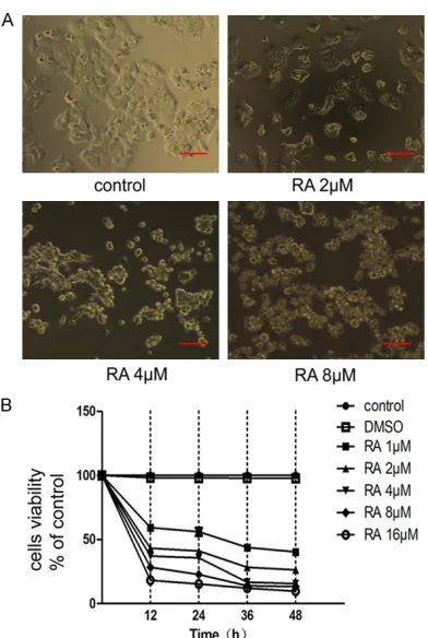
RA treatment inhibited HCT116 cell prolifera-tion
The MTT assay indicated that RA could signifi
-cantly inhibit the proliferation of HCT116 cells. The effect on cellular proliferation different with concentration of RA (1, 2, 4, 8, 16 µM) and length of incubation (12 h, 24 h, 36 h, 48 h). DMSO had the smallest effect on cells viability
(Figure 1B). Observations of HCT116 cells
Figure 1. Raddeanin A (RA) inhibits HCT116 cell
treated with RA using an inverted phase
con-trast microscope showed that the amount of cells treated with RA gradually lessened, and
the cells lost their tentacles to become spheri-cal (Figure 1A).
RA-induced autophagy in HCT116 cells oc-curred via regulation of the PI3K-AKT-mTOR pathway
We observed that RA had a strong effect on the proliferation of HCT116 cells, but the mecha
-nism was not yet clear. Our previous experi
-ments suggested that RA inhibited the prolifer
-ation of gastric cancer cells by inducing apopto
-sis and autophagy. We wanted to investigate whether RA could induce autophagy in HCT116 cells and to this end we took TEM scans of
the cells. The TEM results showed
autophago-somes in the cytoplasm (Figure 2A and 2B),
[image:4.612.94.525.71.471.2]which are indicative of autophagy. For further evidence, we performed RT-PCR and WB. The results of RT-PCR showed that the expression of the autophagy-related genes Beclin-1, ATG5,
Figure 2. Raddeanin A (RA) induced autophagy in HCT116 cells by activating the PI3K-AKT-mTOR pathway. A. Cells were treated with RA (4 µM) for 12 h and transmission electron microscopy (TEM) was used to observe autophago
-somes (2900×). B. A double membrane structure was detected using TEM (18500×). C and D. After being treated with RA (4, 8 µM), reverse transcription-polymerase chain reaction (RT-PCR) and western blot analysis showed the levels of autophagy related genes (Beclin-1, ATG5, ATG12, ATG7) and proteins (Beclin-1, LC3) respectively. Data shown are means ± SD (n = 3, *P<0.05, compared with the control). β-actin was used as an internal control. E.
ATG12, and ATG7 increased with increasing concentrations of RA (Figure 2C). WB indicated
that the expression of Beclin-1 increased with
RA treatment, and that with an increase in RA
concentration, the signal intensity of the pro
-ed (Figure 2D). Furthermore, the classic
[image:5.612.91.523.71.532.2]signal-ing pathway for autophagy regulation is the PI3K-AKT-mTOR pathways. Western blot results also showed downregulation of p-AKT and p-mTOR proteins along with upregulation of
Figure 3. Raddeanin A (RA) induced apoptosis in HCT116 cells via the intrinsic and extrinsic pathways. A. After being treated with RA (2, 4, 8 µM) for 12 h, cells were incubated with Annexin V-FITC and propidium iodide (PI), and the apoptosis rate was analyzed via flow cytometry. Results shown are of an experiment representative of apoptosis. Q1-UL represents necrotic cells, Q1-UR represents cells at a later stage of apoptosis, Q1-LL represents viable cells, Q1-LR represents cells were at an early stage of apoptosis. Data shown are means ± SD (n = 3, *P<0.05, **P<0.01, compared with the control). B. Apoptotic cells were stained with Hoechst 33258 (50 ng/mL) after treatment with RA (2, 4, 8 µM) for 12 h, which was recorded with a Zeiss Axioplan 2 fluorescence microscope (400×). C. Animmunoblot assay shows the expression of apoptosis related proteins (Bcl-2, BAX, caspase-3, cleaved-caspase-3, cleaved-PARP and PARP). D. Cells were treated with RA and levels of apoptosis-related proteins (caspase-9, caspase-8) were de
RA induced apoptosis in HCT116 cells through both intrinsic and extrinsic pathways
The lack of a working apoptosis mechanism is one of the main reasons for the formation of tumors. In this experiment, the MTT assay sh-owed that RA could significantly inhibit the pro
-liferation of HCT116 cells, so we hypothesized
that RA could induce apoptosis in HCT116
cells. The results of the flow cytometry illustrat
-ed that the higher the concentration of RA, the
higher the apoptosis rate (Figure 3A). Hoechst
33258 Staining also confirmed the existence of apoptosis, since we found the broken pieces of the nucleus which are a major symbol of apop -tosis (Figure 3B). Western blot results also
revealed that the expression of the proteins
cleaved-caspase-3, cleaved-PARP, and BAX
increased while the expression of the proteins
Bcl-2, caspase-3, and PARP decreased (Figure 3C). Caspases-8 and 9, proteins critical to the
intrinsic and extrinsic pathways, respectively,
were also upregulated (Figure 3D).
The relationship between autophagy and apop-tosis induced by RA in HCT116 cells
More and more data show that there are
com-plex relationships between autophagy and apoptosis. Hydroxychloroquine (HCQ), which inhibits autophagy, and rapamycin (RAPA), an autophagy agonist, were added to the HCT116
cells along with RA. As shown in Figure 4A,
when treated with HCQ (10 µM) and RA (4 µM)
together, the cellular inhibition decreased com-pared with RA (4 µM) alone. According to the
flow cytometry results, the apoptosis rate of the RA group (4 µM) was higher than that of the HCQ/RA combined group, but was lower than
that of the RAPA (100 nM) and RA (4 µM) com
-bined group (Figure 4B). The WB results also
showed that the density of Beclin-1, LC3,
cleaved-caspase-3, cleaved-PARP, BAX increas- ed in the RA/RAPA group (Figure 4C and 4D).
Discussion
A survey found that with a change in diet, the incidence of colorectal cancer has significantly
increased in developing countries, and that
patients tend to be younger [9]. Chemotherapy is the main treatment method for patients who have no opportunity for surgery [10]. However, with the increase in modern chemotherapy drugs and the frequency of use and length of
treatment, cancer cells are no longer as sensi-tive to chemotherapeutic drugs. Thus,
newanti-tumor drugs must be found as soon as
possible.
RA, which is a type of natural medicine, has been shown to possess a significant anti-tumor effect both in vivo and in vitro [11-14], although
the mechanism is unclear.
As the MTT assay showed, RA could inhibit the growth of HCT116 cells even at low concentra -tions (1 µM), and the inhibition rate was
posi-tively correlation with both RA concentration
and incubation time (Figure 1). We hypothe
-sized that this inhibition might be caused by the induction of autophagy and apoptosis. The involvement of autophagy inhibition is still con -troversial in tumor research. This is because
while autophagy can lead to cell death, it also can protect cells from death in the case of hun
-ger or loss of energy, and makes cells steady by
removing damaged organelles, such as mito-chondria, endoplasmic reticulum, and
peroxi-dases [15].
To verify RA-induce autophagy in HCT116 cells, we observed the cells using TEM, and found the existence of a spherical double layer mem -brane, the autophagosome, considered to be
the golden standard of autophagy determina -tion (Figure 2A and 2B). Moreover, the results
of RT-PCR and WB also showed the upregula
-tion of related genes (Beclin-1, ATG5, ATG7, ATG12) and pro-autophagy proteins. As is
Figure 4. Autophagy induced by Raddeanin A (RA) in HCT116 cells promote apoptosis. A. Prior to treatment with dealt with RA (4 µM), cells were treated with either hydroxychloroquine (HCQ; autophagy inhibitor, 10 µM) or RAPA (autophagy agonist, 100 nM) for 1 h. An MTT assay indicated different rates of cellular inhibition for the different groups. Data shown are means ± SD (n = 3, **P<0.01, compared with the control; ▲P<0.05, compared with the RA
group). B. Cells were incubated with Annexin V-FITC and propidium iodide (PI), and the apoptosis rate was analyzed via flow cytometry. Q1-UL represents necrotic cells, Q1-UR represents cells at a later stage of apoptosis, Q1-LL rep
-resents viable cells, and Q1-LR rep-resents cells at an early stage of apoptosis. Data shown are means ± SD (n =
3, *P<0.05, **P<0.01, compared with the control; ▲P<0.05, compared with the RA group). C and D. Western blot
shown in Figure 2D, with increasing
concentra-tions of RA, LC3I converted to LC3II, which is
required to form the autophagosome mem
-brane. Previous reports have suggested that
PI3K-AKT-mTOR is the major autophagy signal
-ing. The Nobel Prize winner Ohsumi also con
-firmed that the PI3K complex is the key to the process of autophagy [16]. Our experiments also found that the expression of p-AKT and p-mTOR decreased, whilethe expressions of
PI3K, AKT and mTOR increased during autoph
-agy, which supports the supposition that the PI3K-AKT-mTOR pathway is involved in regulat
-ing autophagy (Figure 2E).
With the exception of autophagy, induction of apoptosis the mainmechanism of most
anti-tumor drugs, due to apoptosis being the
earli-est recognized process of tumor cell death. Apoptosis can be divided into three stages [17]: (1) Startup phase: Due to various causes, the cells start the process of apoptosis; (2) Effect
phase: Whether apoptosis occurs is determi-
ned by the presence of apoptotic and anti-apoptotic factors; (3) Execution phase: Apopto-tic cells showed specific morphological and biochemical features, including DNA rupture, nuclearpyknosis, karyorrhexis, karyolysis, the formation of apoptosis bodies, and degrada
-tion by lysosomes. Flowcytometry and Hoechst
33258 staining showed that RA induced
apop-tosis in HCT116 cells, and that the rate of apop -tosis increased with increasing concentrations
of RA (Figure 3A and 3B). In addition, the WB
results also showed that fewer decreased expression of the anti-apoptotic protein Bcl-2 and increased expression of pro-apoptotic pro -tein BAX (Figure 3C) after treatment with RA.
As is well known, apoptosis has biochemical
pathways, namely, the mitochondrial pathway, which is intrinsic pathway, the death receptor signaling pathway, which is extrinsic pathway, and the endoplasmic reticulum stress pathway. Activated caspase-9 is necessary for the intrin
-sic pathway to occur, while activated caspase-8 is necessary for the extrinsic pathway. In this study, we found that the expressions of cas -pase-8 and -9 both increased (Figure 3D), which revealed that RA induced apoptosis in HCT116 cells via intrinsic and extrinsic
pathways.
Autophagy and apoptosis are both types of pro
-grammed cell deaths, but there are many dif
-ferences between them in the cellular morphol
-ogies, molecular mechanisms, and biochemi-cal indicators involved. It has been reported
that in cells with a large number of autophago
-somes, the use of caspase inhibitors does not affect cell death, while the autophagy inhibitor 3-methyl adenine does, which illustrates that the processes of autophagy and apoptosis are different [18]. Nonetheless, there may be a relationship between autophagy and apopto
-sis. Yee et al found that autophagy induced by
PUMA and BAX could repair mitochondrial
damage and reduce progress of apoptosis [19]. Hui L et al found that sodium selenite could promote cellular autophagy while suppressing apoptosis [20]. JingwenY found that 5-Fu com
-bined with an autophagy or apoptosis inhibitor could promote HCT-116 cell proliferation and
inhibit apoptosis, and that 5-Fu combined with
autophagy inhibitor would produce the oppo
-site result [21]. Moreover, under certain condi
-tions, cells can transition between autophagy
and apoptosis and use both processes to regu-late tumor cell death. Forexample, when
apop-tosis was inhibited in HeLa and Chinese ham
-ster ovary (CHO) cells, the cells died via autoph
-agy instead [22]. In order to examine the rela
-tionship between RA-induced autophagy and apoptosis, the autophagy inhibitor HCQ and autophagy agonist RAPA were added to HCT116
cells treated with RA. As is shown in Figure 4,
autophagy could promote RA-induced apopto
-sis in HCT116 cells.
In conclusion, RA can induce autophagy in HCT116 cells by regulating the PI3K-AKT-mTOR pathway and induce apoptosis via the intrinsic and extrinsic pathways. Moreover, autophagy can promote apoptosis, when induced by RA in
HCT116 cells. These findings make RA a prom
-ising candidate for the treatment of colorectal cancer, and also lay the foundation for further
clinical applications. Acknowledgements
This work was supported by the State Admini-stration of Traditional Chinese Medicine of China (grant# JDZX2012087), Jiangsu Provin-cial Commission of Health and Family Planning (grant# BJ14013), Talent Project of Traditional
Chinese Medicine in Jiangsu Province (No.
Programme (FP7/2007-2013/under REA grant
agreement No. PIRSES-GA-2013-61258: China and Europe taking care of healthcare
solu-tions.
Disclosure of conflict of interest
None.
Address correspondence to: Xi Zou and Ruiping
Wang, Department of Oncology, Affiliated Hospital of Nanjing University of Chinese Medicine, Qinhuai District, Nanjing 210029, Jiangsu Province, China.
Tel: +8613705105501; +8613815883181; E-mail:
zxvery@162.com (XZ); wrp61@163.com (RPW)
References
[1] US Cancer Statistics Working Group. United
states cancer statistics USCS: 1999-2008
inci-dence and mortality data. Atlanta: Department of Health and Human Services, CDC, National
Cancer Institute; 2010.
[2] Mokarram P, Albokashy M, Zarghooni M, Moo
-savi MA, Sepehri Z, Chen QM, Hudecki A, Sar
-gazi A, Alizadeh J, Moghadam AR, Hashemi M, Movassagh H, Klonisch T, Owji AA, Łos MJ, Ghavami S. New frontiers in the treatment of colorectal cancer: autophagy and the unfolded
protein response as promising targets.
Autoph-agy 2017; 13: 781-819.
[3] Qian S, Chen QL, Guan JL, Wu Y, Wang ZY. Syn
-thesis and biological evaluation of Raddeanin A, a triterpene saponin isolated from anemone raddeana. Chem Pharm Bull (Tokyo) 2014; 62:
779-785.
[4] Dayou L, Yong L, Bo Z. Studies on the chemical constituents of anemonis raddeanae. Aca
-demic Periodical of Changchun College of Tra -ditional Chinese Medicine 2005; 19: 71.
[5] Xiaoyi W. Studies on the chemical constituents of anemonis raddeanae. Chinese J Anal Chem
2004; 32: 587-592.
[6] Zhongjie Z, Hongxia L, Bo Z. Pharmacological activities of the chemical components of ge -nus anemone. Chinese Pharm J 2004; 39: 493-495.
[7] Xue G, Zou X, Zhou JY, Sun W, Wu J, Xu JL,
Wang RP. Raddeanin A induces human gastric cancer cells apoptosis and inhibits their
inva-sion in vitro. Biochem Biophys Res Commun
2013; 439: 196-202.
[8] Teng YH, Li JP, Liu SL, Zou X, Fang LH, Zhou JY,
Wu J, Xi SY, Chen Y, Zhang YY, Xu S, Wang RP.
Autophagy protects from Raddeanin A-induced apoptosis in SGC-7901 human gastric cancer
cells. Evid Based Complement Alternat Med 2016; 2016: 9406758.
[9] Jemal A, Bray F, Center MM. Global cancer sta -tistics. CA Cancer J Clin 2011; 61: 69-90.
[10] Price TJ. Advanced colorectal cancer treatment
options beyond standard systemic therapy. Lancet Oncol 2017; 18: 157-159.
[11] Guan YY, Liu HJ, Luan X, Xu JR, Lu Q, Liu YR, Gao YG, Zhao M, Chen HZ, Fang C. Raddeanin A, a triterpenoid saponin isolated from anemo -ne raddeana, suppresses the angioge-nesis
and growth of human colorectal tumor by in
-hibiting VEGFR2 signaling. Phytomedicine
2015; 22: 103-110.
[12] Wang MK, Ding LS, Wu FE. Antitumor effects of
Raddeanin A on S180, H22 and U14 cell
xeno-grafts in mice. Ai Zheng 2008; 27: 910-913. [13] Zhang JM. Studies on anticancer activities of
triterpenoid in anemone raddeana regel. Chin
J New Drugs 2003; 12: 191-193.
[14] Liu D, Zhang L, Zhang Y, Yu YE. Studies on anti-tumor activity in vitro of Raddeanemone A. Pharmacology and Clinics of Chinese Materia
Medica 2014; 30: 49-53.
[15] Mathew R, Karantza Wadsworth V, White E. Role of autophagy in cancer. Nat Rev Cancer
2007; 7: 961-967.
[16] Kihara A, Noda T, Ishihara N, Ohsumi Y. Two distinct Vps34 phosphatidylinositol 3-kinase complexes function in autophagy and carboxy
-peptidase-Y sorting in saccharomyces cerevi -siae. Cell Biol 2001; 152: 519-30.
[17] Yoshida H, Kong YY, Yoshida R. Apaf-l is re
-quired for mitochondrial path ways of apopto -sis and brain development. Cell 1998; 94: 739-750.
[18] Li BX, Zhang XS. Mechanism of beclin1 as a favorable molecules marker is that beclin1 in
-hibits tumor proliferation and cell cycle in
stage IIIB colon cancers. Chin J Clinicians 2010; 4: 395-401.
[19] Yee KS, Wilkinson S, James J, Ryan KM, Vous
-den KH. PUMA and Bax induced autophagy contribute to apoptosis. Cell Death Differ
2009; 16: 1135-1145.
[20] Hui L. Research on the effect of AKT/β-catenin/ FoxO3a signaling pathway of apoptosis in
-duced by sodium selenite in colorectal cancer
cells. Peking Union Medical College 2013.
[21] Yang JW. Research on the relationship and the
mechanism of autophagy and apoptosis in
-duced by 5-Fu in HCT-116 cell stem. Tianjin Medical University 2013; 12.
[22] Yanagisawa H, Miyashita T, Nakano Y, Yama
-moto D. HSpin1, a transmembrane protein in