Original Article
Localizing percentages of
interictal 18F-fluorodeoxyglucose
(FDG)-PET and magnetoencephalography
(MEG) in pre-surgical evaluation of 107
consecutive patients with non-lesional epilepsy
Xiufeng Zhang1,2, Huifang Song3, Yongliang Liu4, Fuxing Yang1, Wenjian Shi4, Zhiqiang Liu4, Zhipei Ling1,
Zhiqiang Cui1,4, Bainan Xu1
1Department of Neurosurgery, Chinese PLA General Hospital, Chinese PLA Postgraduate Medical School,
Beijing 100853, P. R. China; 2Medical College, Nankai University, No. 94 Weijin Road, Tianjin 300071, P. R.
China; 3Department of Neurology, Hebei Province Luan County People’s Hospital, Hebei 063700, P. R. China; 4Department of Neurosurgery, Affiliated Tangshan People’s Hospital & Tangshan Cancer Hospital, Hebei United
University, Tangshan 063001, P. R. China
Received July 19, 2016; Accepted July 29, 2016; Epub December 15, 2016; Published December 30, 2016
Abstract: Compare with lesional epilepsy, it is very difficult to localize the epileptogenic zone for non-lesional epi -lepsy. This study was to assess the localizing percentages of interictal 18F-fluorodeoxyglucose (FDG)-positron emis -sion tomography (PET) and magnetoencephalography (MEG) in non-le-sional epilepsy. Eighty-five of one hundred and seven patients who had no detectable lesions via MRIs underwent surgical treatments for intractable epilepsy with good seizure outcomes (Engel class I-III) after a mean postoperative follow-up of 5.22 ± 2.87 (1.5-7.5) years. Most patients underwent several examinations, including invasive monitoring (75 patients), interictal 18F-FDG-PET scans (53 patients), MEG (61 patients), and interictal 18F-FDG-PET with MEG (PET+MEG, 29 patients). A Pearson Chi-square test and Fisher’s exact test was applied for the results of location of epileptogenic zone. Compared with interictal 18F-FDG-PET, MEG and PET+MEG, invasive monitoring had the highest diagnostic sensitivity in localizing epileptogenic foci (Pearson Chi-square test, P=0.00). Interictal 18F-FDG-PET localizing epileptogenic foci was sig -nificantly higher in a single lobe than in multiple lobes of the brain (Pearson Chi-square test, P=0.004). Similarly, PET+MEG showed a higher diagnostic sensitivity in localizing real epileptogenic foci in a single lobe than in multiple lobes (Fisher’s exact test, P=0.008). However, there was no statistical significance in localizing the epileptogenic zone in non-lesional epilepsy among interictal 18F-FDG-PET, MEG, PET+MEG, whether in a single lobe or in multi-lobe brain. PET+MEG had an advantage in localizing the epileptogenic zone in non-lesional epilepsy in a single multi-lobe of the brain. Interictal 18F-FDG-PET showed higher localizing percentages in one lobe than in multi-lobe brain.
Keywords: Non-lesional epilepsy, interictal 18F-fluorodeoxyglucose (FDG)-positron emission tomography (PET), magnetoencephalography (MEG), epileptogenic zone
Introduction
The absence of a lesion on preoperative mag-netic resonance (MR) images is a risk factor for
persistent seizures after surgery for epilepsy. Indeed, the worst postoperative seizures have
been observed when no foci or lesion was
found on MRI [1-4]. Localizing the epileptogenic zone for non-lesional epilepsy is sometimes dif
-ficult, and needs a combination of several tech
-niques such as video-EEG monitoring, intracra
-nial electroencephalography (EEG) monitoring, interictal 18F-fluorodeoxyglucose
(FDG)-posi-tron emission tomography (PET), and
magneto-encephalography (MEG). Although invasive in-tracranial EEG monitoring is the gold standard for localizing the epileptogenic zone [5], interic
assessed the efficacy of interictal 18F-FDG-PET and MEG for localizing the epileptogenic
foci in 16 patients with non-lesional epilepsy [6], and found that they can help in determining the surgical eligibility of a patient, especially
when foci cannot be localized using MRI or vid
-eo-EEG monitoring. The two techniques also
helped with placement of subdural grids and
strips for EEG studies. Despite these findings,
the study was limited by its small cohort of
patients. Here, we analyzed a larger population
with non-lesional epilepsy, and reported the
localizing percentages of epileptogenic zones with interictal 18F-FDG-PET and MEG.
Patients and methods
One hundred and seven consecutive patients who had no detectable lesions via MRI under-went surgical treatments for intractable
epilep-sy in our institution from January 2008 to December 2013. Among the 107 patients, 85 (53 males, 32 females; age: 18.5 ± 4.6 years, range: 15-51 years) with good seizure out -comes (Engel class I-III) were selected for the
study. The seizure history covered 9.3 ± 3.7
years. The duration of the follow-up following
surgery was 5.22 ± 2.87 (1.5-7.5) years. The seizure types included complex partial sei
-zures, generalized sei-zures, simple partial sei
-zures, secondary generalized tonic-clonic sei
-zures, and status epilepticus. All the patients
underwent basic preoperative 1.5 T MRI and
continuous scalp video-EEG monitoring. Interi-ctal 18F-fluorodeoxyglucose (FDG)-PET (53 patients), magnetoencephalography (MEG, 61 patients) or both of them (PET+MEG, 29 patients) were added when the epileptic zones
needed further demonstration. When the non-invasive studies remained inconclusive or the
epileptic zone close to eloquent cortex,
inva-sive monitoring (75 patients), such as subdual grid or depth electrode recording was used. Exclusion criteria included the following lesions that can be detected on MRI: vascular malfor-mations, neoplasms, developmental anoma-lies, hippocampal sclerosis, hypoxic-ischemic changes, and atrophy. The presence of subtle
MRI finding was also eliminated, such as poor
gray-white matter differentiation. Magnetic resonance imaging
All MRI studies were carried out using a stan-dard head coil and a 1.5 T magnetic resonance
scanner (Philips, Intera Achieva, Amsterdam, The Netherlands) (matrix =256 mm × 256 mm,
spatial resolution =1 mm × 1 mm, field of view
=25 cm). The imaging sequences included 1 mm axial T1-weighted images, 2 mm axial and coronal T2-weighted images and 3 mm coro- nal Fluid-attenuated inversion-recovery (FLAIR) images. Coronal imaging was performed per-pendicularly to the long axis of the
hippocam-pus. MRIs were analyzed by two reviewers who
were blinded to all clinical details.
Video-electroencephalography
Every patient underwent continuous scalp
vid-eo-EEG recordings while under observation.
The electrodes were arranged on the scalp according to the standard international 10-20 system, and recordings were made using a 32-channel electroencephalograph (Nicolet- One, VIASYS NeuroCare Inc., Madison, U.S.A). Field plots of ictal onsets or focal interictal
spikes were analyzed to further define the
abnormal regions.
Interictal 18F-FDG positron emission tomogra-phy
Interictal 18F-FDG-PET scans were performed on a Discovery ST PET/CT (General Electric
Healthcare) scanner. The patients fasted from midnight and rested in a darkened, quiet room
for 30 minutefs before FDG administration.
Depending on body weight, the patients were
given an injection of 18F-FDG, intravenously.
After resting with eyes closed for 30 minutes, the patients were scanned and PET images of the complete brain were acquired using 3D serial static scanning and the images were
reconstructed iteratively. EEG monitoring was
not routinely used, however, patients were
asked to report any seizures and they were also
continuously monitored by trained technicians during the scan. Areas of glucose
hypometabo-lism were then identified through analysis as abnormal regions in the 18F-FDG images (hori
-zontal sections) with reduced radioactivity.
Magnetoencephalography
We used a whole-head, 306-channel Neuromag system (Elekta, Helsinki, Finland) to record the
spontaneous MEG activity. Sampling frequency of the MEG was 1000 Hz and band-pass fil
23427 Int J Clin Exp Med 2016;9(12):23425-23432 position indicators (HPI) coils were used to
monitor the continuous head position. Individual spike analysis using Neuromag soft-ware was performed on data segments
con-taining visually identified epileptiform discharg -es. This analysis was conducted without prior knowledge of the clinical history. A minimum
number of six spikes sources were defined as
a cluster if the distance between adjacent sources was no more than 1 cm. And the rest
of the sources were defined as scatter [7]. The MEG image was then displayed on MR-images to evaluate the anatomic localization.
Invasive monitoring
Subdural grid electrodes, depths electrodes were placed according to the hypothesis of
epi-lepsy localization based on the multiple find -ings of the pre-surgical evaluation. Different
grid sizes were used depending on the size of
the region. The sheets in a grid pattern were inserted through either an open craniotomy or a burr-hole. In some cases, the sheets were positioned beyond the borders of the opening to extend the area of investigation. In addition to subdural electrodes, we also use stereo-
electroencephalo-graphy (SEEG) with a stereo
-tactic frame and Robotized Stereo-tactic
Assistant (ROSA, Medtech S.A, Parc de Bellegarde, France) in a minority of patients.
Intracranial EEG abnormalities were used to define the epileptogenic zone, which included the ictal onset zone defined as the region show -ing repetitive spik-ing, bursts of high-frequency discharges, focal transformations into rhythmic activity, or electrodecremental patterns; foci
that consistently activated during a seizure [8].
Definition of PET+MEG/single lobe and multi-lobes
29 patients underwent both interictal 18F-FDG-PET and MEG examination. The concordant
region of PET+MEG in localizing the epilepto
-genic zone was defined as the completely or
partly overlapping region based on the images
of interictal 18F-FDG-PET and MEG, no matter
in a single lobe or in multi-lobes of brain. We
considered a positive result of PET+MEG in localizing the epileptogenic foci if the resected
area which determined by postoperative MRI completely or partly contained the concordant region assessed in the preoperative interictal
18F-FDG-PET and MEG. The epileptic regions in a single lobe of brain defined as follows: frontal
lobe, frontopolar, frontal inferior, frontal superi-or/mesial, parietal lobe, parietal inferior, pari-etal superior/mesial, occipital lobe, occipital mesial, occipital lateral, temporal lobe, tempo-ropolar, temporomesial, temporolateral and insular lobe. The epileptic regions in multiple
lobes of brain defined as the epileptic zones presented in more than two of the five lobes
(frontal, parietal, occipital, temporal and insular lobe).
Statistical analysis
Statistical analysis was performed using SPSS 20.0 for Windows (version 20, IBM Corp., Armonk, New York, USA). Variables are expre-
ssed as mean ± standard deviation, or number
of patients (%). Categorical variables were
analyzed by chi-square test and Fisher’s exact test. Statistical significance was defined
as P<0.05.
Results
Postoperative outcome was graded using the Engel epilepsy surgery outcome scale [9].
Among the 85 patients with good seizure out
-comes (Engel class I-III), 75 (88.2%) participat
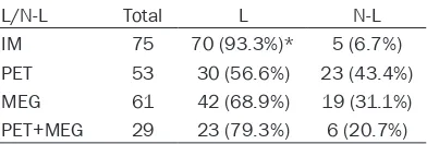
-ed in the invasive monitoring. The localizing per-centages for invasive monitoring, interictal 18F-FDG-PET, MEG, and PET+MEG were 93.3%, 56.6%, 68.9%, and 79.3%, respectively. Com-pared with interictal 18F-FDG-PET, MEG, and PET+MEG, invasive monitoring had the highest diagnostic sensitivity in localizing epileptogenic
foci (Pearson Chi-square test, P=0.00; Table 1).
PET+MEG diagnostic sensitivity tended to be higher than interictal 18F-FDG-PET or MEG,
especially for the temporal non-lesional epilep-sy (Figure 1).
Sometimes the epileptogenic zones found using MEG and interictal 18F-FDG-PET were
[image:3.612.92.288.109.175.2]not limited to a single lobe of the brain (Figure Table 1. Location of epileptogenic foci with
invasive monitoring, PET and MEG in 85
patients (Engel class I~III)
L/N-L Total L N-L
IM 75 70 (93.3%)* 5 (6.7%)
PET 53 30 (56.6%) 23 (43.4%)
MEG 61 42 (68.9%) 19 (31.1%)
PET+MEG 29 23 (79.3%) 6 (20.7%)
2). Localization of epilepto
-genic foci with interictal 18F-FDG-PET had significantly
hi-gher sensitivity when it occ- urred in a single lobe than in multiple lobes (74.1% vs. 34.6%, Pearson Chi-square test, P=0.004; Table 2). When
localizing epileptogenic foci with MEG, no differences
were found between single
and multiple lobes (78.6%
vs. 60.6%, Pearson Chi-squ- are test, P=0.131; Table 2).
PET+MEG showed a higher
diagnostic sensitivity in
local-izing real epileptogenic foci
in a single lobe than in multi-ple lobes (91.3% vs. 33.3%, Fisher’s exact test, P=0.00; Table 2). The percents of
cases that were localized to a
single lobe using interictal
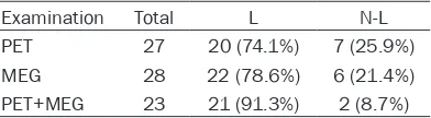
18F-FDG-PET, MEG, and PET+ MEG were 74.1%, 78.6%, and
91.3% respectively (Likeliho- od Ratio test, P=0.249; Table 3). Compared with interictal
18F-FDG-PET, MEG tended to
be higher (Figure 3). There
was no statistical significan-ce among interictal 18F-FDG-PET, MEG, and PET+MEG in localizing epileptogenic foci in
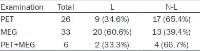
multiple lobes of brain in non-lesional epilepsy (Likelihood Ratio test, P=0.103; Table 4).
Disscussion
Hypometabolism observed
through interictal
18F-FDG-PET is a hallmark of the
sei-zure-onset zone and sur -rounding areas [10], and is commonly assessed when evaluating patients before epilepsy surgery. Lee et al. found that the diagnostic
sen-sitivity of interictal 18F-FDG-PET was 85% for neocortical
[image:4.612.90.379.75.460.2]epilepsies and medial tem- poral, even in patients with bilateral sclerosis, ambiguous Figure 1. A. Lesions in the right temporal lobe were absent in the axial (A1),
[image:4.612.90.380.567.666.2]sagittal (A2), and coronal (A3) FLAIR sequences. B. Hypometabolism in the right temporal lobe is seen in the axial (B1), sagittal (B2), and coronal (B3) interictal FDG-PET scans (the arrows). C. Epileptogenic foci were localized in the right temporal lobe in the axial (C1), sagittal (C2), and coronal (C3) inter-ictal MEG (red dots). D. (D1) No abnormalities in the right temporal cortex. (D2) The anterior temporal lobe and hippocampus were removed. (D3) The CT scan after operation.
23429 Int J Clin Exp Med 2016;9(12):23425-23432 Table 2. The results of location of epileptogenic zone (in one brain
lobe or multi-lobe of brain) with PET, MEG, and PET+MEG in 85
patients (Engel class I~III)
Examination Location of epileptogenic foci Total L N-L valueP PET One brain lobe 27 20 (74.1%)* 7 (17%) 0.004
Multi-lobe of brain 26 9 (34.6%) 17 (65.4%) MEG One brain lobe 28 22 (78.6%) 6 (21.4%) 0.131
Multi-lobe of brain 33 20 (60.6%) 13 (39.4%) PET+MEG One brain lobe 23 21 (91.3%)† 2 (8.7%) 0.008
Multi-lobe of brain 6 2 (33.3%) 4 (66.7%)
L, localizing; N-L, non-localizing; PET, positron emission tomography; MEG,
magnetoencephalo-graphy; *: Pearson Chi-square test, P<0.05; †: Fisher’s exact test, P<0.05.
sclerosis, atrophy, or unremarkable MRI find -ings [11]. Other studies have reported that the
sensitivity of interictal 18F-FDG-PET images in lateralizing temporal lobe epilepsy (TLE) in
patients without a discrete neocortical mass lesion was between 60% and 90% [12-16].
Similar to interictal 18F-FDG-PET, MEG is also a non-invasive method used for localizing sei
-zure foci. As reported in Ray et al. [17], MEG can be used to complement EEG for localiza
-tion of seizure foci, and provides a combina-tion
of noninvasiveness with very high spatial and temporal resolutions. They concluded that the
overall accuracy of MEG in source localization is better than that of EEG.Other studies
com-paring the ability of MEG, scalp video-EEG, and brain MRI to localize the epileptic focus have suggested that MEG is a useful technique for
pre-surgical evaluation because its sensitivity
(approximately 80%) in detecting clinically sig
-nificant epileptiform activity is relatively high [18]. However, a few studies have investigated
which of these two non-invasive methods
(interictal 18F-FDG-PET or MEG) is better at
visualization techniques used to study the relationship between MEG and interictal 18F-FDG-PET metabolism for medical refracto -ry partial epilepsy in a series of 12 patients
[19]. They found that MEG and interictal 18F-FDG-PET provided the highest correlation
of any combination of non-invasive method, and when concordant could accurately predict
the epileptogenic zone. Similarly, Lamusuo et al. evaluated combined interictal 18F-FDG-PET and MEG for preoperative localization of the epileptogenic zone in 9 patients and found concordant interictal 18F-FDG-PET and MEG results in 78% of cases [20]. For non-lesional
epilepsy, Knowlton and colleagues obtained
localizing values of association between MSI
(magnetic source imaging) and interictal
18F-FDG-PET in 51 patients who were free of seizures after surgery [21]. They found that the
combined sensitivity of both MSI and interictal
18F-FDG-PET was only 25%, however, the diag
-nostic specificity was high: 95% for MSI+PET
compared with 79% for MSI or interictal
18F-FDG-PET alone. In the present study of
non-lesional epileptic patients, we found no
sig-nificant difference between PET+MEG and interictal 18F-FDG-PET or MEG in localizing epi
-leptogenic foci, though PET+MEG trended to
have a higher diagnostic sensitivity. However,
PET+MEG was especially helpful in non-lesional
temporal lobe epilepsy of our studies. The tem-poral resection can be determined if both
inter-ictal 18F-FDG-PET and MEG localized the epi
-leptogenic zones to the ipsilateral temporal
lobe, and if this is consistent with the interictal
and ictal scalp EEG, combined with typical types of seizure video monitoring. Ten patients
Table 3. The results of location of
epilepto-genic zone in one brain lobe with PET, MEG, and PET+MEG in 85 patients (Engel class
I~III)
Examination Total L N-L
PET 27 20 (74.1%) 7 (25.9%)
MEG 28 22 (78.6%) 6 (21.4%)
PET+MEG 23 21 (91.3%) 2 (8.7%)
L, localizing; N-L, non-localizing; PET, positron emission tomography; MEG, magnetoencephalo-graphy; Likelihood
Ratio test, P=0.249.
localizing seizure foci in
non-lesional epilepsy. Here, we fo-
und no significant difference
in their sensitivities, although
MEG trended to be more sensi
-tive (interictal 18F-FDG-PET, 56.6%; MEG, 68.9%).
Although the sensitivities of the two techniques were not
significantly different when
conducted separately, perha- ps combining them together
could significantly increase
[image:5.612.92.288.315.369.2]PET and MEG located epileptogenic zones in
a single lobe, most cases turned out to be tem-poral lobe epilepsy, and a smaller number were frontal lobe epilepsy. Additionally, the results
for interictal 18F-FDG-PET and MEG among
patients with multi-lobes non-lesional epilepsy were always inconsistent.
With regard to the interictal 18F-FDG-PET and MEG used in isolation, interictal 18F-FDG-PET
[image:6.612.90.377.72.460.2]showed a higher diagnostic sensitivity in the Figure 3. A. Lesions in the left parietal lobe were absent in the axial (A1),
sagittal (A2), and coronal (A3) FLAIR sequences. B. Hypometabolism in the left parietal lobe is absent in the axial (B1), sagittal (B2), and coronal (B3) interictal FDG-PET scans. C. The epileptogenic foci were localized in the left central area in the axial (C1), sagittal (C2), and coronal (C3) interictal MEG (red dots). D. (D1) No abnormalities in the left parietal cortex. (D2) The results of invasive monitoring showed epileptogenic foci in the left central area. (D3) The non-functional cortex was removed and the central gyrus was coagulated.
Table 4. The results of location of
epilepto-genic zone in multi-lobes of brain with PET, MEG, and PET+MEG in 85 patients (Engel
class I~III)
Examination Total L N-L
PET 26 9 (34.6%) 17 (65.4%)
MEG 33 20 (60.6%) 13 (39.4%)
PET+MEG 6 2 (33.3%) 4 (66.7%)
L, localizing; N-L, non-localizing; PET, positron emission tomography; MEG, magnetoencephalo-graphy; Likelihood
Ratio test, P=0.103.
(11.8%) in our group had go-od seizures outcomes (Engel
class I-III) after temporal re- sections that were performed directly without intracranial
EEG monitoring.
Both interictal 18F-FDG-PET and MEG sometimes located epileptogenic zones in multi -ple lobes of the brain. For some patients several brain
lobes were localized, and we
even saw cases in which the two techniques contradicted each other and indicated the
epileptogenic zones were in
opposite hemispheres. In our study, the highest diagnostic
sensitivity in localizing real
epileptogenic foci was achi- eved when both interictal
18F-FDG-PET and MEG local
-ized the epileptogenic zone in
the same brain lobe, or the volume of the potential epi-leptogenic foci partly or com-pletely overlapped. Conver- sely, locating the real
epilep-togenic foci was very difficult when both interictal 18F-FDG-PET and MEG localized multi
-ple epileptogenic zones in dif -ferent lobes of the brain, or when the potential epilepto-genic foci provided by the two methods did not overlap. In these cases, the epileptogen-ic foci appear to be constantly multi-focal, even with invasive monitoring. We also found
[image:6.612.92.287.628.678.2]18F-FDG-23431 Int J Clin Exp Med 2016;9(12):23425-23432
localization of epileptogenic foci in a single
brain lobe than in multiple brain lobes (74.1%
vs. 34.6%, P=0.004), while localization of epi
-leptogenic foci with MEG did not differ between single and multiple brain lobes (78.6% vs.
60.6%, P=0.131). Compared with interictal
18F-FDG-PET, our findings suggest that MEG localized zones were more diffuse and were
located more extensively throughout the
cor-tex. Although interictal 18F-FDG-PET showed a
higher diagnostic sensitivity for single-lobed
localization, this could be explained by the epi -lepsy types, for example, this study included more cases of temporal lobe non-lesional epi-lepsy. As Henry reported that in pure temporal lobe epilepsy, regional glucose hypometabo-lism was typically presented in the temporal
lobe ipsilateral to the EEG seizure onset loca -tion [22].
Additionally, in the present study, we found that
there was no statistical significance among interictal 18F-FDG-PET, MEG, and PET+MEG in localizing non-lesional epileptogenic foci in a
single lobe of brain and in multiple lobes of
brain. Compared with interictal 18F-FDG-PET or MEG, PET+MEG had the highest diagnostic sensitivity in the localization of epileptogenic
foci in a single brain lobe, while in multiple
lobes, MEG trended to be higher. This could be explained that both interictal 18F-FDG-PET and MEG had their own interpretations of the epi
-leptic zone and their limitations. For example, for the epileptogenic zones located in mesial temporal lobe epilepsy with interictal 18F-FDG-PET and MEG, when most of the two volumes overlapped, the interictal 18F-FDG-PET usually
showed hypometabolism in the temporal pole,
while the MEG showed epileptic-form discharg -es on the outer side of the medium temporal lobe. The latter can be attributed to the
limita-tions of MEG, which is insensitive to exclusively
radially oriented sources, such as those found at the depth of sulci or top of gyri [23, 24].
Nevertheless, both interictal FDG-PET and MEG
(either in isolation or combination) were helpful
in localizing non-lesional epileptogenic zone,
the main role of which were to help doctors determine where to place subdural grids and strips.
Several limitations of this study should be con-sidered. First, this is a single center study with a retrospective design. Second, the positive
results of localizing non-lesional epileptogenic zone by interictal 18F-FDG-PET, MEG, or PET+ MEG are evaluated in qualitative, not in quanti -tative. Further studies are necessary to accu-rately calculate the volume of the epileptogenic
zone by MEG images and precisely delineate the epileptogenic foci by interictal
18F-FDG-PET images.
We found that combining interictal 18F-FDG-PET with MEG had an advantage in localizing the epileptogenic zone in non-lesional epilepsy,
especially in a single lobe of the brain.
Additionally, when interictal 18F-FDG-PET anal -ysis showed hypometabolism in single lobes of the brain, it had a higher diagnostic
sensitiv-ity in localizing epileptogenic foci than when
hypometabolism was seen in multiple lobes. However, despite these positive results, the two techniques consistently revealed a minority of patients who could have resections direc-
tly performed without intracranial EEG
moni-toring.
Disclosure of conflict of interest
None.
Address correspondence to: Zhiqiang Cui and Bai- nan Xu, Department of Neurosurgery, Chinese PLA General Hospital, Chinese PLA Postgraduate Med-ical School, No. 28, Fuxing Road, Haidian District, Beijing 100853, P. R. China. Tel: +86 10 66938340; Fax: +86 10 66938038; E-mail: zhiqiangcui2008@ 163.com (ZQC); Tel: +86 10 66938439; Fax: +86 10 66938038; E-mail: bnx301hos@163.com (BNX)
References
[1] Cascino GD, Jack CR Jr, Parisi JE, Marsh WR, Kelly PJ, Sharbrough FW, Hirschorn KA and Trenerry MR. MRI in the presurgical evalua- tion of patients with frontal lobe epilepsy and children with temporal lobe epilepsy: patho-logic correlation and prognostic importance. Epilepsy Res 1992; 11: 51-59.
[2] Lorenzo NY, Parisi JE, Cascino GD, Jack CR Jr, Marsh WR and Hirschorn KA. Intractable fron-tal lobe epilepsy: pathological and MRI fea-tures. Epilepsy Res 1995; 20: 171-178. [3] Mosewich RK, So EL, O’Brien TJ, Cascino GD,
[4] Tebo CC, Evins AI, Christos PJ, Kwon J and Schwartz TH. Evolution of cranial epilepsy sur -gery complication rates: a 32-year systematic review and meta-analysis. J Neurosurg 2014; 120: 1415-1427.
[5] Lawson JA, Cook MJ, Vogrin S, Litewka L, Strong D, Bleasel AF and Bye AM. Clinical, EEG, and quantitative MRI differences in pediatric frontal and temporal lobe epilepsy. Neurology 2002; 58: 723-729.
[6] Wang Y, Liu B, Fu L and Cui Z. Use of interictal (18)F-fluorodeoxyglucose (FDG)-PET and mag -netoencephalography (MEG) to localize epilep -togenic foci in non-lesional epilepsy in a cohort of 16 patients. J Neurol Sci 2015; 355: 120-124.
[7] Widjaja E, Otsubo H, Raybaud C, Ochi A, Chan D, Rutka JT, Snead OC 3rd, Halliday W, Sakuta R, Galicia E, Shelef I and Chuang SH. Characteristics of MEG and MRI between Taylor’s focal cortical dysplasia (type II) and other cortical dysplasia: surgical outcome af-ter complete resection of MEG spike source and MR lesion in pediatric cortical dysplasia. Epilepsy Res 2008; 82: 147-155.
[8] Jayakar P, Dunoyer C, Dean P, Ragheb J, Resnick T, Morrison G, Bhatia S and Duchowny M. Epilepsy surgery in patients with normal or nonfocal MRI scans: integrative strategies of-fer long-term seizure relief. Epilepsia 2008; 49: 758-764.
[9] Eliashiv DS, Elsas SM, Squires K, Fried I and Engel J Jr. Ictal magnetic source imaging as a localizing tool in partial epilepsy. Neurology 2002; 59: 1600-1610.
[10] Mauguiere F and Ryvlin P. The role of PET in presurgical assessment of partial epilepsies. Epileptic Disord 2004; 6: 193-215.
[11] Lee DS, Lee SK and Lee MC. Functional neuro-imaging in epilepsy: FDG PET and ictal SPECT. J Korean Med Sci 2001; 16: 689-696. [12] Engel J Jr, Kuhl DE, Phelps ME and Mazziotta
JC. Interictal cerebral glucose metabolism in partial epilepsy and its relation to EEG chang -es. Ann Neurol 1982; 12: 510-517.
[13] Ho SS, Berkovic SF, Berlangieri SU, Newton MR, Egan GF, Tochon-Danguy HJ and McKay WJ. Comparison of ictal SPECT and interictal PET in the presurgical evaluation of temporal lobe epilepsy. Ann Neurol 1995; 37: 738-745. [14] Henry TR. PET: cerebral blood flow and
glu-cose metabolism--presurgical localization. Adv Neurol 2000; 83: 105-120.
[15] O’Brien TJ, Hicks RJ, Ware R, Binns DS, Murphy M and Cook MJ. The utility of a 3-dimensional, large-field-of-view, sodium iodide crystal-based PET scanner in the presurgical evaluation of partial epilepsy. J Nucl Med 2001; 42: 1158-1165.
[16] Theodore WH, Newmark ME, Sato S, Brooks R, Patronas N, De La Paz R, DiChiro G, Kessler RM, Margolin R, Manning RG, et al. [18F]fluo -rodeoxyglucose positron emission tomography in refractory complex partial seizures. Ann Neurol 1983; 14: 429-437.
[17] Ray A and Bowyer SM. Clinical applications of magnetoencephalography in epilepsy. Ann Indian Acad Neurol 2010; 13: 14-22.
[18] Stefan H, Hummel C, Scheler G, Genow A, Druschky K, Tilz C, Kaltenhauser M, Hopfen-gartner R, Buchfelder M and Romstock J. Magnetic brain source imaging of focal epilep-tic activity: a synopsis of 455 cases. Brain 2003; 126: 2396-2405.
[19] Wong STC, Hoo KS Jr and Knowlton RC. Image coregistration and visualization techniques to study relationships between MEG neuro -physiology and FDG-PET metabolism in epilep -sy imaging. Pro.SPIE2709, Medical Imaging 1996: Physiology and Function From Multidi- mensional Images 1996; 2709: 280-290. [20] Lamusuo S, Forss N, Ruottinen HM, Bergman
J, Makela JP, Mervaala E, Solin O, Rinne JK, Ruotsalainen U, Ylinen A, Vapalahti M, Hari R and Rinne JO. [18F]FDG-PET and whole-scalp MEG localization of epileptogenic cortex. Epilepsia 1999; 40: 921-930.
[21] Knowlton RC, Elgavish RA, Bartolucci A, Ojha B, Limdi N, Blount J, Burneo JG, Ver Hoef L, Paige L, Faught E, Kankirawatana P, Riley K and Kuzniecky R. Functional imaging: II. Prediction of epilepsy surgery outcome. Ann Neurol 2008; 64: 35-41.
[22] Henry TR, Mazziotta JC and Engel J Jr. Interictal metabolic anatomy of mesial temporal lobe epilepsy. Arch Neurol 1993; 50: 582-589. [23] Stefan H. Magnetic source imaging. Rev Neurol
(Paris) 2009; 165: 742-745.