Original Article
A comparative study of open reduction with internal
fixation and percutaneous poking reduction fixation for
the treatment of Sanders type II calcaneal fractures
Junfeng Zhan, Nan Zhu, Wang Fang, Juehua Jing
Department of Orthopaedics, The Second Hospital of Anhui Medical University, Hefei 230601, Anhui Province, P.R. China
Received March 13, 2016; Accepted May 10, 2016; Epub June 15, 2016; Published June 30, 2016
Abstract: Objective: To compare and analyze the clinical efficacy of open reduction with internal fixation and per
-cutaneous poking reduction fixation for Sanders type II calcaneal fractures. Methods: A total of 57 patients with
calcaneal fractures were randomly divided into the poking group (27 cases, underwent percutaneous poking
reduc-tion) and the incision group (30 cases, underwent open reduction with internal fixareduc-tion). The operation time, drain -age volume, intraoperative blood loss, and hospitalization days were recorded. During the postoperative follow-up, fracture healing and incidence of complications were observed and recorded for both groups. At the last follow-up, Visual Analog Scale (VAS), American Orthopaedic Foot and Ankle Society (AOFAS) score, and the MOS item short
form health survey (SF-36) were used to evaluate the clinical efficacy. Results: The operation time, drainage volume, intraoperative blood loss and hospitalization days in poking group were significantly less than those in the incision group, with statistically significant differences (P<0.05). In the postoperative follow-up, it was found that there
was no significant difference in fracture healing time between the two groups. The incidence of complications was 3.70% in poking group, significantly lower than 10.00% in incision group (P<0.05). The Böhler and Gissane angles
were significantly improved after surgery in both groups (P<0.05), but there was no significant difference between
the two groups after surgery (P>0.05). At the last follow-up, VAS and SF-36 scores in the poking group were signifi -cantly higher than those in the incision group (P<0.05). There was no significant difference in excellent and good
ratebetween the poking group and the incision group (P>0.05). Conclusion: Percutaneous poking reduction fixation can effectively reduce the incidence of postoperative complications and significantly improve the clinical efficacy and outcomes in treatment of Sanders II calcaneal fractures, so it is an efficient treatment method for calcaneal
fractures.
Keywords: Calcaneal fractures, percutaneous poking reduction fixation, open reduction with internal fixation
Introduction
Calcaneal fracture is a common trauma in Department of Orthopedics, mostly caused by traffic accident, falling injury and so on [1]. At present, surgical treatment is mainly used clini-cally, including open reduction with internal fix -ation (ORIF) and percutaneous poking reduc-tion fixareduc-tion, etc [2]. Open reducreduc-tion with inter -nal fixation (ORIF) is a traditio-nal surgical treat -ment for calcaneal fractures, with advantages of simple operation and adequate exposure, which can effectively reset the fracture site and offer secured fixation [3]. However, ORIF also has some limitations such as large surgical
calca-neal fractures by percutaneous poking reduc-tion fixareduc-tion have not been massively reported both in domestic and abroad. The objective of this study was to compare and analyze the clini-cal efficacy of open reduction with internal fixa -tion and percutaneous poking reduc-tion fixa -tion for the treatment of Sanders type II calca-neal fractures.
Materials and methods
General information
57 patients with Sanders type II calcaneal frac-tures in Department of Orthopedics in our hos-pital from February 2013 to December 2014 were selected in this study. These 57 patients were randomly divided into the poking group and incision group. In the poking group, there were 27 patients, including 14 males and 13 females, aged from 22 to 65 years old, with an average age of 41.77±4.17 years old; 15 cases had fractures in the left, and 12 cases in the right; body mass index (BMI): 21-31.5 kg/m2,
with an average value of 24 kg/m2; the cause of
injury: traffic injury in 12 cases, falling injury in 15 cases; Böhler angle (14.4±4.5)°, Gissane angle (87.1±5.7)°; the average time from injury to surgery was (12.8±2.8) d. In the incision group, there were 30 patients, including 15 males and 15 females, aged from 20 to 67 years, with an average age of 42.57±5.57 years; 17 cases had fractures in the left and 13 cases in the right; body mass index (BMI): 20.5-32 kg/m2, with an average value of 23.8 kg/m2;
cause of injury: traffic injury in 20 cases, and falling injury in 10 cases; Böhler angle (15.1±3.4)°, Gissane angle (88.2±5.5)°; the average time from injury to surgery was (11.8±2.7) d. There were no significant differ -ences in gender, age, body mass index, lateral, fracture side, Böhler angles, Gissane angles, the time from injury to surgery and other gen-eral information between two groups (P>0.05), so these two groups were comparable.
Treatment method
The patients in poking group received the sur-gery treatment of percutaneous poking reduc-tion fixareduc-tion: after the satisfacreduc-tion of patients with subarachnoid block anesthesia, patients took the normal lateral position, with conven-tional disinfection and draping. One Kirschner wire was drilled on the edge of achilles tendon.
If the X-ray results confirmed that the Kirschner wire had reached the bottom of posterior artic-ular calcaneal, upward poking was performed to reset the posterior articular surface and the calcaneal at the same time. When necessary, two Kirschner wires could be drilled in for fixa -tion. Upon satisfaction with the reduction in imaging examination, the wound was washed and plaster fixation was performed.
The patients in incision group received open reduction with internal fixation: its preoperative preparation was the same as the poking group. Then incision (“L” shaped) was made at about 5cm above the lateral calcaneal of lateral supramalleolar. After separating soft tissues, traction was performed in the nodules and the calcaneal was reset after poking the collapsed calcaneal. Suitable steel plate was used for internal fixation after the reduction. As the imaging results showed satisfied reduction and fixing effects, conventional negative pressure drainage was performed, incision was sutured, and plaster fixation was implemented.
Postoperative management
Antibiotics were postoperatively used in a rou-tine manner, combined with ice compress, pressure dressing, and raising the affected limb for treatment. Negative pressure drainage was removed at 48 h postoperative, with ankle joint activities from postoperative day 2, and the stitches were taken out three weeks after the operation. In the regular follow-up in outpa-tient clinic, the occurrence of complications was recorded, Böhler angles and Gissane angles of the patients were measured, and radiography was performed to evaluate the healing of fractures. In the last follow-up, the ankle joint scoring systems of Visual Analogue Scale (VAS), the Short Form-36 Health Survey (SF-36), and Maryland Foot Score standard by American Orthopaedic Foot and Ankle Society (AOFAS) were used to evaluate the final clinical efficacy.
Statistical treatment
All the data were statistically analyzed by SPSS 17.0 software. Measurement data were expressed with _X±S, and t test was used for comparison between groups; the enumeration data were expressed with percentage, and χ2
There was statistically significant difference when P<0.05.
Results
Comparison of operation time, intraoperative blood loss and hospitalization days between two groups
The operation time was 11~27 min (mean value of 19.4±7.2 min) in the poking group and 62~115 min (mean value of 71.5±10.1 min) in the incision group, with significant difference between the two groups (P<0.05). The drainage volume was 12~21 ml (mean value of 15.4±1.1 ml) in the poking group, lower than 18~25 ml
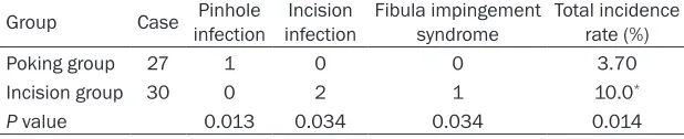
[image:3.612.87.401.97.162.2]sion infection was present in 2 patients, and fibula impingement syndrome was observed in 1 patient, with an incidence rate of 10.00%. The incidence of complications in the incision group was significantly higher than that in the poking group, with statistically significant differ -ence between the two groups (P<0.05). See Table 2 for details.
Comparison of Böhler angles and Gissane angles between two groups of patients with calcaneal fractures
The preoperative Böhler angles were (14.4± 4.5)° and (15.1±3.4)° respectively in the pok-ing group and incision group; and the postop-Table 1. Comparison of operation-related indexes and hospitalization
duration between two groups
Group Case Operation time (min) volume (ml)Drainage blood loss (ml)Intraoperative Hospitalization duration Poking group 27 19.4±7.2 15.4±1.1 17.8±6.5 5.6±1.5 Incision group 30 71.5±10.1* 21.1±1.2* 66.5±16.4* 18.1±5.2*
P value 0.015 0.034 0.034 0.024
[image:3.612.90.399.209.272.2]*P<0.05, compare with poking group.
Table 2. Comparison of postoperative complications between two groups
Group Case Pinhole infection infectionIncision Fibula impingement syndrome Total incidence rate (%)
Poking group 27 1 0 0 3.70
Incision group 30 0 2 1 10.0*
P value 0.013 0.034 0.034 0.014
[image:3.612.89.401.329.407.2]*P<0.05, compare with poking group.
Table 3. Comparison of the Böhler angles and Gissane angles between two groups
Group Case
Böhler angles Gissane angles Preoperative
(°) Postoperative (°) Preoperative (°) Postoperative (°) Poking group 27 14.4±4.5 26.6°±4.7* 87.1°±5.7 135.7°±9.1*
Incision group 30 15.1±3.4 25.1°±3.5* 88.2°±5.5 136.6°±11.5*
P value 0.078 0.032 0.013 0.012
[image:3.612.90.395.449.506.2]*P<0.05, compare with preoperative value.
Table 4. Comparison of AOFAS and VAS scores between two groups
Group Case AOFAS VAS SF-36
Poking group 27 92.2±7.3 2.5±0.1 81.5±8.1 Incision group 30 91.8±6.7 1.1±0.2* 64.2±6.5*
P value 0.153 0.0124 0.0147
*P<0.05, compare with poking group.
(mean value of 21.1± 1.2 ml) in the incision group, with statistical- ly significant difference (P<0.05). The mean in- traoperative blood loss was 66.5±16.4 ml in the incision group, and 17.8±6.5 ml in the pok-ing group, with signifi -cant difference bet- ween the two groups. The mean hospitaliza-tion durahospitaliza-tion was (5.6± 1.5) d in the poking group, and (18.1±5.2) d in the incision group, with statistically signifi -cant difference between the two groups. See Table 1 for details. Comparison of postop-erative complications between two groups
inci-nificantly improved as compared with preopera -tive Böhler angles (P<0.05); but there was no significant difference between two groups after operation (P>0.05). The preoperative Gissane angles were (87.1±5.7)° and (88.2±5.5)° respectively in of the poking group and incision group; and the postoperative Gissane angles were (135.7±9.1)° and (136.6±11.5)° respec-tively. It was also significantly improved in both groups as compared with preoperative condi-tions, with statistically significant difference (P<0.05); but there was no statistically signifi -cant difference between two groups after oper-ation (P>0.05). See Table 3 for details.
Comparison of AOFAS, VAS and SF-36 scores between two groups
The patients were followed up for 22-49 months in the poking group (with a mean value of 36.2±12.1 months) and 23-48 months in the incision group (with a mean value of 36.7±12.3 months). There was no significant difference in the fracture healing time between the poking group (4.9±0.7 months) and incision group (5.1±0.8 months) (P>0.05). The postoperative AOFAS, VAS and SF-36 scores were 92.2±7.3, 2.5±0.1, and 81.5±8.1 respectively in the pok-ing group, and 91.8±6.7, 1.1±0.2, and 64.2±6.5 in the incision group. VAS and SF-36 scores in the poking group were significantly higher than those in the incision group, with statistically sig-nificant difference (P<0.05), but there was no significant difference in AOFAS scores between the two groups. See Table 4 for details.
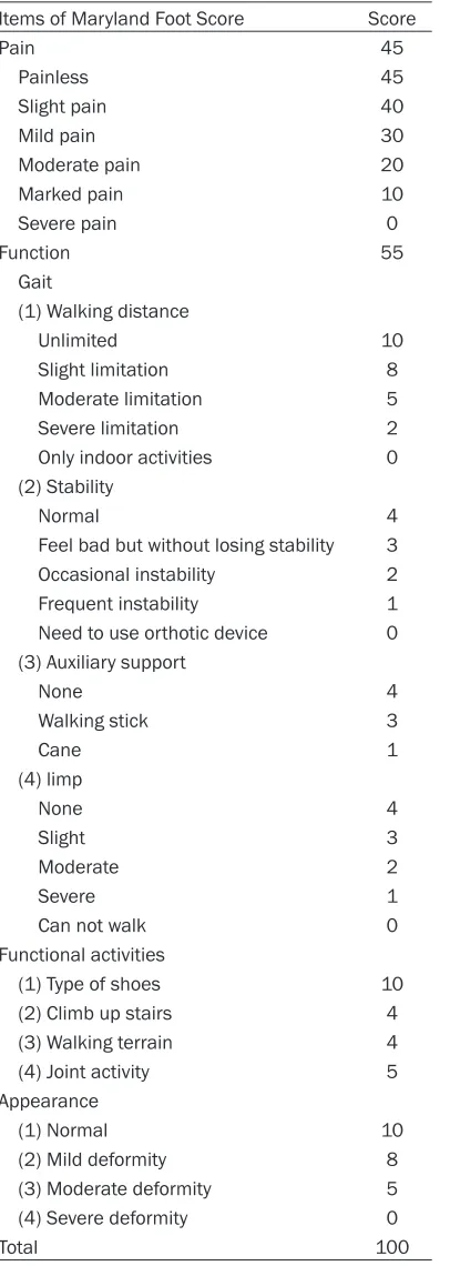
Comparison of postoperative Maryland Foot Scores between two groups
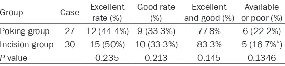
In the 27 patients of poking group, 12 patients achieved “excellent”, and 9 patients achieved “good”, with an excellent and good rate of 77.8%. In the 30 patients of incision group, 15 patients achieved “excellent”, and 10 patients achieved “good”, with an excellent and good rate of 83.3%. There was no statistically signifi -cant difference in excellent and good rate between these two groups (P>0.05). See Tables 5 and 6 for details.
Discussion
[image:4.612.93.296.83.653.2]Calcaneal fracture is a common trauma in Department of Orthopedics, mostly caused by Table 5. Maryland Foot Score
Items of Maryland Foot Score Score
Pain 45
Painless 45
Slight pain 40
Mild pain 30
Moderate pain 20
Marked pain 10
Severe pain 0
Function 55
Gait
(1) Walking distance
Unlimited 10
Slight limitation 8 Moderate limitation 5 Severe limitation 2 Only indoor activities 0 (2) Stability
Normal 4
Feel bad but without losing stability 3 Occasional instability 2 Frequent instability 1 Need to use orthotic device 0 (3) Auxiliary support
None 4
Walking stick 3
Cane 1
(4) limp
None 4
Slight 3
Moderate 2
Severe 1
Can not walk 0 Functional activities
(1) Type of shoes 10 (2) Climb up stairs 4 (3) Walking terrain 4 (4) Joint activity 5 Appearance
(1) Normal 10
(2) Mild deformity 8 (3) Moderate deformity 5 (4) Severe deformity 0
Total 100
Note: the patients with total score of 90-100 points are graded as “excellent”, 75-89 points as “good”, 50-75 points as “available”, and those under 50 points are graded as “poor”.
Calcaneal is a very important bearing bone of the human body, so it is very easy to affect the daily life and work of patients in case of improp-er treatment for calcanea fractures. At present, surgical treatment is mainly used clinically, including open reduction with internal fixation (ORIF) and percutaneous poking reduction fixa -tion, etc. Open reduction with internal fixation (ORIF) is a traditional surgical treatment for cal-caneal fractures, with advantages of restoring the anatomic calcaneal morphology and talo-calcaneal joints matching, which could achieve good clinical effect and reduce the incidence of traumatic arthritis. However, incision complica -tions with varying degrees of severity are often found after ORIF, even chronic calcaneal osteo-myelitis may be present [8, 9]. The results of this study showed that the incidence of compli-cations in patients undergoing ORIF in the inci-sion group was up to 10%. The complications such as incision infections and pain would seri-ously affect the prognosis and long-term clini-cal efficacy for patients.
To make up for these limitations and disadvan-tages, some scholars recommended percuta-neous poking reduction fixation for the treat -ment of calcaneal fracture. It was to pry up the collapsed articular surface by using the lever principle, combined with manual reduction of the calcaneal height and articular surface [10]. Studies showed that open reduction with inter-nal fixation and percutaneous poking reduction fixation can obtain satisfactory therapeutic effect [11]. Moreover, percutaneous poking reduction fixation has low incidence of postop -erative complications, less trauma, fast recov-ery of postoperative functions and other advan-tages that are nonexistent in other methods [12]. In this study, the operation time was (19.4±7.2) min in the poking group, significantly shorter than (71.5±10.1) min in the incision group; the drainage volume was (15.4±1.1) ml in the poking group, significantly lower than (21.1±1.2) ml in the incision group; the intraop-erative blood loss was (17.8±6.5) ml in the
pok-indices have a P value less than 0.05 that stands for statistically significant differences between two groups.
In addition, the incidence of complications was 3.70% in the poking group, significantly lower than 10% in the incision group, with significant differences (P<0.05). The postoperative Böhler angles and the Gissane angles were significant -ly improved in both groups (P<0.05), but there was no significant difference between two groups after operation (P>0.05). This showed that the percutaneous poking reduction fixation was equally effective with the conventional ORIF in the correction of Böhler angles and Gissane angles. Besides, at the last follow-up, AOFAS, VAS, SF-36, and Maryland Foot Scores in two groups were measured and recorded. VAS and SF-36 scores in the poking group were significantly higher than those in the incision group, with statistically significant difference (P<0.05). This indicated that the patients undergoing percutaneous poking reduction fix -ation could have better long-term curative effect and higher quality of life. The results of Maryland Foot Score system showed that the excellent and good rate was 77.8% in the pok-ing group and 83.3% in the incision group, with no statistically significant difference (P>0.05). This indicated that both percutaneous poking reduction fixation and conventional ORIF have very high excellent and good rate as well as sat-isfactory clinical efficacy.
[image:5.612.93.373.87.152.2]Poking reduction fixation has many advantages in the treatment of calcaneal fractures, but there are also some defects [13, 14]. Previous studies showed that poking reduction fixation can often achieve more satisfactory efficacy for the fractures where the articular surface destruction and displacement are not very seri-ous, such as Sanders type I fractures [15]. However, with the continuous increase of Sanders type, the patients would have corre-spondingly increased proportion of postopera-tive pain and dysfunction [16]. Analysis showed Table 6. Comparison of Maryland Foot Score between two groups
Group Case Excellent rate (%) Good rate (%) and good (%)Excellent or poor (%)Available Poking group 27 12 (44.4%) 9 (33.3%) 77.8% 6 (22.2%) Incision group 30 15 (50%) 10 (33.3%) 83.3% 5 (16.7%*)
P value 0.235 0.213 0.145 0.1346
that it may be because the poking reduction was difficult to achieve complete anatomical reduction for the fractures where the articular surface is seriously destroyed and displaced [17]. In addition, the minimally invasive poking reduction is difficult to completely remove the small pieces of broken bones in the articular cavity, resulting in the uneven surface of subta-lar joint and leading to traumatic arthritis [18]. It also seriously affects the long-term efficacy and prognosis. The clinical efficacy of poking reduction fixation for Sanders type II fractures was studied for the first time in this trial, and it was comprehensively analyzed and compared with the traditional ORIF method. X-ray was used to make sure that after the Kirschner wire had reached the bottom of posterior articular calcaneal, upward poking could be conducted to reset the posterior articular surface and cal-caneal at the same time, and 2 Kirschner wires can be drilled in when necessary. The incision was sutured after satisfactory with the reduc-tion results in imaging examinareduc-tion, ensuring the complete reduction of the articular surface, long-term efficacy and prognosis [19, 20]. This type of research has not been widely reported both at home and abroad.
In summary, poking reduction fixation for Sanders type II calcaneal fractures can achieve the same excellent and good rate with tradition-al ORIF, obtain satisfactory efficacy, effectively shorten the operation time, reduce intraopera-tive blood loss and postoperaintraopera-tive drainage vol-ume, shorten the hospitalization duration, reduce the incidence of postoperative compli-cations, and improve functional recovery, prog-nosis and quality of life, which is worthy of pop-ularization in clinical application.
Declaration of conflict of interest None
Address correspondence to: Juehua Jing, Depart-
ment of Orthopaedics, The Second Hospital of Anhui
Medical University, No.678 Furong Road, Economic
and Technological Development Zone, Hefei
230-601, Anhui Province, P.R. China. Tel: +86-5516- 3869506. E-mail: juehua_jing@sina.cn.
References
[1] Schepers T. The sinus tarsi approach in dis-placed intra-articular calcaneal fractures: a systematic review. Int Orthop 2011; 35: 697-703.
[2] DeWall M, Henderson CE, McKinley TO, Phelps
T, Dolan L and Marsh JL. Percutaneous
reduc-tion and fixareduc-tion of displaced intra-articular
calcaneus fractures. J Orthop Trauma 2010; 24: 466-472.
[3] Rammelt S, Amlang M, Barthel S, Gavlik JM
and Zwipp H. Percutaneous treatment of less
severe intraarticular calcaneal fractures. Clin Orthop Relat Res 2010; 468: 983-990.
[4] Woon CY, Chong KW, Yeo W, Eng-Meng Yeo N
and Wong MK. Subtalar arthroscopy and fluro
-socopy in percutaneous fixation of intra-articu -lar calcaneal fractures: the best of both worlds. J Trauma 2011; 71: 917-925.
[5] Mostafa MF, El-Adl G, Hassanin EY and
Abdellatif MS. Surgical treatment of displaced intra-articular calcaneal fracture using a single small lateral approach. Strategies Trauma Limb Reconstr 2010; 5: 87-95.
[6] Femino JE, Vaseenon T, Levin DA and Yian EH. Modification of the sinus tarsi approach for open reduction and plate fixation of intra-artic -ular calcaneus fractures: the limits of proximal extension based upon the vascular anatomy of the lateral calcaneal artery. Iowa Orthop J 2010; 30: 161-167.
[7] Besch L, Waldschmidt JS, Daniels-Wreden-
hagen M, Varoga D, Mueller M, Hilgert RE,
Mathiak G, Oestern S, Lippross S and Seekamp A. The treatment of intra-articular calcaneus fractures with severe soft tissue damage with
a hinged external fixator or internal stabiliza -tion: long-term results. J Foot Ankle Surg 2010; 49: 8-15.
[8] Kissel CG, Husain ZS, Cottom JM, Scott RT and
Vest J. Early clinical and radiographic out-comes after treatment of displaced intra-artic-ular calcaneal fractures using delta-frame
ex-ternal fixator construct. J Foot Ankle Surg
2011; 50: 135-140.
[9] Mehta S, Mirza AJ, Dunbar RP, Barei DP and Benirschke SK. A staged treatment plan for the management of Type II and Type IIIA open cal-caneus fractures. J Orthop Trauma 2010; 24: 142-147.
[10] Goldzak M, Mittlmeier T and Simon P. Locked nailing for the treatment of displaced articular fractures of the calcaneus: description of a new procedure with calcanail(®). Eur J Orthop Surg Traumatol 2012; 22: 345-349.
[11] Jacquot F and Atchabahian A. Balloon
reduc-tion and cement fixareduc-tion in intra-articular cal -caneal fractures: a percutaneous approach to intra-articular calcaneal fractures. Int Orthop 2011; 35: 1007-1014.
[12] Tomesen T, Biert J and Frolke JP. Treatment of displaced intra-articular calcaneal fractures with closed reduction and percutaneous screw
fixation. J Bone Joint Surg Am 2011; 93:
[13] Dayton P, Feilmeier M and Hensley NL.
Technique for minimally invasive reduction of calcaneal fractures using small bilateral
exter-nal fixation. J Foot Ankle Surg 2014; 53:
376-382.
[14] Illert T, Rammelt S, Drewes T, Grass R and
Zwipp H. Stability of locking and non-locking
plates in an osteoporotic calcaneal fracture model. Foot Ankle Int 2011; 32: 307-313.
[15] Hammond AW and Crist BD. Percutaneous
treatment of high-risk patients with intra-artic-ular calcaneus fractures: a case series. Injury 2013; 44: 1483-1485.
[16] Schepers T. The primary arthrodesis for se-verely comminuted intra-articular fractures of the calcaneus: a systematic review. Foot Ankle Surg 2012; 18: 84-88.
[17] Pelliccioni AA, Bittar CK and Zabeu JL. Surgical treatment of intraarticular calcaneous frac-tures of sanders’ types II and III. Systematic review. Acta Ortop Bras 2012; 20: 39-42.
[18] Gurkan V, Dursun M, Orhun H, Sari F, Bulbul M
and Aydogan M. Long-term results of conserva-tive treatment of Sanders type 4 fractures of the calcaneum: a series of 64 cases. J Bone Joint Surg Br 2011; 93: 975-979.
[19] Badillo K, Pacheco JA, Padua SO, Gomez AA, Colon E and Vidal JA. Multidetector CT evalua-tion of calcaneal fractures. Radiographics 2011; 31: 81-92.
[20] Schepers T, van Lieshout EM, Ginai AZ, Mulder
PG, Heetveld MJ and Patka P. Calcaneal frac
-ture classification: a comparative study. J Foot