Original Article
Effects of long non-coding RNA MALAT1 on prognosis of
various tumors: a meta-analysis of cohort studies
Jiayuan Wu1*, Liren Hu2*, Fenping Wu3, Gaohua Zhang2, Taiping He4
1Departmentof Nutritional, The Affiliated Hospital of Guangdong Medical College, Zhanjiang, China; 2Department of Epidemiology and Health Statistics, School of Public Health, Guangdong Medical College, Zhanjiang, China; 3Department of Radiotherapy, The Seventh People’s Hospital of Chengdu, The Oncology Hospital of Chengdu, Chengdu, China; 4School of Public Health, Guangdong Medical College, Zhanjiang, China. *Equal contributors.
Received September 29, 2015; Accepted December 19, 2015; Epub February 15, 2016; Published February 29, 2016
Abstract: Background: Metastasis-associated lung adenocarcinoma transcript 1 (MALAT1) is involved in tumor pro-gression and may serve as a prognostic biomarker for various cancers. Objective: This meta-analysis aimed to reveal the association between MALAT1 expression and survival in solid tumors. Methods: A literature search was performed via electronic retrieval until August 2015. Different clinical outcomes of overall survival (OS) and disease-free survival (DFS) were analyzed. Pooled hazard ratios (HRs) or odds ratios (ORs) and 95% confidence intervals (CIs) were calculated to evaluate the relationship of high MALAT1 expression with survival rates and clinicopatho-logical characteristics. Results: Fourteen studies with 1468 patients were included in this meta-analysis. MALAT1 overexpression was highly associated with OS of 1.64 (95% CI: 1.29-2.10) and DFS of 2.26 (95% CI: 1.66-3.08). MALAT1 overexpression was also significantly associated with tumor size (OR = 2.34; 95% CI = 1.14-4.79), tumor stage (OR = 1.48; 95% CI = 1.09-2.01), depth of invasion (OR = 1.49; 95% CI = 1.05-2.11), and lymph node metas-tasis (OR = 2.06; 95% CI = 1.19-3.58). Conclusion: MALAT1 overexpression is obviously ascribed to poor prognosis in numerous cancers, and MALAT1 may serve as a biomarker for the progression of solid tumors.
Keywords: MALAT1, lncRNA, solid tumor, prognosis, meta-analysis
Introduction
Dysregulation of gene expression plays a criti-cal role in carcinogenesis and metastasis. With the development of sequencing and microarray for whole genome and transcriptome, at least 90% of the human genome has been actively transcribed into non-coding RNAs (ncRNAs); more than 80% of the transcribed RNAs did not code for proteins in mammalians, and the pro-tein-coding genes account for only 2% of the gene sequences [1, 2]. Although ncRNAs have been described as “noise” in the transcription-al process or “garbage” in the human body, substantial evidence has proven that ncRNAs also demonstrate important physiological func-tions in cell metabolism and play significant regulatory roles in some diseases [3-5].
Increasing numbers of long ncRNAs (lncRNAs) with length of more than 200 nt but frequently up to 100 kb are found comprising 80% of
ncRNAs, and have become the focus of recent studies. To date, lncRNAs are defined as “RNA molecules that may function as either primary or spliced transcripts and do not fit into known classes of small RNAs or into classes of struc-tural RNAs” [4], suggesting that lncRNAs par-ticipate in multiple gene-regulating processes, such as chromosome silencing, genomic imprinting, transcriptional activation, post-tran-scriptional interference, and nuclear-cytoplas-mic trafficking at various levels, which are involved in almost all physiological and patho-logical processes [6, 7]. However, current stud-ies on lncRNAs remain at initial stage, and only few lncRNAs have been well characterized. Thus, further studies are needed to expand this research field and elucidate the functions and mechanisms of lncRNAs.
discovered; MALAT1 exhibits a length of 8000 nt and is also known as nuclear-enriched abun-dant transcript 2 [8]. MALAT1 cannot be trans-lated into a protein because of its nuclear local-ization and the lack of an open coding frame with sufficient length [9]. Furthermore, MALAT1 gene is located in human chromosome 11q13.1 with a highly conserved and homologous sequence in evolution of various species, which indicates that this gene may potentially influ-ence several physiological functions. Since the discovery of MALAT1 in 2003, several data have clarified the influence of this transcript on the progression or metastasis of different malignancies, such as lung cancer [10, 11], gastric cancer (GC) [12], hepatocellular carci-noma (HCC) [13], and gallbladder cancer [14]; data suggest that MALAT1 may serve as an independent factor for tumor prognosis. However, some studies reported that MALAT1 overexpression contributes to a poor survival outcome for non-small-cell lung cancer (NSCLC) [11], colorectal cancer [15], and HCC [16]; by contrast, several studies indicated that the high expression of MALAT1 is not associated with cancer prognosis [17, 18] or even predict-ing a better cancer prognosis [19]. Therefore, the real value of MALAT1 on predicting the prognosis of solid tumors remains contradicto-ry, and a meta-analysis is necessary to evalu-ate the relationship between MALAT1 expres-sion and solid tumor prognosis.
Materials and methods
Literature search
This meta-analysis was conducted according to the guidelines of the Preferred Reporting Items for Systematic Reviews and Meta-analyses [20]. A systematic, computerized searching was performed through the PubMed, Embase, and Web of Science databases, as well as the China National Knowledge Infrastructure by using the following terms: “MALAT1 or Metastasis Associated Lung Adenocarcinoma Transcript 1”, “Tumor or Cancer or Carcinoma”, and “Prognosis or Survival or Outcome”. No lan-guage restrictions were imposed, and literature search was conducted until August 5, 2015. Lists of references of retrieved articles and reviews were also checked to identify addition-al relevant studies.
Inclusion and exclusion criteria
Studies were eligible if they met the following criteria: 1) Studies reported the relationship between MALAT1 expression and tumor prog-nosis outcomes [i.e., overall survival (OS), or disease-free survival (DFS)]. 2) Studies used a cohort design. 3) Hazard ratios (HRs) and 95% confidence intervals (CIs) can be directly obtained or indirectly calculated from the origi-nal data. Studies were ineligible if they were reviews, conference abstracts, editorials or case reports, or non-human research, articles with insufficient data to estimate HRs and 95% CIs. If more than one publication with the same study population was identified, only the most recent data were included in the final analysis.
Data extraction
Information was carefully and independently extracted by two investigators (WJY and HLR) based on the inclusion and exclusion criteria stipulated above. Any disagreement was resolved through consensus. The following data were collected from each study: first author’s name, year of publication, recruitment time, country of the studied population, sample size, tumor type, follow-up period, testing meth-od of MALAT1, cut-off value, numbers of high/ low MALAT1 expression, and HRs and 95% CIs for survival outcomes as applicable. Stratifi- cation into subgroups will be conducted if at least two studies reported the same outcome for the same tumor type; otherwise, they will be assigned into a subgroup named “Others.” HRs and 95% CIs were preferentially obtained from the outcomes of multivariable analysis followed by univariate analysis. If no direct data were available, the HRs and 95% CIs were calculated in each study from the numbers of patients at risk and events, as well as the P values of log-rank statistics, or from the survival plots of Kaplan-Meier curves [21].
Quality assessment
study groups (one item, up to two stars), and outcome of interest (three items, one star each). Stars were then added up to a total score ranging from 0 to 9. We considered studies as of high quality if they met six scores or more.
Statistical analysis
All statistical analyses were performed using STATA software version 11.0 (STATA Corporation, College Station, TX, USA). All statistical tests were two-sided.For the pooled analysis of the correlation between MALAT1 overexpression and clinicopathological parameters (age, sex, tumor size, histological grade, tumor stage, depth of invasion, lymph node metastasis, and distant metastasis), odds ratios (ORs) with their corresponding 95% CIs were combined to esti-mate the effects.Combined HRs and 95% CIs were used to assess the strength of the asso-ciation between MALAT1 expression and differ-ent prognostic outcomes. We classified the studies into two subgroups based on different survival results (OS and DFS) to separately evaluate the effects of MALAT1 overexpression
and survival. HR > 1 indicated poor prognosis for patients with MALAT1 high expression when the 95% CI was also > 1.Statistical significance of the pooled HR was determined by Z-test, in which P < 0.05 was considered statistically significant.
Heterogeneity assumption was examined by chi-square test based on Q statistic and I2
met-ric [23]. Heterogeneity was considered statisti-cally significant when P < 0.10, which promoted the use of a random-effects model; otherwise, a fixed-effects model was used [24]. The degree of heterogeneity was quantified by the I2 metric
(I2 < 25%, no heterogeneity; I2 = 25%-50%,
moderate heterogeneity; I2 > 50%, extreme
heterogeneity).
Sensitivity analysis was performed to validate the credibility of these meta-analysis out-comes. If the results did not significantly change when one study was removed, the sensitivity is low and the results are robust.Potential publi-cation bias was evaluated statistically using Begg’s and Egger’s asymmetry tests [25] and visually with funnel plots. Statistical signifi-cance of Egger’s test results was defined as P < 0.10.
Results
Characteristics of included studies
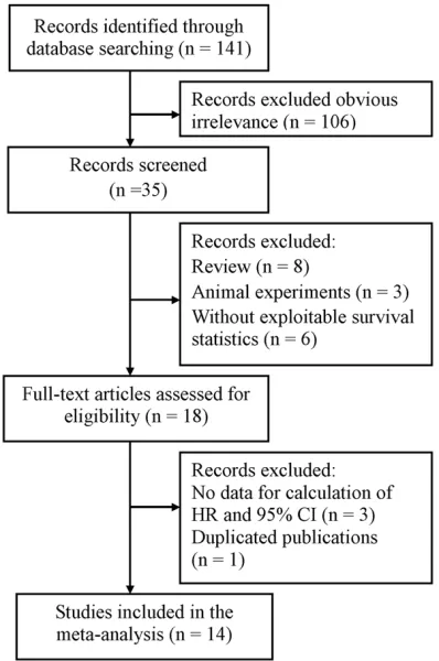
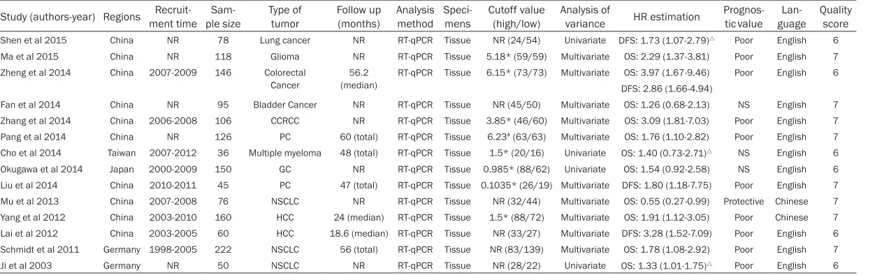
[image:3.612.91.290.71.371.2]Out of the initial number of 141 studies, 14 were found eligible for this meta-analysis. The processes of identifying and selecting studies are presented in Figure 1. Majority the 14 stud-ies [8, 11, 15-19, 26-32], with a total sample of 1468 patients, were almost published in 2011 or later and mainly conducted in China, where-as the others were conducted in Germany [8, 32], Japan [18], or Taiwan [29]. Twelve studies were published in English, whereas the other two were in Chinese [19, 31]. Various cancer types were recorded in our meta-analysis, including GC, HCC, and NSCLC. Quantitative real-time polymerase chain reaction was used to detect MALAT1 in all the 14 studies, and the tested specimens were all extracted from human tissues. HR estimations in 11 studies were directly extracted from original data, and three were extrapolated from survival curves [8, 11, 29]. Eleven studies reported OS as the primary outcome, whereas four trials reported data DFS [11, 15, 16, 30]. The main character-istics of these 14 studies are listed in Table 1. Figure 1. Flow diagram of study selection process
Table 1. Main characteristics of 11 eligible studies in the meta-analysis
Study (authors-year) Regions ment timeRecruit- ple sizeSam- Type of tumor Follow up (months) Analysis method Speci-mens Cutoff value (high/low) Analysis of variance HR estimation Prognos-tic value guageLan- Quality score
Shen et al 2015 China NR 78 Lung cancer NR RT-qPCR Tissue NR (24/54) Univariate DFS: 1.73 (1.07-2.79)△ Poor English 6
Ma et al 2015 China NR 118 Glioma NR RT-qPCR Tissue 5.18* (59/59) Multivariate OS: 2.29 (1.37-3.81) Poor English 7 Zheng et al 2014 China 2007-2009 146 Colorectal
Cancer (median)56.2 RT-qPCR Tissue 6.15* (73/73) Multivariate DFS: 2.86 (1.66-4.94)OS: 3.97 (1.67-9.46) Poor English 6 Fan et al 2014 China NR 95 Bladder Cancer NR RT-qPCR Tissue NR (45/50) Multivariate OS: 1.26 (0.68-2.13) NS English 7 Zhang et al 2014 China 2006-2008 106 CCRCC NR RT-qPCR Tissue 3.85* (46/60) Multivariate OS: 3.09 (1.81-7.03) Poor English 7 Pang et al 2014 China NR 126 PC 60 (total) RT-qPCR Tissue 6.23# (63/63) Multivariate OS: 1.76 (1.10-2.82) Poor English 7
Cho et al 2014 Taiwan 2007-2012 36 Multiple myeloma 48 (total) RT-qPCR Tissue 1.5* (20/16) Univariate OS: 1.40 (0.73-2.71)△ NS English 6
Table 2. Meta-analysis of Rsf-1 overexpression and clinicopathological features in solid tumors pa-tients
Categories Studies (no. of patients) OR (95% CI) I2 P
h Z P
Age 9 (922) 1.01 (0.77-1.34) 0.0% 0.496 0.10 0.922
Gender 9 (922) 1.04 (0.68-1.58)R 49.7% 0.044 0.17 0.868
Tumor size 7 (681) 2.34 (1.14-4.79)R 76.6% < 0.001 2.33 0.020
Histological grade 6 (548) 0.87 (0.58-1.31) 29.5% 0.214 0.66 0.510 Tumor stage 7 (712) 1.48 (1.09-2.01)R 76.1% < 0.001 2.53 0.012
Depth of invasion 5 (542) 1.49 (1.05-2.11) 49.0% 0.098 2.23 0.026 Lymph node metastasis 7 (744) 2.06 (1.19-3.58)R 65.4% 0.008 2.57 0.010
Distant metastasis 7 (744) 1.23 (0.59-2.57)R 61.8% 0.015 0.56 0.575
All pooled HRs were calculated from fixed-effect model except for cells marked with (randomR). P
h denotes P value for
heteroge-neity based on Q test; P denotes P value for statistical significance based on Z test.
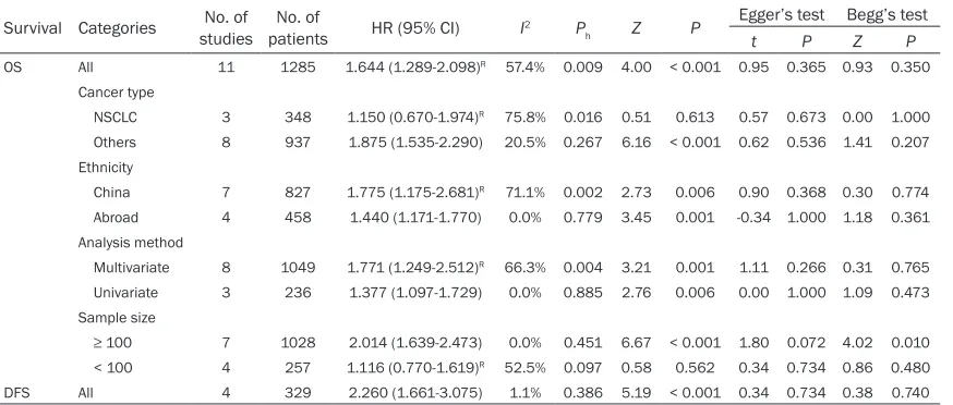
Table 3. Main results of the meta-analysis
Survival Categories studiesNo. of patientsNo. of HR (95% CI) I2 P
h Z P
Egger’s test Begg’s test
t P Z P
OS All 11 1285 1.644 (1.289-2.098)R 57.4% 0.009 4.00 < 0.001 0.95 0.365 0.93 0.350
Cancer type
NSCLC 3 348 1.150 (0.670-1.974)R 75.8% 0.016 0.51 0.613 0.57 0.673 0.00 1.000
Others 8 937 1.875 (1.535-2.290) 20.5% 0.267 6.16 < 0.001 0.62 0.536 1.41 0.207 Ethnicity
China 7 827 1.775 (1.175-2.681)R 71.1% 0.002 2.73 0.006 0.90 0.368 0.30 0.774
Abroad 4 458 1.440 (1.171-1.770) 0.0% 0.779 3.45 0.001 -0.34 1.000 1.18 0.361 Analysis method
Multivariate 8 1049 1.771 (1.249-2.512)R 66.3% 0.004 3.21 0.001 1.11 0.266 0.31 0.765
Univariate 3 236 1.377 (1.097-1.729) 0.0% 0.885 2.76 0.006 0.00 1.000 1.09 0.473 Sample size
≥ 100 7 1028 2.014 (1.639-2.473) 0.0% 0.451 6.67 < 0.001 1.80 0.072 4.02 0.010 < 100 4 257 1.116 (0.770-1.619)R 52.5% 0.097 0.58 0.562 0.34 0.734 0.86 0.480
DFS All 4 329 2.260 (1.661-3.075) 1.1% 0.386 5.19 < 0.001 0.34 0.734 0.38 0.740
All pooled HRs were calculated from fixed-effect model except for cells marked with (randomR). P
h denotes P value for heterogeneity based on Q test; P denotes P value
for statistical significance based on Z test.
Correlation of MALAT1 expression with clinico-pathological parameters
The correlations of MALAT1 expression with clinicopathological characteristics are present-ed in Table 2. Relationships existed between MALAT1 overexpression and some phenotypes of tumor progression, such as tumor size (pooled OR = 2.34; 95% CI = 1.14-4.79; P = 0.020; random effects), tumor stage (pooled OR = 1.48; 95% CI = 1.09-2.01; P = 0.012; ran-dom effects), depth of invasion (pooled OR = 1.49; 95% CI = 1.05-2.11; P = 0.026; fixed effects), and lymph node metastasis (pooled OR = 2.06; 95% CI = 1.19-3.58; P = 0.010; ran-dom effects), which suggested that MALAT1 may demonstrate a promoting effect on tumor progression. However, when age (pooled OR = 1.01; 95% CI = 0.77-1.34; P = 0.922; fixed
effects), gender (pooled OR = 1.04; 95% CI = 0.68-1.58; P = 0.868; random effects), histo-logical grade (pooled OR = 0.87; 95% CI = 0.58-1.31; P = 0.510; fixed effects), and distant metastasis (pooled OR = 1.23; 95% CI = 0.59-2.57; P = 0.575; random effects) were consid-ered, no significant association existed.
Effect of MALAT1 expression on survival
[image:5.612.91.528.274.460.2]Figure 2. Forest plots of overall association between MALAT1 expression and survival in solid tumors. A.Forest plot for pooled OS estimation. B. Forest plot for pooled DFS estimation.
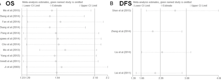
Figure 3. Effects of individual studies on pooled hazard ratios (HRs) for MALAT1 expression and survival in solid tumors. A.Result of sensitivity analysis for pooled OS estimation. B. Result of sensitivity analysis for pooled DFS estimation.
0.001; random effects) with a moderate het-erogeneity (I2 = 57.4%, P
h = 0.009). When the
eligible studies were stratified into subgroup analyses, a significant correlation was observed in studies published in China (HR = 1.78; 95% CI, 1.18-2.68; P = 0.006; random effects) or those published abroad (HR = 1.44; 95% CI, 1.17-1.77; P = 0.001; fixed effects), as well as in multivariate analysis (HR = 1.77; 95% CI, 1.25-2.51; P = 0.001; random effects) and univariate analysis (HR = 1.38; 95% CI, 1.10-1.73; P = 0.006; fixed effects). However, when the sub-group analyses were conducted in terms of tumor types and sample sizes, the negative role of MALAT1 in predicting cancer prognosis was
obvious in other cancer types (HR = 1.88; 95% CI, 1.54-2.29; P < 0.001; fixed effects), and studies with number of cases ≥ 100 (HR = 2.01; 95% CI, 1.64-2.47; P < 0.001; fixed effect), but not in NSCLC (HR = 1.15; 95% CI, 0.67-1.97; P
= 0.613; random effects), nor those with num-ber of cases < 100 (HR = 1.12; 95% CI, 0.77-1.62; P = 0.562; random effects).
Four studies comprising 329 patients reported DFS as the primary endpoint, upregulation of MALAT1 was associated with worse DFS (HR = 2.26; 95% CI, 1.66-3.08; P < 0.001; fixed effects), and significant heterogeneity did not exist (I2 = 1.1%, P
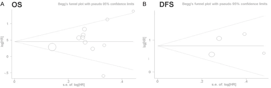
[image:6.612.98.520.320.476.2]Sensitivity analysis and publication bias
Sensitivity analysis of pooled OS and pooled DFS is presented in Figure 3. Notably, the cor-responding overall HR estimated by OS or DFS did not change significantly when each study was omitted individually. These results suggest that no individual study affected the anal-ysis results, and the outcomes of this meta-analysis were robust.
Neither Egger’s test nor Begg’s test showed obvious publication bias for the pooled HR esti-mations of OS (Egger’s test, t = 0.95, P = 0.365; Begg’s test, Z = 0.93, P = 0.350) or DFS (Egger’s test, t = 0.34, P = 0.734; Begg’s test, Z = 0.38, P = 0.740) (Table 3). The shapes of the funnel plots (Figure 4) also did not show apparent evi-dence of asymmetry, indicating that our results were statistically credible.
Discussion
The invasion and metastasis of solid tumors comprise an extremely complex process involv-ing an interplay among multiple cytokines, sig-nal pathways, and other factors; invasion and metastasis of solid tumors mainly cause of death in patients. Thus, searching for sensitive and specific biomarkers for early tumor detec-tion and accurate prognosis, as well as targets for more efficient treatment, is valuable. MALAT1 was upregulated in many solid tumors, including lung cancer [8], bladder cancer [17], HCC [33], and colorectal cancer [34]. MALAT1 can promote tumor cell proliferation and migra-tion, which implies its participation in human cancer development [9]. High level expression of MALAT1 in tissues with cancer metastasis
suggested that this transcript may significantly affect tumor progression [35]. However, the prognostic role of MALAT1 on solid tumors remains uncertain. Considering meta-analysis can provide an overall and precise evaluation of several individual studies for a specified out-come, we conducted this first meta-analysis to explore the prognostic values of lncRNA MALAT1 in solid tumors.
[image:7.612.92.524.73.214.2]ed as a tumor suppressor [19]. Thus, the influ-ence of MALAT1 on NSCLC remains con- troversial, and conclusion should be made with caution because only 348 patients were includ-ed in these studies.
Moreover, significant correlations were found between MALAT1 high expression and some clinicopathological features, such as tumor size, tumor stage, depth of invasion, and lymph node metastasis, which revealed that MALAT1 may boost tumor progression and aggressive-ness. MALAT1 was originally found as a metas-tasis-related gene in NSCLC. Tano et al. [36] found that after MALAT1 was interfered by siRNA, four different genes (CTHRC1, CCT4, HMMR, and ROD1) related to cell mobility were found by comparing the pre- and post-interfer-ence gene expression screening, and knock-down of any of the above-mentioned genes can significantly suppress the migration of lung cancer cells; this indicated that MALAT1 can control the migration ability of lung cancer cells by regulating mobility-related genes. Moreover, epithelial-mesenchymal transition (EMT) is a key step of tumor metastasis, and loss of expression of E-cadherin is one of the land-mark events [37]. In bladder cancer, MALAT1 can suppress the expression of E-cadherin, promote EMT, and finally assist tumor metasta-sis [17, 38]. Furthermore, MALAT1 advances the cell differentiation and the cell proliferation in gastric cancer by recruiting and regulating SF2/ASF which plays a vital role in inflammato-ry diseases and human tumors by alternative splicing [12]. Above all, we can learn that MALAT1 can affect the occurrence and devel-opment of different tumors by a variety of path-ways; however, the mechanisms remain un- clear. Thus, more studies should be conducted to explore its biological functions.
Although this meta-analysis showed some advantages through an overall and consistent estimation, a few limitations should be acknowl-edged. First, the heterogeneities of some pooled results were moderate or even extreme, and subgroup analyses cannot identify the source of heterogeneity. Second, the number of included studies and the total sample size were relatively small. Third, prognosis is a com-prehensive final result reflected by multiple fac-tors, for instance, tumor types, therapeutic regimen, tumor location, and histological types. Nevertheless, we failed to assess these
poten-tial confounders in individual studies. Fourth, the inconsistence in cut-off values and experi-mental designs may in part a source of the inter-study heterogeneity. Finally, the HRs in some studies in our meta-analysis was calcu-lated from the survival curves, which may lead to some minor differences from the actual HRs [22].
In conclusion, lncRNA MALAT1 overexpression is associated with a poor survival rate on OS as well as DFS in many cancer types, and MALAT1 may be an independent biomarker for indicat-ing aggressive tumor development and poor prognosis in solid tumors. However, one should take caution to interpreting these results due to the limitations in this current meta-analysis, and large scale, high-quality clinical investiga-tions are still needed to further confirm these results.
Acknowledgements
This study was funded by the Scientific and Technological Innovation Project of Educational Commission of Guangdong Province (Grant No. 2013KJCX0092).
Disclosure of conflict of interest
None.
Address correspondence to: Dr. Jiayuan Wu, De- partment of Nutritional, The Affiliated Hospital of Guangdong Medical University, Zhanjiang, China. E-mail: [email protected]; Dr. Taiping He, School of Public Health, Guangdong Medical University, No. 2 Wenming Road, Zhanjiang 524023, China. E-mail: [email protected]
References
Rey-mond A, Kapranov P, Rozowsky J, Zheng D, Castelo R, Frankish A, Harrow J, Ghosh S, San-delin A, Hofacker IL, Baertsch R, Keefe D, Dike S, Cheng J, Hirsch HA, Sekinger EA, Lagarde J, Abril JF, Shahab A, Flamm C, Fried C, Hacker-müller J, Hertel J, Lindemeyer M, Missal K, Tan-zer A, Washietl S, Korbel J, Emanuelsson O, Pedersen JS, Holroyd N, Taylor R, Swarbreck D, Matthews N, Dickson MC, Thomas DJ, Weir-auch MT, Gilbert J, Drenkow J, Bell I, Zhao X, Srinivasan KG, Sung WK, Ooi HS, Chiu KP, Fois-sac S, Alioto T, Brent M, Pachter L, Tress ML, Valencia A, Choo SW, Choo CY, Ucla C, Manza-no C, Wyss C, Cheung E, Clark TG, Brown JB, Ganesh M, Patel S, Tammana H, Chrast J, Hen-richsen CN, Kai C, Kawai J, Nagalakshmi U, Wu J, Lian Z, Lian J, Newburger P, Zhang X, Bickel P, Mattick JS, Carninci P, Hayashizaki Y, Weiss-man S, Hubbard T, Myers RM, Rogers J, Stadler PF, Lowe TM, Wei CL, Ruan Y, Struhl K, Gerstein M, Antonarakis SE, Fu Y, Green ED,Karaöz U, Siepel A, Taylor J, Liefer LA, Wetterstrand KA, Good PJ, Feingold EA, Guyer MS, Cooper GM, Asimenos G, Dewey CN, Hou M, Nikolaev S, Montoya-Burgos JI, Löytynoja A, Whelan S, Par-di F, Massingham T, Huang H, Zhang NR, Holmes I, Mullikin JC, Ureta-Vidal A, Paten B, Seringhaus M, Church D, Rosenbloom K, Kent WJ, Stone EA; NISC Comparative Sequencing Program; Baylor College of Medicine Human Genome Sequencing Center; Washington Uni-versity Genome Sequencing Center; Broad In-stitute; Children’s Hospital Oakland Research Institute, Batzoglou S, Goldman N, Hardison RC, Haussler D, Miller W, Sidow A, Trinklein ND, Zhang ZD, Barrera L, Stuart R, King DC, Ameur A, Enroth S, Bieda MC, Kim J, Bhinge AA, Jiang N, Liu J, Yao F, Vega VB, Lee CW, Ng P, Shahab A, Yang A, Moqtaderi Z, Zhu Z, Xu X, Squazzo S, Oberley MJ, Inman D, Singer MA, Richmond TA, Munn KJ, Rada-Iglesias A, Wallerman O, Ko-morowski J, Fowler JC, Couttet P, Bruce AW, Dovey OM, Ellis PD, Langford CF, Nix DA, Eu-skirchen G, Hartman S, Urban AE, Kraus P, Van Calcar S, Heintzman N, Kim TH, Wang K, Qu C, Hon G, Luna R, Glass CK, Rosenfeld MG, Al-dred SF, Cooper SJ, Halees A, Lin JM, Shulha HP, Zhang X, Xu M, Haidar JN, Yu Y, Ruan Y, Iyer VR, Green RD, Wadelius C, Farnham PJ, Ren B, Harte RA, Hinrichs AS, Trumbower H, Clawson H, Hillman-Jackson J, Zweig AS, Smith K,Thakkapallayil A, Barber G, Kuhn RM, Karol-chik D, Armengol L, Bird CP, de Bakker PI, Kern AD, Lopez-Bigas N, Martin JD, Stranger BE, Woodroffe A, Davydov E, Dimas A, Eyras E, Hallgrímsdóttir IB, Huppert J, Zody MC, Abeca-sis GR, Estivill X, Bouffard GG, Guan X, Hansen NF, Idol JR, Maduro VV, Maskeri B, McDowell JC, Park M, Thomas PJ, Young AC, Blakesley
RW, Muzny DM, Sodergren E, Wheeler DA, Wor-ley KC, Jiang H, Weinstock GM, Gibbs RA, Graves T, Fulton R, Mardis ER, Wilson RK, Clamp M, Cuff J, Gnerre S, Jaffe DB, Chang JL, Lindblad-Toh K, Lander ES, Koriabine M, Nefe-dov M, Osoegawa K, Yoshinaga Y, Zhu B, de Jong PJ. Identification and analysis of function-al elements in 1% of the human genome by the ENCODE pilot project. Nature 2007; 447: 799-816.
[2] Esteller M. Non-coding RNAs in human dis-ease. Nat Rev Genet 2011; 12: 861-874. [3] Ebisuya M, Yamamoto T, Nakajima M, and
Nishida E. Ripples from neighbouring tran-scription. Nat Cell Biol 2008; 10: 1106-1113. [4] Mercer TR, Dinger ME, and Mattick JS. Long
non-coding RNAs: insights into functions. Nat Rev Genet 2009; 10: 155-159.
[5] Deng Q, Sun H, He B, Pan Y, Gao T, Chen J, Ying H, Liu X, Wang F, Xu Y, Wang S. Prognostic val-ue of long non-coding RNA HOTAIR in various cancers. PLoS One 2014; 9: e110059. [6] Kung JT, Colognori D and Lee JT. Long
noncod-ing RNAs: past, present, and future. Genetics 2013; 193: 651-669.
[7] Yoon JH, Abdelmohsen K, and Gorospe M. Posttranscriptional gene regulation by long noncoding RNA. J Mol Biol 2103; 425: 3723-3730.
[8] Ji P, Diederichs S, Wang W, Boing S, Metzger R, Schneider PM, Tidow N, Brandt B, Buerger H, Bulk E, Thomas M, Berdel WE, Serve H, Müller-Tidow C. MALAT-1, a novel noncoding RNA, and thymosin beta4 predict metastasis and surviv-al in early-stage non-smsurviv-all cell lung cancer. Oncogene 2003; 22: 8031-8041.
[9] Gutschner T, Hammerle M, and Diederichs S. MALAT1-a paradigm for long noncoding RNA function in cancer. J Mol Med 2013; 91: 791-801.
[10] Gutschner T, Hammerle M, Eissmann M, Hsu J, Kim Y, Hung G, Revenko A, Arun G, Stentrup M, Gross M, Zörnig M, MacLeod AR, Spector DL, Diederichs S. The noncoding RNA MALAT1 is a critical regulator of the metastasis phenotype of lung cancer cells. Cancer Res 2013; 73: 1180-1189.
[11] Shen LQ, Chen L, Wang YS, Jiang XC, Xia HP, and Zhuang ZX. Long noncoding RNA MALAT1 promotes brain metastasis by inducing epithe-lial-mesenchymal transition in lung cancer. J Neurooncol 2015; 121: 101-108.
[12] Wang J, Su L, Chen X, Li P, Cai Q, Yu B, Liu B, Wu W, Zhu Z. MALAT1 promotes cell prolifera-tion in gastric cancer by recruiting SF2/ASF. Biomed Pharmacother 2014; 68: 557-564. [13] Li G, Zhang H, Wan X, Yang X, Zhu C, Wang A,
progno-sis of hepatocellular carcinoma. Biomed Res Int 2014; 2014: 780521.
[14] Wu XS, Wang XA, Wu WG, Hu YP, Li ML, Ding Q, Weng H, Shu YJ, Liu TY, Jiang L, Cao Y, Bao RF, Mu JS, Tan ZJ, Tao F, Liu YB. MALAT1 promotes the proliferation and metastasis of gallbladder cancer cells by activating the ERK/MAPK path-way. Cancer Biol Ther 2014; 15: 806-814. [15] Zheng HT, Shi DB, Wang YW, Li XX, Xu Y,
Tripa-thi P, Gu WL, Cai GX, Cai SJ. High expression of lncRNA MALAT1 suggests a biomarker of poor prognosis in colorectal cancer. Int J Clin Exp Pathol 2014; 7: 3174-3181.
[16] Lai MC, Yang Z, Zhou L, Zhu QQ, Xie HY, Zhang F, Wu LM, Chen LM, Zheng SS. Long non-cod-ing RNA MALAT-1 overexpression predict tumor recurrence of hepatocellular carcinoma after liver transplantation. Med Oncol 2012; 29: 1810-1816.
[17] Fan Y, Shen B, Tan MY, Mu XY, Qin Y, Zhang F, Liu Y. TGF-b-induced upregulation of MALAT1 promotes bladder cancer metastasis by asso-ciating with suz12. Clin Cancer Res 2014; 20: 1531-1541.
[18] Okugawa Y, Toiyama Y, Hur K, Toden S, Saigusa S, Tanaka K, Inoue Y, Mohri Y, Kusunoki M, Bo-land CR, Goel A. Metastasis-associated long non-coding RNA drives gastric cancer develop-ment and promotes peritoneal metastasis. Carcinogenesis 2014; 35: 2731-2739. [19] Mu YY. Expression of the long noncoding
MALAT-1 RNA in NSCLC tissues and its clinical significance, MS, Dissertation, Henan Univer-sity, 2013.
[20] Liberati A, Altman DG, Tetzlaff J, Mulrow C, Gøtzsche PC, Ioannidis JP, Clarke M, Devereaux PJ, Kleijnen J, Moher D. The PRISMA statement for reporting systematic reviewsand meta-analyses of studies that evaluate health care interventions: explanation and elaboration. PLoS Med 2009; 6: e1000100.
[21] Tierney JF, Stewart LA, Ghersi D, Burdett S, Sydes MR. Practical methods for incorporating summary time-to-event data into meta-analy-sis. Trials 2007; 8: 16.
[22] Wells GA, Shea B, O’Connell D, Peterson J, Welch V, Losos M. The Newcastle-Ottawa Scale(NOS) for assessing the quality of non-randomised studies in meta-analyses, Ottawa Health Research Institute Wed site, 2012. [23] Higgins JP, Thompson SG, Deeks JJ and Altman
DG, Measuring inconsistency in meta-analy-ses. BMJ 2003; 327: 557-560.
[24] Mantel N and Haenszel W. Statistical aspects of the analysis of data from retrospective stud-ies of disease. J Natl Cancer Inst 1959; 22: 719-748.
[25] Egger M, Davey SG, Schneider M and Minder C. Bias in meta-analysis detected by a simple, graphical test. BMJ 1997; 315: 629-634.
[26] Ma KX, Wang HJ, Li XR, Li T, Su G, Yang P, Wu JW. Long noncoding RNA MALAT1 associated with the malignant status and poor prognosis in glioma. Tumor Biol 2015; 36: 3355-3359. [27] Zhang HM, Yang FQ, Chen SJ, Che JP and
Zheng JH. Upregulation of long non-coding RNA MALAT1 correlates with tumor progres-sion and poor prognosis in clear cell renal cell carcinoma. Tumor Biol 2014; 36: 2947-2955. [28] Pang EJ, Yang R, Fu XB and Liu YF. Overexpres-sion of long non-coding RNA MALAT1 is corre-lated with clinical progression and unfavorable prognosis in pancreatic cancer. Tumor Biol 2014; 36: 2403-2407.
[29] Cho SF, Chang YC, Chang CS, Lin SF, Liu YC, Hsiao HH, Chang JG, Liu TC. MALAT1 long non-coding RNA is overexpression in multiple my-eloma and may serve as a marker to predict disease progression. BMC Cancer 2104; 14: 809.
[30] Liu JH, Chen G, Dang YW, Li CJ and Luo DZ. Expression and prognostic significance of ln-cRNA MALAT1 in pancreatic cancer tissues. Asian Pac J Cancer Prev 2104; 15: 2971-2977. [31] Yang Z. Molecular biomarkers for predicting
tu-mor recurrence and prognosis in hepatocellu-lar carcinoma after liver transplantation. Ph. D. Dissertation, Zhejiang University 2012. [32] Schmidt LH, Spieker T, Koschmieder S,
Hum-berg J, Jungen D, Bulk E, Hascher A, Wittmer D, Marra A, Hillejan L, Wiebe K, Berdel WE, Wiewrodt R, Muller-Tidow C. The long noncod-ing MALAT1 RNA indicates a poor prognosis in non-small cell lung cancer and induces migra-tion and tumor growth. J Thorac Oncol 2011; 6: 1984-1992.
[33] Lin R, Maeda S, Liu C, Karin M and Edgington TS. A large noncoding RNA is a marker for mu-rine hepatocellular carcinoma and a spectrum of human carcinomas. Oncogene 2007; 26: 851-858.
[34] Ji Q, Zhang L, Liu X, Zhou L, Wang W, Han Z, Sui H, Tang Y, Wang Y, Liu N, Ren J, Hou F, Li Q. Long noncoding RNA MALAT1 promotes tumor growth and metastasis in colorectal cancer through binding to SFPQ and releasing onco-gene PTBP2 from SFPQ/PTBP2 complex. BJC 2014; 111: 736-748.
[35] Tseng JJ, Hsieh YT, Hsu SL, and Chou MM. Me-tastasis associated lung adenocarcinoma transcript 1 is up-regulated in placenta previa increta/percreta and strongly associated with trophoblast-like cell invasion in vitro. Mol Hum Reprod 2009; 15: 725-731.
[37] Chua HL, Bhat-Nakshatri P, Clane SE, Morimiya A, Badve S and Nakshatri H. NF-kappaB re-presses E-cadherin expression and enhances epithelial to mesenchymal transition of mam-mary epithelial cells: potential involvement of ZEB-1 and ZEB-2. Oncogene 2007; 26: 711-724.