Quantum Chemical Calculations of Tryptophan
fi
Heme
Electron and Excitation Energy Transfer Rates in
Myoglobin
Christian J. Suess, Jonathan D. Hirst
, and Nicholas A. Besley
*
The development of optical multidimensional spectroscopic techniques has opened up new possibilities for the study of bio-logical processes. Recently, ultrafast two-dimensional ultraviolet spectroscopy experiments have determined the rates of trypto-phan!heme electron transfer and excitation energy transfer for the two tryptophan residues in myoglobin (Consani et al., Science, 2013, 339, 1586). Here, we show that accurate predic-tion of these rates can be achieved using Marcus theory in con-junction with time-dependent density functional theory. Key intermediate residues between the donor and acceptor are iden-tified, and in particular the residues Val68 and Ile75 play a critical
role in calculations of the electron coupling matrix elements. Our calculations demonstrate how small changes in structure can have a large effect on the rates, and show that the different rates of electron transfer are dictated by the distance between the heme and tryptophan residues, while for excitation energy transfer the orientation of the tryptophan residues relative to the heme is important.VC2017 The Authors Journal of Computa-tional Chemistry Published by Wiley Periodicals, Inc.
DOI: 10.1002/jcc.24793
Introduction
Electron transfer (ET) and the transfer of excitation energy are fun-damental processes in biological systems. The efficient and con-trolled movement of electrons is one of the primary regulation mechanisms in biology and critical for the existence of living organisms,[1,2]and excitation energy transfer (EET) is important in light harvesting systems.[3]This has motivated the development of experimental and computational approaches to characterize the mechanisms of ET and EET processes. However, these studies are challenging due to the fast time-scale of ET and EET and the complexity of biological systems. Two-dimensional ultraviolet (2D-UV) spectroscopy has emerged as a powerful technique for the study of biological systems, as many biological chromo-phores, such as tryptophan, absorb in this region of the spec-trum.[4]Alongside these experimental techniques complementary theoretical approaches have been developed,[5]which can play an important role in interpreting spectra measured in experiment. Here, we focus on tryptophan!heme ET and EET in myoglobin. Myoglobin, an oxygen carrier in muscle tissue, comprises a single polypeptide chain of 153 amino acids arranged in eighta-helices with an iron porphyrin active site and has been described as the hydrogen atom of biology and a paradigm of complexity.[6]
Myoglobin has two tryptophan residues (Trp7 and Trp14), and the fluorescence decay times of these residues have been mea-sured in several myoglobin complexes and are approximately 120 ps for Trp7 and 20 ps for Trp14.[7–10]Recently, Chergui and coworkers studied myoglobin with ultrafast 2D-UV spectroscopy providing insight into the ET and EET processes from the trypto-phan residues to the heme.[8]The tryptophan residues are locat-ed in an a helix separated from the heme by the E helix, of which several amino acids (Val68, Leu69, Thr70, Gly74, Ile75, and Leu76) lie within the direct path to the heme. The experiments showed that Trp14, which is closer to the heme, decays predom-inantly by ET, whereas Trp7 relaxes by EET. The rate of ET was
quantified, with the relaxation time for Trp14 determined to be 40 ps, with a much slower time of>40 ns for Trp7. Further-more, a time of 140 ps was measured for EET from Trp7. The dis-tance between the tryptophan and heme suggests the EET occurs via the Forster mechanism. Subsequent work on deoxy-€ myoglobin indicated that a similar ET process is present in ligat-ed ferrous myoglobins.[9] It has been suggested that the ET pathway involves the Leu69 residue which is in van der Waals contact with the Trp14 and Val68 residues,[9] although other work has found a glutamic acid residue to be important.[11]
It is important that computational modeling of ET and EET develops alongside advances in experiments to predict rates of ET and EET with sufficient accuracy to elaborate on the detailed underlying mechanisms. In particular, computational modeling of these processes can provide an understanding of the different ET and EET properties of the two tryptophan residues. The theo-retical treatment of ET usually follows Marcus theory[12–14] wherein for weak coupling the rate is expressed as
kET5 jVDAj2
h
ffiffiffiffiffiffiffiffiffiffi
p kkBT r
exp 2ðk1DGÞ 2
4kkBT
" #
(1)
whereDG is the change in free energy between the final and initial states, k is the reorganization energy and corresponds
This is an open access article under the terms of the Creative Commons Attribution License, which permits use, distribution and reproduction in any medium, provided the original work is properly cited.
C. J. Suess, J. D. Hirst, N. A. Besley
School of Chemistry, University of Nottingham, University Park, Nottingham, NG7 2RD, United Kingdom E-mail: nick.besley@nottingham.ac.uk
Contract grant sponsor: University of Nottingham High Performance Computing Facility and the MidPlus Regional Centre of Excellence for Computational Science, Engineering and Mathematics under Engineering and Physical Sciences Research Council; Contract grant number: EP/K000128/1
to the energy for ET in the absence of a change in structure and VDA is the diabatic electron coupling matrix element between the two electronic states. Within this formalism the rate can be estimated once the key quantities, DG,k, andVDAhave been determined, and this approach has been successfully applied to many systems.[15–17]Many heme proteins, such as cytochrome c, are involved in biologically important ET reactions. In contrast to cytochrome c, the heme group in myoglobin is not covalently bound and metal substitution is easier to effect, which has moti-vated many experiments on myoglobin as an exemplar of ET in heme proteins.[18]The quantitative determination of the rates of ET and EET in myoglobin provides an opportunity to assess com-putational models for determination of these rates. This can establish the important criteria for calculation of these rates which can then be applied in studies of ET and EET of biological-ly important processes.
A range of computational methods of varying degrees of sophistication have been proposed for the study of ET, and comprehensive review articles on this subject are avail-able.[19–21]One relatively simple but successful approach is the pathway tunneling model of Beratan and coworkers.[22,23] This model provides a framework to characterize the influence of the protein structure onVDAin biological ET reactions. We use this approach here as a starting point for more sophisticated time-dependent density functional theory (TDDFT) calculations and as a benchmark. The model assumes a donor-acceptor complex mechanism and the ET is mediated by consecutive interactions between atoms connecting the donor and accep-tor. The steps taken are characterized by decay factors or “penalties” associated with covalent bonds, hydrogen bonds, or through-space jumps. Covalent-bond mediated steps are deemed to have a low tunneling barriers and hence assigned a low penalty, whilst higher penalties are applied to those with high tunneling barriers such as a through-space jump. Using a graph-search algorithm[24] to determine all potential pathways, the overall penalty for ET is the product of each penalty throughout every step[25]
VDA5A Y
i
eC i
Y
j
eH j
Y
k
eS
k (2)
where eC
i is the penalty for propagation through covalent bond i,eH
j is the penalty for propagation through a hydrogen bond jandeS
k is the penalty for the through-space jump k. A is the pre-factor, and in this work we take A51 eV.[26] Using this approach, it is possible to identify the strongest ET path-ways between the donor and the acceptor, estimate partial electronic couplings mediated by each pathway, calculate the importance of individual protein groups for mediating ET, and ultimately determine the most dominate ET pathway between the donor and acceptor.
The direct calculation of the electron coupling with quan-tum chemical methods provides a potentially more accurate means for determining the coupling strength. There is a wide range of approaches to this problem including different schemes to characterize the diabatic states[19,27–29]along with the choice of electronic structure method. The coupling
elements for ET can be evaluated using the Generalized Mulliken-Hush (GMH) scheme[30]
VETDA5 ffiffiffiffiffiffiffiffiffiffiffiffiffiffiffiffiffiffiffiffiffiffiffiffiffiffiffiffiffiffiffiffiffiffiffiffiffiffiffiffiffiðE22E1Þj~l12j ð~l112~l22Þ214j~l12j2
q (3)
where~l11; ~l22, and ~l12 represent the permanent and transi-tion dipole moments for the two states.
An advantage of a quantum chemistry-based approach is that it is also possible to study the rates of EET, and an over-view of the theoretical treatment of EET is available.[31] The coupling elements associated with EET can be calculated through the fragment excitation difference (FED) approach.[27]
VDAEET5 ffiffiffiffiffiffiffiffiffiffiffiffiffiffiffiffiffiffiffiffiffiffiffiffiffiffiffiffiffiffiffiffiffiffiffiffiffiffiffiffiffiffiffiffiðE22E1ÞDx12 ðDx112Dx22Þ214Dx
2 12
q (4)
where Dx represents elements of the excitation difference matrix. Here, we are concerned with EET occurring over long range through dipole-dipole coupling in a Forster mechanism.€ Computational methods for the calculation of EET via a Dexter mechanism, which requires overlap of the wavefunctions of the donor and acceptor, have been described elsewhere.[32]
Evaluating the terms in eq. (1) requires excited state electronic structure methods, including structural optimization of the rele-vant states. This is commonly done using methods such as sin-gle excitation configuration interaction (CIS) or TDDFT.[19,33–39] The study of large biological systems is challenging, as the size of the system usually precludes a full treatment with quantum chemical methods, necessitating some further approximations. Despite this inherent complexity, several studies have reported calculations of ET in proteins based on Marcus theory.[40] One approach has been to compute coupling matrix elements for structures extracted from a molecular dynamics simula-tion.[41–43] Recent work has studied the ultrafast ET in crypto-chromes based on GMH coupling strength computed using TDDFT.[44]Furthermore, the absorption and fluorescence spectra of tryptophan residues embedded in the protein environment have been studied using TDDFT calculations.[45,46]Studies of EET of large biological systems have also been reported.[47–49]This includes application of the FED approach with TDDFT to study the electronic energy transfer pathways in cyanobacteria phyco-cyanin[48]and cyanobacteria allophycocyanin.[49]
In this study, we aim to provide insight into the structural factors that affect the rates of both ET and EET in the protein myoglobin. The availability of experimental data for the rates of tryptophan! heme ET and EET allows the accuracy of TDDFT-based calculations to be assessed. Treating ET and EET in an equivalent way, and achieving good agreement with experiment observations, pro-vides a sound basis for the physical basis for the ET and EET rates observed in experiment to be explored with confidence.
Methodology
using the pathways tunneling method based on the coordi-nates of the crystal structure (PDB: 1YMB) using the software of Balabin et al.[24] The electron coupling was also evaluated using TDDFT with the GMH scheme for ET and the FED approach for EET. The TDDFT calculations, using the long-range corrected CAM-B3LYP exchange-correlation functional,[50] were performed on reduced structural models based on the crystal structure coordinates. The location of hydrogen atoms were constructed using the IQMOL software.[51] Key interven-ing residues that were identified based on the pathways tunneling calculations were included in the TDDFT calcula-tions. To explore the sensitivity of the computed coupling to fluctuations in structure, further couplings were computed for a range of crystal structures (PDB: 1BZR, 2MB4, 4MBN, 1YMB) including those recorded in time-resolved serial femtosecond crystallography experiments (PDB: 5CN4 at 0, 50, and 150 ps).[52]
In the 2D-UV experiment, ET or EET transfer occurs following the initial electronic excitation of the tryptophan. Here, we study the ET from the tryptophan residues to the porphyrin ring to form an Fe(II)-porphyrinp-anion radical. Subsequent ET from the ring to iron to form the ferric heme is expected to be sufficiently fast that it does not affect the overall rate. A schematic of the electronic states involved is given in Figure 1. It shows that to describe the ET and EET processes requires calculations on the ground state, local excited state of the tryptophan, local excited state of the heme and a tryptophan !heme charge transfer (CT) excited state. The accurate description of the electronic structure of metal-porphyrin sys-tems is a challenge for computational methods, where the prediction of the correct ground state electronic configuration is debated.[56–58] Multiconfigurational perturbation theory-based calculations in conjunction with large basis sets repre-sent the most reliable methods, however, the computational cost of these methods makes them unsuitable for the current study and the calculations presented here are based on DFT and TDDFT. These calculations find the lowest energy spin state of the iron porphyrin ring to be a triplet state, which is computed in our work to lie about 1.8 eV lower in energy than the singlet state at the TDDFT level of theory. This is con-sistent with previous DFT studies.[58] The excited states arise from local excitations between the highest occupied molecular
orbital (HOMO) and lowest unoccupied molecular orbital (LUMO) of the tryptophan and a CT excitation from the HOMO of tryptophan to the heme. These orbitals correspond to p
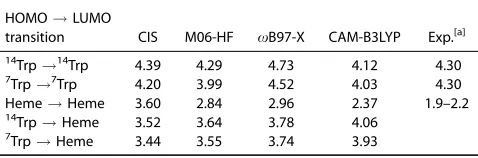
and p orbitals localized on the tryptophan residues or the heme. Table 1 shows excitation energies for the relevant states computed with CIS and a range of exchange-correlation func-tionals that are suitable for describing CT excitations for a model system that includes the two tryptophan residues and the heme in their geometries taken directly from the crystal structure. The CAM-B3LYP functional predicts excitation ener-gies for the HOMO!LUMO transitions of Trp14 and Trp7 to occur at 4.12 eV and 4.03 eV, respectively, which are consistent with the value of 4.3 eV measured for tryptophan in the gas phase.[53] The HOMO ! LUMO transition localized on the heme lies at 2.37 eV. This transition corresponds to the Q band in porphyrin related compounds, which lies in the region 1.9–2.2 eV.[54,55] CAM-B3LYP performs best of the functionals considered, particularly for Q band excitation of the heme.
All calculations were performed with Q-CHEM[59] and used the 31G* basis set. Calculations using larger basis set 6-311G* did not show significant difference in the computed val-ues for the smaller systems studied here, and hence, the less demanding basis set was used. This observation is consistent with previous simulations of the electronic spectra of porphyrin.[60]
[image:3.612.65.292.64.192.2]One of the most computationally challenging aspects of the application of Marcus theory is the structural optimization of the initial and final states. These states are electronically excit-ed states, and in our calculations we assessexcit-ed optimizing the structures using several methods: CIS, TDDFT, and the Maxi-mum Overlap Method (MOM).[61]The S1 excited state geome-tries for the heme predicted by the three excited state methods, CIS, TDDFT, and MOM, show a largest RMSD between any two structures of 0.04 A˚ . MOM provides accurate predictions for excited state structures[62] and was chosen as the preferred method of excited state optimization, as it was the most straightforward to apply. When optimizing the struc-ture of high-lying excited states with TDDFT or CIS, in many instances the order of the roots changed between optimiza-tion cycles making it necessary to monitor the nature of the excited states constantly to ensure that the correct state was being optimized. An unconstrained optimization of the reduced model of the protein would yield an unphysical struc-ture. Consequently, it is necessary to constrain the optimiza-tion in some way to maintain a biologically realistic geometry Figure 1. Schematic of the electronic configurations for ET and EET
pro-cesses from theS1state of a tryptophan residue to the heme.
Table 1.Computed excitation energies in eV with CIS and TDDFT with different exchange-correlation functionals.
HOMO!LUMO
transition CIS M06-HF xB97-X CAM-B3LYP Exp.[a] 14
Trp!14
Trp 4.39 4.29 4.73 4.12 4.30
7Trp!7Trp 4.20 3.99 4.52 4.03 4.30
Heme!Heme 3.60 2.84 2.96 2.37 1.9–2.2
14Trp!Heme 3.52 3.64 3.78 4.06
7
[image:3.612.316.555.652.731.2]for the residues within the protein while allowing for some relaxation of the structure. These constraints represent the physical constraints that the surrounding protein environment would impose on the fragments. During the optimization a subset atoms, shown in Figure 2 and listed in Table 2, were kept frozen in position. Under the constraint of these frozen atoms, the two tryptophans are kept a fixed distance from the heme consistent with the crystal structure, but allowed suffi-cient freedom for relaxation in the optimization process. The constraints also restrict the macrocyclic of the heme from becoming unduly distorted while allowing porphyrins to dome and ruffle in their excited states.[63,64] The methodology described does not take into account the effects of entropy in
DGand neglects the role of solvent. These effects may be sig-nificant, but to describe them accurately at a quantum chemi-cal level is beyond our current capabilities.
[image:4.612.139.480.63.257.2]Results and Discussion
Figure 3 shows the strongest ET pathways predicted by the pathways tunneling model for ET between the tryptophan res-idues and the heme. The computed pathways for the two tryptophans pass through different parts of the E helix, where key amino acids are Val68 for Trp14 and Ile75 for Trp7. The computed values of the electron coupling term ðjVDAjÞ are
8:8231023 eV and 4:2731024 eV for Trp14 and Trp7, respec-tively. These values are consistent with the observation from experiment that ET from Trp14 occurs on a faster timescale than for Trp7.
[image:4.612.328.541.566.708.2]Tables 3 and 4 show the computed coupling elements for structural models that incorporate different components of the ET pathway that were identified by the pathways tunneling model calculation. The TDDFT coupling strengths are comput-ed using the GMH scheme, and correspond to the initial state being the S1 excited state of the tryptophan residue. The tunneling pathways model used here makes no distinction between the strength of coupling between the donor in differ-ent electronic states, and we will examine the significance of this later. For some of the reduced structural models, the dom-inant pathway is broken resulting in a vacuum tunneling path-way with no significant coupling. The simplest structural model includes only the donor tryptophan and heme. Howev-er, intervening residues are likely to affect the computed value for the coupling. We study the importance of the intervening residues by including a single residue (Val68 for Trp14 and Ile75 for Trp7), three residues (Val68, Leu69 and Thr70 for Figure 2. Atoms highlighted in red are held fixed in position during the optimization of the structure of the heme, intervening residue and tryptophan. [Color figure can be viewed at wileyonlinelibrary.com]
Table 2.Atoms frozen in the geometry optimizations.
Residue Index in PDB (1YMB)
Trp7 41,44,45,53
Trp14 103,106,107,115
Val68 534–540
Leu69 541–548
Thr70 549–555
Ile75 577–584
Phe137 1060–1067
Phe138 1068–1078
Heme 1207,1208,1209,1210
[image:4.612.60.299.631.742.2]Trp14; Ile75, Leu137, and Phe138 for Trp7) and the full E helix with the additional Leu137 and Phe138 residues between the tryptophan and heme. These residues are shown in Figure 4. The calculations show that the inclusion of just a single key residue, Val68 for Trp14 and Leu137 for Trp7, (denoted Trp1Heme11AA in the table) has a significant affect on the computed coupling, with a three- to five-fold increase in the strength of coupling. This indicates that Val68 and Ile75 are important in facilitating ET to the heme. The inclusion of fur-ther structural elements leads to much smaller additional increases in the coupling.
The computed TDDFTjVDAj for the structural models with three intervening residues are 6:9231023 eV and 4:8731024 eV for Trp14 and Trp7, respectively. When the donor is the tryptophan in its ground (S0) state, the corresponding values computed with the same structural model are 6:7531023 eV and 4:1031024 eV. Thus, there is an increase in jVDAj in the excited state compared to the ground state. The calculation of jVDAjfor the excited state adds significantly to the cost of the TDDFT calculation, as it requires higher energy roots in the TDDFT calculation to be computed. These results suggest that using the coupling value for the ground state is a reasonable approximation for the excited state, but is likely to underesti-mate the value forjVDAj.
The calculated coupling strengths can be sensitive to changes in the structure. Average values for the coupling com-puted over seven different crystal structures reported in the
literature with the Trp1Heme13AA structural model are also shown. The magnitude of the computed couplings vary by at most a factor of two, and the average values are reasonably close to the single structure values. For all structures, the qual-itative difference between the computed couplings for the two tryptophan residues is observed, and average values of 8:1431023 eV and 6:6231024 eV are obtained for the TDDFT calculations. The ratio jVDAj(Trp14):jVDAj(Trp7) is found to be 12.3 from the TDDFT calculations, consistent with the value of 13.0 from the Beratan model.
It is common to describe the strength of electronic coupling for ET as an exponential dependence on the distance between the donor and acceptor[65]
VDAðrÞ5VDA0 ðr0Þexp 2
b
2ðr2r0Þ
(5)
where r0 is the van der Waals contact distance, and b is a parameter reflecting the effectiveness of the protein in mediat-ing ET and typically ranges from 1.10 to 1.65 A˚21 for con-densed phase systems and from 3 to 5 A˚21 for electron tunneling across a vacuum.[1] Using the values ofjVTDDFT
DA jfor Trp14 and Trp7 computed here gives a value forbof 0.8 A˚21. This is close to typical values for this parameter for condensed phase systems, and suggests that the protein is effectively mediating ET and the slower rate of ET for Trp7 is associated largely with its greater distance from the heme (22.6 A˚ com-pared with 15.9 A˚ for Trp14 for the 1YMB crystal structure).
jVDAj for EET have been computed using the FED scheme with three intervening residues (Trp1Heme13AA) for both theS0andS1initial states of tryptophan. These values and the corresponding values for ET are summarized in Table 5. The key change for EET compared with ET is thatjVEET
[image:5.612.315.555.64.205.2] [image:5.612.60.300.100.180.2]DAjfor Trp7 is larger than the value for Trp14. If the intervening residues are removed from the calculation, the computed couplings are 5.68 3 1023 eV and 2.56 3 1024 eV for Trp7 and Trp14, respectively. This represents only a modest change in the rela-tive coupling strengths, whereby the strength of coupling for Trp7 remains about 23 times larger than for Trp14. This sug-gests that the intervening residues do not play a key role in the qualitative difference in the rates of EET between the two Figure 4. The reduced myoglobin system consisting of heme, Trp14, Trp7, and the E-Helix. Different colors represent each amino acid. [Color figure can be viewed at wileyonlinelibrary.com]
Table 3.Computed pathways model (jVBeratanDA j) and TDDFT (jV TDDFT DA j)
cou-pling values in eV for Trp14!heme electron transfer for different struc-tural models.
Model jVTDDFT
DA j jVBeratanDA j
Full Protein 8:8231023
Trp141Heme1E helix 7:9231023 8:7231023
Trp141Heme13AA (Average) 8:1431023 7
:7131023
Trp141Heme13AA 6:9231023 6:1631023
Trp141Heme11AA 6:9131023 3:6531023
Trp141Heme 2:2931023 –
3AA and 1AA indicate three and one intervening amino acid residue included in the calculation, see text for details. The average values are evaluated using seven different crystal structures with the Trp141 He-me13AA structural model.
Table 4.Computed pathways model (jVBeratan
DA j) and TDDFT (jV TDDFT DA j)
cou-pling values in eV for Trp7!heme electron transfer for different struc-tural models.
Model jVTDDFT
DA j jVBeratanDA j
Full Protein 4:2731024
Trp71Heme1E helix 4:9431024 3:6931024
Trp71Heme13AA (Average) 6:6231024 5:9131024
Trp71Heme13AA 4:8731024 3
:2931024
Trp71Heme11AA 4:8231024 –
Trp71Heme 9:4631025 –
[image:5.612.57.300.623.701.2]tryptophan residues. Similar to ET, there is an increase in the strength of coupling for theS1state compared to theS0state for EET.
Another potentially important factor that determines the strength of the coupling is the orientation of the tryptophan residues with respect to the heme. To investigate this, the strength of coupling has been computed with the Trp7 rotat-ed such that it has the orientation of Trp14 and conversely Trp14 was rotated to have the orientation of Trp7. This is illus-trated in Figure 5. This increases the coupling of “Trp14” from 2.5631024eV to 2.8531022eV, and decreases the coupling of “Trp7” from 5.68 3 1023 eV to 7.80 3 1025 eV, and thus accounts for the qualitative difference in the EET of the two residues. There is an increase in the modified coupling strengths indicating that distance from the heme does play a role, but the effect of orientation is orders of magnitude great-er. This sensitivity to the orientation of the donor residues is consistent with EET occurring by a F€orster mechanism.
To compute the rates ET and EET and allow a direct compar-ison with experiment, the reorganization energy (k) and change in free energy (DG) need to be determined [eq. (1)]. This requires structural optimization of the excited states cor-responding to the final and initial states of the ET and EET pro-cesses. Based on optimizations of the excited states using the MOM approach with CAM-B3LYP/6-31G*, we computed values ofDG andkof20.16 eV and 0.69 eV for Trp14 and20.06 eV and 0.55 eV for Trp7 for ET, and values ofDGandkof 21.46
eV and 0.83 eV for Trp14 and21.38 eV and 0.72 eV for Trp7 for EET. The calculated values ofkare of similar magnitude to those reported for other related systems.[66]
Through combining the computedDG andkwith the cou-pling strengthsjVDAj, the rates and relaxation times for ET and EET (sET51=kET and sEET51=kEET) can be evaluated, and three sets of relaxation times are given in Table 6. Overall, the calcu-lation that most closely corresponds to experiment evaluated the relaxation time through combining the computedDGand
kwith jVDAj computed with TDDFT for the S1 state, and this relaxation time is denoted sES. The remaining two relaxation times correspond to whereDGandkare combined withjVDAj computed with TDDFT for theS0state of tryptophan (denoted
[image:6.612.55.299.103.142.2]sGS) and with jVDAj evaluated using the pathways tunneling model (denoted shybrid). sGS also reproduce the experimental rates well and shows that the additional computational effort to evaluate the excited state coupling strengths could be avoided. The computed relaxation times reproduce the key observations made in the experiment. For ET, the relaxation time for Trp14 is much faster than for Trp7, and for EET the relaxation time for Trp7 is much faster than for Trp14. The qualitative description of the computed rate is heavily influ-enced byVDA, and it is possible to account for the experimen-tal observations based solely on comparing coupling values, kET/ jVDAj2. Addressing some of the approximations made in Table 5.Computed TDDFT (CAM-B3LYP/6-31G*) coupling values in eV for
ET and EET for the Trp1Heme with three intervening amino acid resi-dues model.
Trp14ðS0Þ Trp14ðS1Þ Trp7ðS0Þ Trp7ðS1Þ jVET
DAj 6:7531023 6:9231023 4:1031024 4:8731024 jVEET
DAj 2:73310
[image:6.612.315.555.113.172.2]24 3:1331024 6:2531023 7:0831023
Figure 5. Modified heme and tryptophan system. Original tryptophan orientations shown in green and modified orientations shown in red. [Color figure can be viewed at wileyonlinelibrary.com]
Table 6.Calculated relaxation times in ps. ForsESthejVDAjis computed
using TDDFT for theS1state, forsGSjVDAjis computed using TDDFT for
theS0state and forshybridjVDAjis evaluated using the pathways
tunnel-ing model.
System sES sGS shybrid Exp.
ET: Trp14 42 60 47 34
ET: Trp7 12000 32000 15000 40000 EET: Trp14 54000 70000 – –
[image:6.612.103.518.507.719.2]the calculations primarily the structural models used, neglect of entropy in DG and the neglect of solvent could lead to a more precise quantitative agreement with experiment.
Conclusions
The rates of tryptophan!heme ET and EET in myoglobin have been studied using a combination of DFT and TDDFT. These rates have been measured in recent 2D-UV spectroscopic experiments by Chergui and coworkers[8]providing an oppor-tunity to assess the accuracy of different computational mod-els and probe structural factors that affect the rates. Application of the tunneling pathways model shows that the important intermediate residues for ET are Val68 and Leu69 for Trp14 and Ile75 for Trp7, and inclusion of these residues is important in TDDFT calculations of the coupling matrix ele-ments. Both the pathways tunneling model and TDDFT calcu-lations correctly predict diabatic electron coupling matrix elements consistent with the rate of ET for Trp14 being greater than for Trp7. The predicted rate is greater for an initial S1 electronic state of the tryptophan donor compared to the ground state. With TDDFT it is possible to extend the study to consider EET, and the calculations correctly predict that the rate for EET is greater for Trp7.
Marcus theory calculations using the computed electron coupling elements for ET and EET combined with k and DG evaluated from quantum chemical calculations of the appropri-ate excited stappropri-ates gives relaxation times in good agreement with experimental measurements. Subsequent analysis of the structure shows that the different rates of ET from the two tryptophan residues can be associated with the distance between the heme and tryptophan residues, while for EET the orientation of the tryptophan residues relative to the heme is important.
Keywords: myoglobin
electron transferexcitation energy transferTDDFTHow to cite this article: C. J. Suess, J. D. Hirst, N. A. Besley. J. Comput. Chem.2017, DOI: 10.1002/jcc.24793
[1] H. B. Gray, J. R. Winkler,Q. Rev. Biophys.2003,36, 341. [2] G. McLendon, R. Hake,Chem. Rev.1992,92, 481.
[3] R. J. Cogdell, T. D. Howard, R. Bittl, E. Schlodder, I. Geisenheimer, W. Lubitz,Philos. Trans. R. Soc. Lond. B. Biol. Sci.2000,355, 1345. [4] B. A. West, A. M. Moran,J. Phys. Chem. Lett.2012,3, 2575.
[5] D. Abramavicius, J. Jiang, B. M. Bulheller, J. D. Hirst, S. Mukamel,J. Am. Chem. Soc.2010,132, 7769.
[6] H. Frauenfelder, B. H. McMahon, P. W. Fenimore,Proc. Natl. Acad. Sci. USA2003,100, 8615.
[7] C. Shih, A. K. Museth, M. Abrahamsson, A. M. Blanco-Rodriguez, A. J. Di Bilio, J. Sudhamsu, B. R. Crane, K. L. Ronayne, M. Towrie, A. Vicˇek, J. H. Richards, J. R. Winkler, H. B. Gray,Science2008,320, 1760. [8] C. Consani, G. Aubock, F. van Mourik, M. Chergui,€ Science2013, 339,
1586.
[9] R. Monni, A. Al Haddad, F. van Mourik, G. Aub€ock, M. Chergui,Proc. Natl. Acad. Sci. USA2015,112, 5602.
[10] B. Nolting, N. Salimi, U. Guth,€ J. Theor. Biol.2008,251, 331.
[11] W. Qiu, T. Li, L. Zhang, Y. Yang, Y.-T. Kao, L. Wang, D. Zhong,Chem. Phys.2008,350, 154.
[12] R. A. Marcus,J. Chem. Phys.1956,24, 966. [13] R. A. Marcus,J. Chem. Phys.1965,43, 679. [14] R. A. Marcus,Angew. Chem. Int. Ed.1993,32, 1111.
[15] M. D. Newton, N. Sutin,Annu. Rev. Phys. Chem.1984,35, 437. [16] R. A. Marcus, N. Sutin,Biochim. Biophys. Acta1985,811, 265. [17] G. de la Torre, F. Giacalone, J. L. Segura, N. Martın, D. M. Guldi,
Chemis-try2005,11, 1267.
[18] J. N. Onuchic, D. N. Beratan, J. R. Winkler, H. B. Gray,Annu. Rev. Bio-phys. Biomol. Struct.1992,21, 349.
[19] C. P. Hsu,Acc. Chem. Res.2009,42, 509.
[20] T. Koslowski, F. Burggraf, S. Krapf, T. Steinbrecher, C. Wittekindt, Bio-chim. Biophys. Acta2012,1817, 1955.
[21] J. Blumberger,Chem. Rev.2015,115, 11191.
[22] D. N. Beratan, J. N. Betts, J. N. Onuchic,Science1991,252, 1285. [23] S. S. Skourtis, D. N. Beratan,J. Biol. Inorg. Chem.1997,2, 378. [24] I. A. Balabin, X. Hu, D. N. Beratan,J. Comput. Chem.2012,33, 906. [25] D. N. Beratan, J. N. Onuchic, J. R. Winkler, H. B. Gray,Science 1992,
258, 1740.
[26] S. Keinan, J. M. Nocek, B. M. Hoffman, D. N. Beratan, Phys. Chem. Chem. Phys.2012,14, 13881.
[27] C. P. Hsu, Z. Q. You, H. C. Chen,J. Phys. Chem. C2008,112, 1204. [28] A. A. Voityuk,J. Phys. Chem. C2014,118, 1478.
[29] A. A. Voityuk,Chem. Phys. Lett.2006,427, 177. [30] R. J. Cave, M. D. Newton,Chem. Phys. Lett.1996,249, 15. [31] A. Olaya-Castro, G. M. Scholes,Int. Rev. Phys. Chem.2011,30, 49. [32] S. S. Skourtis, C. Liu, P. Antoniou, A. M. Virshup, D. N. Beratan,Proc.
Natl. Acad. Sci. USA2016,113, 8115.
[33] H.-C. Chen, C.-P. Hsu,J. Phys. Chem. A2005,109, 11989.
[34] J. E. Subotnik, J. Vura-Weis, A. J. Sodt, M. A. Ratner,J. Phys. Chem. A 2010,114, 8665.
[35] J. E. Subotnik,J. Chem. Phys.2011,135, 071104.
[36] C. Leng, H. Qin, Y. Si, Y. Zhao,J. Phys. Chem. C2014,118, 1843. [37] X. Yang, E. R. Bittner,J. Phys. Chem. A2014,118, 5196.
[38] B. S. Veldkamp, X. Liu, M. R. Wasielewski, J. E. Subotnik, M. A. Ratner,J. Phys. Chem. A2015,119, 253.
[39] E. A. Briggs, N. A. Besley,J. Phys. Chem. A2015,119, 2902.
[40] C. Narth, N. Gillet, F. Cailliez, B. Levy, A. de la Lande,Acc. Chem. Res. 2015,48, 1090.
[41] A. de la Lande, S. Martı, O. Parisel, V. Moliner,J. Am. Chem. Soc.2007,
129, 11700.
[42] E. E. Hammi, C. Houee-Levin, J. Rezac, B. Levy, I. Demachy, L. Baciou, A. de la Lande,Phys. Chem. Chem. Phys.2012,14, 13872.
[43] C. Melia, S. Ferrer, J. Rezac, O. Parisel, O. Reinaud, V. Moliner, A. de la Lande,Chem. Eur. J.2013,19, 17328.
[44] F. Cailliez, P. Muller, M. Gallois, A. de la Lande,€ J. Am. Chem. Soc.2014,
136, 12974.
[45] D. M. Rogers, N. A. Besley, P. O’Shea, J. D. Hirst,J. Phys. Chem. B2005,
109, 23061.
[46] D. Robinson, N. A. Besley, P. O’Shea, J. D. Hirst,J. Phys. Chem. B2009,
113, 14521.
[47] K. J. Fujimoto, S. Hayashi,J. Am. Chem. Soc.2009,131, 14152. [48] Y. Ren, B. Chi, O. Melhem, K. Wei, L. Feng, Y. Li, X. Han, D. Li, Y. Zhang,
J. Wan, X. Xin,J. Comput. Chem.2013,34, 1005.,
[49] Y. Ren, O. Melhem, Y. Li, B. Chi, X. Han, H. Zhu, L. Feng, J. Wan, X. Xu,
J. Comput. Chem.2015,36, 137.
[50] T. Yanai, D. P. Tew, N. C. Handy,Chem. Phys. Lett.2004,393, 51. [51] IQmol: A molecular editor and visualization package. Available at:
http://iqmol.org.
[52] T. R. M. Barends, L. Foucar, A. Ardevol, K. Nass, A. Aquila, S. Botha, R. B. Doak, K. Falahati, E. Hartmann, M. Hilpert, M. Heinz, M. C. Hoffmann, J. Kofinger, J. E. Koglin, G. Kovacsova, M. Liang, D. Milathianaki, H. T. Lemke, J. Reinstein, C. M. Roome, R. L. Shoeman, G. J. Williams, I. Burghardt, G. Hummer, S. Boutet, I. Schlichting,Science2015,350, 445. [53] T. R. Rizzo, Y. D. Park, L. A. Peteanu, D. H. Levy,J. Chem. Phys.1986,
84, 2534.
[54] M. Gouterman, G. H. Wagnire, L. C. Snyder,J. Mol. Spectrosc.1963,11, 108.
[56] Y.-K. Choe, T. Hashimoto, H. Nakano, K. Hirao,Chem. Phys. Lett.1998,
295, 380.
[57] Y.-K. Choe, T. Nakajima, K. Hirao, R. Lindh, J. Chem. Phys.1999, 111, 3837.
[58] M.-S. Liao, S. Scheiner,J. Chem. Phys.2002,117, 205.
[59] Y. Shao, Z. Gan, E. Epifanovsky, A. T. B. Gilbert, M. Wormit, J. Kussmann, A. W. Lange, A. Behn, J. Deng, X. Feng, D. Ghosh, M. Goldey, P. R. Horn, L. D. Jacobson, I. Kaliman, R. Z. Khaliullin, T. Kus, A. Landau, J. Liu, E. I. Proynov, Y. Min Rhee, R. M. Richard, M. A. Rohrdanz, R. P. Steele, E. J. Sundstrom, H. L. Woodcock III, P. M. Zimmerman, D. Zuev, B. Albrecht, E. Alguire, B. Austin, G. J. O. Beran, Y. A. Bernard, E. Berquist, K. Brandhorst, K. B. Bravaya, S. T. Brown, D. Casanova, C.-M. Chang, Y. Chen, S. Hung Chien, K. D. Closser, D. L. Crittenden, M. Diedenhofen, R. A. DiStasio Jr., H. Do, A. D. Dutoi, R. G. Edgar, S. Fatehi, L. Fusti-Molnar, A. Ghysels, A. Golubeva-Zadorozhnaya, J. Gomes, M. W.D. Hanson-Heine, P. H.P. Harbach, A. W. Hauser, E. G. Hohenstein, Z. C. Holden, T.-C. Jagau, H. Ji, B. Kaduk, K. Khistyaev, J. Kim, J. Kim, R. A. King, P. Klunzinger, D. Kosenkov, T. Kowalczyk, C. M. Krauter, K. U. Lao, A. D. Laurent, K. V. Lawler, S. V. Levchenko, C. Y. Lin, F. Liu, E. Livshits, R. C. Lochan, A. Luenser, P. Manohar, S. F. Manzer, S.-P. Mao, N. Mardirossian, A. V. Marenich, S. A. Maurer, N. J. Mayhall, E. Neuscamman, C. M. Oana, R. Olivares-Amaya, D. P. O’Neill, J. A. Parkhill, T. M. Perrine, R. Peverati, A. Prociuk, D. R. Rehn, E. Rosta, N. J. Russ, S. M. Sharada, S. Sharma, D. W. Small, A. Sodt, T. Stein, D. St€uck, Y.-C. Su, A. J.W. Thom, T. Tsuchimochi, V. Vanovschi, L. Vogt, O. Vydrov, T. Wang, M. A. Watson, J. Wenzel, A. White, C. F. Williams, J. Yang, S.
Yeganeh, S. R. Yost, Z.-Q. You, I. Y. Zhang, X. Zhang, Y. Zhao, B. R. Brooks, G. K. L. Chan, D. M. Chipman, C. J. Cramer, W. A. Goddard III, M. S. Gordon, W. J. Hehre, A. Klamt, H. F. Schaefer III, M. W. Schmidt, C. D. Sherrill, D. G. Truhlar, A. Warshel, X. Xu, A. Aspuru-Guzik, R. Baer, A. T. Bell, N. A. Besley, J.-D. Chai, A. Dreuw, B. D. Dunietz, T. R. Furlani, S. R. Gwaltney, C.-P. Hsu, Y. Jung, J. Kong, D. S. Lambrecht, W. Z Liang, C. Ochsenfeld, V. A. Rassolov, L. V. Slipchenko, J. E. Subotnik, T. Van Voorhis, J. M. Herbert, A. I. Krylov, P. M. W. Gill, M. Head-Gordon,Phys. Chem. Chem. Phys.2006,8, 3172.
[60] P. V. Solntsev, J. R. Sabin, S. J. Dammer, N. N. Gerasimchuk, V. N. Nemykin,Chem. Commun.2010,46, 6581.
[61] A. T. B. Gilbert, N. A. Besley, P. M. W. Gill,J. Phys. Chem. A2008,112, 13164. [62] M. W. D. Hanson-Heine, M. W. George, N. A. Besley, J. Chem. Phys.
2013,138, 064101.
[63] T. G. Spiro, J. M. Burke,J. Am. Chem. Soc.1976,98, 5482.
[64] Z. Zhou, M. Shen, C. Cao, Q. Liu, Z. Yan,Chem. Eur. J.2012,18, 7675. [65] J. J. Hopfield,Proc. Nat. Acad. Sci. USA1974,71, 3640.
[66] C. A. Bortolotti, M. E. Siwko, E. Castellini, A. Ranieri, M. Sola, S. Corni,J. Phys. Chem. Lett.2011,2, 1761.
Received: 12 January 2017 Revised: 1 March 2017 Accepted: 3 March 2017
![Figure 2. Atoms highlighted in red are held fixed in position during the optimization of the structure of the heme, intervening residue and tryptophan.[Color figure can be viewed at wileyonlinelibrary.com]](https://thumb-us.123doks.com/thumbv2/123dok_us/8587443.370554/4.612.139.480.63.257/figure-highlighted-position-optimization-structure-intervening-tryptophan-wileyonlinelibrary.webp)


