R E S E A R C H
Open Access
Positive end-expiratory airway pressure does not
aggravate ventilator-induced diaphragmatic
dysfunction in rabbits
Catherine SH Sassoon
1,2*, Ercheng Zhu
1,2, Liwei Fang
1,2, Gary C Sieck
3and Scott K Powers
4Abstract
Introduction:Immobilization of hindlimb muscles in a shortened position results in an accelerated rate of inactivity-induced muscle atrophy and contractile dysfunction. Similarly, prolonged controlled mechanical ventilation (CMV) results in diaphragm inactivity and induces diaphragm muscle atrophy and contractile dysfunction. Further, the application of positive end-expiratory airway pressure (PEEP) during mechanical ventilation would result in shortened diaphragm muscle fibers throughout the respiratory cycle. Therefore, we tested the hypothesis that, compared to CMV without PEEP, the combination of PEEP and CMV would accelerate CMV-induced diaphragm muscle atrophy and contractile dysfunction. To test this hypothesis, we combined PEEP with CMV or with assist-control mechanical ventilation (AMV) and determined the effects on diaphragm muscle atrophy and contractile properties.
Methods:The PEEP level (8 cmH2O) that did not induce lung overdistension or compromise circulation was determined.In vivosegmental length changes of diaphragm muscle fiber were then measured using sonomicrometry. Sedated rabbits were randomized into seven groups: surgical controls and those receiving CMV, AMV or continuous positive airway pressure (CPAP) with or without PEEP for 2 days. We measuredin vitro diaphragmatic force, diaphragm muscle morphometry, myosin heavy-chain (MyHC) protein isoforms, caspase 3, insulin-like growth factor 1 (IGF-1), muscle atrophy F-box (MAFbx) and muscle ring finger protein 1 (MuRF1) mRNA. Results:PEEP shortened end-expiratory diaphragm muscle length by 15%, 14% and 12% with CMV, AMV and CPAP, respectively. Combined PEEP and CMV reduced tidal excursion of segmental diaphragm muscle length; consequently, tidal volume (VT) decreased. VT was maintained with combined PEEP and AMV. CMV alone decreased maximum tetanic force (Po) production by 35% versus control (P< 0.01). Combined PEEP and CMV did not decrease Po further. Po was preserved with AMV, with or without PEEP. Diaphragm muscle atrophy did not occur in any fiber types. Diaphragm MyHC shifted to the fast isoform in the combined PEEP and CMV group. In both the CMV and combined PEEP and CMV groups compared to controls, IGF-1 mRNAs were suppressed, whereas Caspase-3, MAFbx and MuRF1 mRNA expression were elevated.
Conclusions:Two days of diaphragm muscle fiber shortening with PEEP did not exacerbate CMV-induced diaphragm muscle dysfunction.
* Correspondence:[email protected] 1
Department of Medicine, Section of Pulmonary and Critical Care Medicine, VA Long Beach Healthcare System, 5901 East 7th Street, Long Beach, CA 90822, USA
2Department of Medicine, University of California, 101 The City Drive South,
Orange, Irvine, CA 92868, USA
Full list of author information is available at the end of the article
Introduction
Studies in both animals [1,2] and humans [3,4] have demonstrated that complete diaphragm muscle inactivity due to controlled mechanical ventilation (CMV) induces a rapid decline in diaphragm muscle function associ-ated with muscle fiber injury [1] and fiber atrophy [3,5]. This phenomenon is commonly termed ventilator-indu-ced diaphragmatic dysfunction (VIDD) [6]. Conversely, partial diaphragm muscle activation with assist-control mechanical ventilation (AMV) [7] or adaptive servo-ventilation (ASV) [8] preserves diaphragmatic function and mitigates CMV-induced diaphragm muscle injury [9] and atrophy [8].
In addition to CMV-induced diaphragm muscle in-activity, it is unknown whether shortening of diaphragm muscle fiber from the application of positive-end expira-tory airway pressure (PEEP) during mechanical ventilation influences diaphragm muscle structural and contractile properties. This is important because, in rat hindlimb skel-etal muscle immobilized with the muscle in a shortened position, protein synthesis has been observed to decrease more rapidly than when the muscle was stretched beyond its resting length [10]. Further, in other studies of hind-limb immobilization with plaster cast [11] or suspension [12], researchers have reported that greater muscle at-rophy occurs when muscles are immobilized at a fiber length less than the resting length.
In the diaphragm, PEEP application increases lung volume above its equilibrium volume and shortens dia-phragm muscle fiber [13]. PEEP is an important compo-nent in the management of patients receiving mechanical ventilation. In patients with acute respiratory distress syndrome (ARDS), PEEP improves oxygenation and mitigates lung injury by preventing the closing and opening of the collapsed alveoli [14,15]. In patients with chronic obstructive pulmonary disease, PEEP decreases the work of breathing by counteracting the threshold load [16]. However, the benefits of PEEP might be offset by its possible deleterious effect of shortened diaphragm muscle fiber. When PEEP is applied with CMV, the dia-phragm constantly cycles passively from its shortened muscle fiber length. With AMV, the application of PEEP leads to diaphragm muscle contractions, also from a shortened muscle fiber length above its resting lung volume.
The mechanisms responsible for CMV-induced diaphrag-matic dysfunction are complex. Nonetheless, CMV-induced oxidative stress increases in the diaphragm [17-20], leading to myonuclear apoptosis [21,22] and protein degradation via numerous proteolytic systems, including autophagy [23], calpains [4,5,24], caspases [24] and the ubiquitin proteasome system [4,25,26]. CMV-induced molecular changes in the diaphragm include the overexpression of genes associated with protein degradation—muscle
atrophy F-box (MAFbx or atrogin 1) and muscle ring finger protein 1 (MuRF1)—are accompanied by simultaneous suppression of genes associated with protein synthesis (insulin-like growth factor 1 (IGF-1)) [25,27].
It is established that immobilization of hindlimb skeletal muscles in a shortened position accelerates fiber atrophy; however, it is unclear whether the application of PEEP will exaggerate the diaphragm muscle changes induced by CMV or AMV alone. Therefore, in this investigation, we tested the hypothesis that, compared to CMV or AMV without PEEP, the combination of PEEP and CMV or AMV would accelerate ventilator-induced activation of proteolytic activity, resulting in a more rapid deve-lopment of diaphragm muscle atrophy and contractile dysfunction.
Material and methods
Subjects, animal preparation, surgical procedures and experimental protocol
The study received approval of the Research and Develop-ment Subcommittee on Animal Studies of the Veterans Affairs Long Beach Healthcare System. A total of 50 patho-gen-free male New Zealand White rabbits were studied. Four animals were employed for PEEP titration, four ani-mals for determination of segmental length changes of diaphragm muscle fiber with PEEP application and forty-two animals for 2 days of mechanical ventilation with or without PEEP.
Surgeries were performed while the animals were under general anesthesia with an intramuscular mixture of ketamine hydrochloride 35 mg/kg and xylazine 5 mg/ kg. Maintenance of anesthesia (intramuscular ketamine hydrochloride 2 mg/kg and xylazine 0.2 mg/kg) was pro-vided every 20 minutes as needed. Monitoring of the depth of anesthesia was based on the absence of jaw tone. The trachea was cannulated with a tracheostomy tube (4 mm inner diameter, 6 cm long). The external jugular vein was cannulated for continuous infusion of fluids and intravenous medications. The common carotid artery was cannulated for blood pressure monitoring (Grass model P23XL; Astro-Med, West Warwick, RI, USA) and for heart rate monitoring and arterial blood sampling, as previously reported [1,7]. For animals re-ceiving 2 days of mechanical ventilation, a feeding tube was inserted into the stomach via a small incision in the esophagus.
with set inspiratory pressure (IP) to deliver a very small tidal volume (VT) of about 4 ml/kg (animal resting breathing VT; see Table 1). VT was obtained by integrat-ing the flow signal, measured usintegrat-ing a heated pneumo-tachograph (model 8300B; Hans Rudolph, Shawnee, KS, USA) connected to the tracheostomy tube. The inspira-tory time (TI) was set to approximately that of an average TI of spontaneous breaths, and the ventilator rate was set at 40 breaths/min. PEEP was applied at increasing incre-ments of 2 cmH2O for 5 minutes each until static
compli-ance of the respiratory system (CstRS) decreased below
that of control (Figure 1A). CstRSwas calculated as the
ratio of changes in volume to changes in airway plateau pressure (Pplat) minus applied PEEP. Pplat was obtained
using the end-inspiratory occlusion technique held for 1 second by pressing the inspiratory hold button of the ventilator. An average of three Pplat values obtained at
1-minute intervals and average VTs measured prior to airway occlusion were used for CstRS calculation. As
shown in Figure 1A, CstRS decreased significantly at
PEEP of 10 cmH2O; therefore, for the subsequent
exper-iments carried out to assess the effects of PEEP on dia-phragm muscle function (see below), we applied a PEEP of 8 cmH2O combined with CMV or AMV for 2 days,
taking into account the set VT above PEEP of 6 to 8 ml/kg body weight, similar to that used in clinical practice but higher than that used for PEEP titration. Mean arterial pressure (MAP) was maintained at 70 mmHg, if necessary by boluses of intravenous fluid infusion.
Determination ofin vivosegmental length changes of diaphragm muscle fiber
In vivo end-expiratory and tidal excursion of segmental length changes of diaphragm muscle fibers at various levels of applied PEEP were estimated using sonomicro-metry (model 120; Triton Technology, San Diego, CA, USA). While the animals were under general anesthesia, a pair of wire electrodes were implanted into the left costal hemidiaphragm using a midline laparotomy to monitor diaphragm muscle electrical activity (Grass; Astro-Med) to ensure its complete suppression during CMV. A pair of piezoelectric crystals (1 mm in diameter; Triton Technology) were positioned about 1 cm apart in the midcostal region of the right hemidiaphragm. Care-fully aligned along the longitudinal axis of muscle fi-bers, the crystals were held in place with purse-string sutures. After implantation of the length transducers, the abdomen was tightly sutured in two layers [28]. The distance between the transducer pair was sampled at a rate of 1,537 Hz using a sonomicrometer. The output from the sonomicrometer was a DC signal proportional to length [29].
For three modes of ventilation (spontaneous breathing (CPAP), AMV or CMV), PEEP levels of 0, 2, 4, 6 and 8 cmH2O were applied in random order for 5 minutes
[image:3.595.58.539.472.682.2]each. Ventilator settings were as previously described [1,7]. Briefly, a pressure-cycled ventilator (840 Puritan Bennett; Covidien) equipped with a neonatal ventilator circuit and humidified inspired gas was employed. The
Table 1 Peak inspiratory airway pressure, applied positive end-expiratory airway pressure, breathing pattern and timing on various modes of mechanical ventilationa
Control CPAP CPAP-8 AMV AMV-8 CMV CMV-8
Peak inspiratory airway pressure (cmH2O) above PEEP
−0.8 ± 0.2 3.2 ± 0.3 3.7 ± 0.6 8.2 ± 0.3b 7.7 ± 0.3b 8.1 ± 0.2b 7.4 ± 0.3b
Applied positive end-expiratory airway pressure (cmH2O)
N/A N/A 8.3 ± 0.2 N/A 8.1 ± 0.1 N/A 8.4 ± 0.3
Tidal volume (ml/kg)
3.6 ± 0.1b 5.8 ± 0.6 6.4 ± 0.6 7.5 ± 0.3 7.4 ± 0.5 7.5 ± 0.4 5.1 ± 0.2c
Respiratory frequency (breaths/min)
51.6 ± 4.8b 36.6 ± 2.2 29.8 2.8 30.4 ± 2.9 28.7 ± 2.9 40.1 ± 0.1 40.0 ± 0.1
Minute ventilation (L/min)
0.6 ± 0.1 0.8 ± 0.1 0.7 ± 0.1 0.8 ± 0.1 0.7 ± 0.1 1.2 ± 0.1c 0.7 ± 0.0
Inspiratory time (seconds)
0.40 ± 0.04 0.58 ± 0.02c 0.51 ± 0.03 0.47 ± 0.01 0.48 ± 0.01 0.48 ± 0.01 0.47 ± 0.01
Duty cycle
0.35 ± 0.07 0.35 ± 0.02 0.25 ± 0.03 0.24 ± 0.02 0.23 ± 0.03 0.32 ± 0.01 0.32 ± 0.01
a
Values are mean ± SE average of those after 1 and 2 days of various modes of mechanical ventilation (n= 6 animals in each group). AMV, Assist-control mechanical ventilation; CMV, Controlled mechanical ventilation (numerical value of 8 next to the ventilatory mode denotes the set CPAP or PEEP of 8 cmH2O); CPAP, Continuous positive airway pressure; N/A, Not applicable; PEEP, Positive end-expiratory airway pressure; VT, Tidal volume. Peak inspiratory airway pressure:b
P< 0.01 compared with CPAP and CPAP-8; VT:b
P< 0.01 compared with CPAP, CPAP-8, AMV, AMV-8 and CMV.c
P< 0.05 compared with CMV, AMV-8 and AMV. Respiratory frequency:b P< 0.01 compared with CPAP, CPAP-8, AMV and AMV-8. Total minute ventilation:cP
< 0.05 compared with control, CPAP-8, AMV-8 and CMV-8. Ti:cP
inspired oxygen fraction was set to maintain arterial oxygen tension of 60 mmHg or greater. With CMV and AMV, with and without PEEP, the IP was set to produce a volume of approximately 6 to 8 ml/kg. TI was set
approximate to that of spontaneous breathing. For CMV, the respiratory rate was adjusted to completely suppress spontaneous diaphragm muscle contractions based on monitoring of electrical activity. With AMV, the respira-tory rate was set at 4 breaths/min. Flow trigger sensitivity was set at 1 L/min. For a given PEEP, end-expiratory and tidal excursion of segmental diaphragm muscle fiber length were expressed as the percentage of that measured at PEEP of 0 cmH2O (LZEEP) (Figures 1B and 1C).
Mechanical ventilation: positive end-expiratory pressure
Animals were randomized into seven groups with six animals in each group: CMV and AMV with and with-out PEEP of 8 cmH2O to contrast between complete
and partial diaphragm inactivation, respectively; CPAP of 0 and 8 cmH2O as controls, respectively; and a sham
surgical control group. The ventilator settings for CMV and AMV were identical to those employed for determin-ation of segmental length changes of diaphragm muscle fiber. The surgical control mice were killed on the day of surgical procedures, and the other mice were killed after 2 days of ventilation.
Animal monitoring during mechanical ventilation
In those animals that underwent 2 days of mechanical ventilation, a continuous sedation with intravenous diaze-pam was administered [1]. In the CMV group, suppression of diaphragm muscle activity was monitored via airway pressure and flow waveform as previously described [1] and reconfirmed in sonomicrometry experiments in which diaphragm electrical activity was monitored. Ringer lactate (80 ml/kg/day) was infused intravenously; however, with the application of 8 cmH2O PEEP, administration of
additional fluids was necessary to maintain a MAP of 70 mmHg or greater. The lungs of animals without PEEP application were inflated with a set of IP to pro-duce a volume of 12 ml/kg for five consecutive breaths every 15 minutes to prevent atelectasis. Liquid nutrition (100 kcal/kg/day, F3978SP; BioServ, Frenchtown, NJ, USA) was administered via a feeding tube. Blood pres-sure, heart rate and rectal temperature, as well as airway pressure, flow and volume, were monitored continuously as previously reported [1,7]. End-tidal partial pressure of carbon dioxide (PaCO2) was monitored at the proximal
[image:4.595.56.291.86.480.2]end of the tracheostomy tube (Capstar-100; CWE, Ardmore, PA, USA). At the end of 24 hours and 48 hours, breathing pattern and timing were measured and averaged over 30 seconds. Respiratory timing was obtained from the flow signal. Respiratory frequency was calculated as 60 divided by total breath cycle (Ttot) duration, total minute ventilation (VE) as the product of VT and respiratory frequency, and duty cycle as the ratio of TI to Ttot. Arterial blood was withdrawn for blood gas analyses every 12 hours and as needed as dictated by the end-tidal PaCO2. A
Figure 1Data gathered at varying positive end-expiratory pressure levels. (A)Static compliance of the respiratory system (CstRS) and volume normalized for body weight.(B)Changes of
costal segmental diaphragm muscle length at end expiration expressed as a percentage of that at positive end-expiratory airway pressure (PEEP) of 0 cmH2O (LZEEP).(C)Tidal excursion of segmental
diaphragm muscle length expressed as a percentage of that at LZEEP.
AMV, Assist-control mechanical ventilation; CMV, Controlled mechanical ventilation; CPAP, Continuous positive airway pressure. Paralysis was incorporated only in experimental results shown in (A). Values are mean ± SE (n= 4 animals in each experiment). (A) *P< 0.05 PEEP of 10 cmH2O compared with 8, 6, 4 and 2 cmH2O. (B)‡P< 0.01 CPAP of 8
cmH2O compared with CPAP of 6, 4, 2 and 0 cmH2O;†P< 0.01 AMV
with PEEP of 8 cmH2O compared with PEEP of 6, 4, 2 and 0 cmH2O;
*P< 0.01 CMV with PEEP of 8 cmH2O compared with PEEP of 6, 4, 2
and 0 cmH2O. (C) *P< 0.01 CMV with PEEP of 8 cmH2O compared with
AMV with PEEP of 8 cmH2O and with various PEEP and CPAP levels
physician or research scientist provided round-the-clock observation and animal care for the duration of the study.
In vitromeasurements of diaphragm muscle contractile properties
At the end of the experiments, the animals were killed with an overdose of pentobarbital sodium (100 mg/kg intravenously). The diaphragm muscle was rapidly ex-cised from the midcostal region, with the insertion of fibers at the ribs and central tendon intact for isometric contractile measurements. Briefly, a muscle strip (approxi-mately 5 mm wide) was obtained and mounted vertically between two platinum plate electrodes that covered the entire length of the muscle strip in a 26°C bath containing Rees-Simpson solution, through which 95% O2/5% CO2
was continuously aerated, maintaining a pH of 7.40 [1]. The rib end of the muscle was clamped, and its central tendon was attached to the lever arm of a Cambridge sys-tem (model 300B; Aurora Scientific, Aurora, ON, Canada) to allow adjustment of muscle length. The muscle was stimulated using 1.5-ms monophasic rectangular pulses delivered via a current amplifier (Mayo Foundation, Engineering Section, Rochester, MN, USA) controlled by a Grass stimulator (model S88; Astro-Med). Once the maximal stimulus intensity and optimal muscle length (Lo) for force production were determined (at 50 Hz and train of 500 milliseconds), peak twitch tension (Ptw), time-to-peak twitch tension (TPT) and RT½ (the time for Ptw to relax to one-half Ptw) were determined at Lo from a series of contractions induced by single-pulse stimuli. Isometric forces were determined using the Cambridge system, and the signals were acquired with a data acquisi-tion system (MetraByte DAC-16; Keithley Instruments, Cleveland, OH, USA). Force outputs were sampled at a frequency of 1,000 Hz/channel. To determine maximum isometric tetanic force (Po), the muscle strip was stimu-lated using 1-second trains at 10, 20, 40, 50, 75 and 100 Hz with at least a 2-minute interval between each stimulus train. Forces were normalized for muscle cross-sectional area (CSA), which was estimated using the fol-lowing formula:
Muscle mass gð Þ Lo cmð Þ Muscle density g=cm3;
using 1.056 g/cm3for muscle density.
Determination of myofiber cross-sectional area and contractile proteins
Myofiber cross-sectional area
A diaphragm muscle strip was prepared and stretched to 1.5 times the resting excised muscle length and rap-idly frozen in isopentane cooled to its melting point by liquid nitrogen. Tenμm serial muscle sections were cut by using a cryostat kept at -20°C (model CM1850; Leica
Microsystems, Buffalo Grove, IL, USA). Immunohisto-chemistry was performed to identify muscle fiber types [1]. Fiber CSAs of each fiber type were measured using image-processing software (Image Pro Plus v. 4.0; Media Cybernetics, Rockville, MD, USA). Fiber proportions were then calculated for specific MyHC.
Contractile protein fractions
An additional segment of diaphragm muscle strip was homogenized and denatured. The homogenates were loaded into the wells of a 4% to 15% gradient Tris-HCl polyacrylamide gels (Bio-Rad Laboratories, Hercules, CA, USA) at a protein concentration of 2μg/well. The relative proportions of myosin and actin were obtained by meas-uring the density of each contractile protein band and expressing it as a percentage of the total density of all proteins in that sample [7].
Myosin heavy-chain isoform expression
MyHC protein isoforms were separated using techniques described previously [7]. This method separates fast, MyHC2A, MyHC2X(MyHC2Bwas not identified in rabbits
diaphragm muscle) and slow (MyHCslow) isoforms. At the
completion of electrophoresis, gels were stained for 1 hour with R-250 Coomassie Blue and then destained using a 40% methanol/10% acetic acid solution. Gels were then digitally imaged and analyzed using ImageQuant software (Molecular Dynamics, Sunnyvale, CA, USA).
Molecular analysis of genes associated with protein degradation
Using reverse transcription coupled with quantitative RT-PCR, we measured caspase 3, IGF-1, MAFbx, and MuRF1 mRNA together with 18S as an alternate housekeeping gene with the Bio-Rad iQ cycler system (Bio-Rad Labora-tories). The sequences, respectively, for primers of rabbit oligonucleotide (Eurofins MWG Operon) for 3′-region sense and 5′-region antisense were as follows: GCT GGA CAG TGG CAT CGA GA and AAT TCA AGG GAC GGG TCA TG for caspase 3; CCG GAG CTG TGA TCT GAG GAG and CTG AGT CTT GGG CAT GTC GGT GTG for IGF-1; CAG AAC AGC AAA ACC AAA ACT CAG and GCG ATG CCA CTCA GGG ATG T for MAFbx; and GAC TCG TGC AGA GTG ACC AA and GTG GAC TTT TCC AGC TGC TC for MuRF1.
Statistical analysis
are expressed as mean ± SE, unless specifically indicated. Two-way ANOVA was used to compare diaphragmatic force, MAP and arterial blood gas (ABG) among groups using the appropriate grouping variables of frequency of stimulation (diaphragmatic force) or timing (MAP, ABG) with modes of ventilation (CPAP, AMV, CMV with and without PEEP). For other variables, one-way ANOVA was used for comparisons between groups. When theF-value was significant,post hocanalysis was performed using the Tukey test for pairwise multiple comparisons. Group dif-ferences were considered significant at P≤0.05.
Results
Diaphragmatic segmental length changes with positive end-expiratory application
The application of 8 cmH2O PEEP or CPAP shortened
end-expiratory diaphragm muscle fiber significantly, by 15%, 14% and 12% from LZEEPwith CMV, AMV and CPAP,
respectively (Figure 1B). When PEEP was applied to CMV (CMV-8), it markedly reduced tidal excursion of segmental diaphragm muscle length (Figure 1C). Consequently, at a set peak inspiratory airway pressure similar to that used with CMV, VT was significantly reduced with CMV-8 (Table 1). In contrast, VT was preserved with AMV-8 (Table 1). The maintenance of tidal excursion of segmen-tal diaphragm muscle length with AMV-8 is likely due to abdominal muscle recruitment to optimize diaphragmatic function, a form of load compensation to defend VT [30,31]. In controls, the small VT was accompanied by an elevated respiratory frequency to maintain a constant VE. With CMV-8, the preset frequency was adequate to main-tain a constant VE, despite the reduced VT. In contrast, with CMV alone, the preset frequency increased VE significantly because, without PEEP, tidal excursion of segmental diaphragm muscle length was unaffected, and therefore its delivered VT (Table 1).
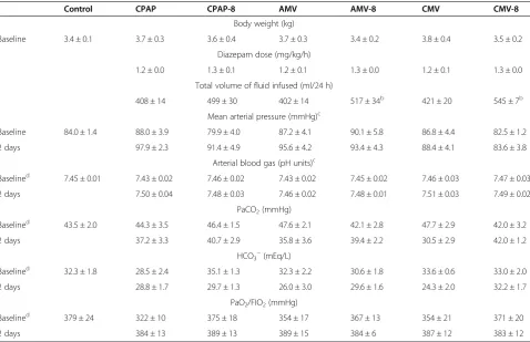
As shown in Table 2, to maintain the target MAP, the groups with CMV and AMV combined with PEEP received significantly larger amounts of intravenous fluids compared with the corresponding mode of ven-tilation without PEEP. In fact, blood pressure and gas exchange were stable throughout the 2-day study (Table 2).
Isometric diaphragm muscle contractile properties
After 2 days of CMV or CMV-8, Po was significantly reduced compared with AMV or CPAP without or with PEEP (AMV-8, CPAP-8), respectively (Table 3). The combined CMV and PEEP did not aggravate the already-reduced Po with CMV alone. Also, PEEP did not have any effects on diaphragmatic force-generating capacity when combined with AMV or CPAP. Likewise, at all stimulation frequencies, PEEP had no additional effects
on the already-reduced force with CMV alone or on the force preserved with AMV and CPAP (Figure 2).
Fiber cross-sectional area, and contractile proteins
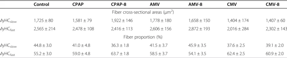
Table 4 shows that after 2 days of CMV or AMV, with or without applied PEEP, fiber CSA was unchanged com-pared with CPAP or CPAP-8, respectively, or with control, although a trend of reduced CSA for all fiber types was demonstrated with CMV and CMV-8. The proportion of all fiber types was not significantly different between groups (Table 4).
The average total myosin pool with various modes of mechanical ventilation was similar to control, except for the CMV and AMV-8 groups, in which myosin was reduced. With the combined CMV and PEEP, MyHC isoform expressions were altered with a shift toward fast myosin (MyHC2X) compared with control, AMV-8 and
CPAP-8 (Table 5).
Levels of transcripts associated with protein degradation and synthesis
Caspase 3 mRNA tended to be elevated for all modes of mechanical ventilation, but attained statistical significance only with CMV and CMV-8 (fivefold increase compared with control). IGF-1 mRNA, transcripts of protein synthe-sis, were markedly reduced with CMV and CMV-8. On the contrary, molecular markers of protein degradation, MAFbx (tenfold and sevenfold increases for CMV-8 and CMV, respectively) and MuRF1 (a fivefold increase for both CMV-8 and CMV) mRNA, were markedly elevated with CMV, with and without PEEP (Figure 3).
Discussion
PEEP is an important treatment modality in acute hypoxic respiratory failure. Most patients receiving me-chanical ventilation for ARDS require PEEP of 5 to 12 cmH2O combined with a lung-protective ventilatory
strategy to improve oxygenation and to prevent atelec-trauma, which is the repetitive opening and closing of lung units [32,33]. The results of the present study con-firm the rapid and profound diaphragm muscle force loss with short-term CMV, and our novel results reveal that the use of PEEP combined with CMV or AMV does not exacerbate VIDD. More specifically, our major find-ings are that (1) the application of 8 cmH2O PEEP does
Positive end-expiratory pressure did not exacerbate diaphragm muscle dysfunction induced by controlled mechanical ventilation
Similar to previous studies [1-4], the present study confirms the deleterious effects of CMV on diaphragm contractile properties. Potential explanation for the lack
[image:7.595.59.538.121.430.2]of interactive effects of PEEP and CMV on diaphrag-matic force production is that diaphragm muscle fiber length was not sufficiently shortened with PEEP applied or that CMV exerted maximal deleterious effects on the diaphragm, such that the use of PEEP application had no additional effects. To our knowledge, we are the first
Table 3In vitrodiaphragm muscle contractile properties with various modes of mechanical ventilation, with and without applied positive end-expiratory airway pressurea
Control CPAP CPAP-8 AMV AMV-8 CMV CMV-8
Lo (cm) 3.2 ± 0.1 3.1 ± 0.1 3.4 ± 0.1 3.2 ± 0.1 3.2 ± 0.2 2.9 ± 0.2 3.2 ± 0.1
TPT (ms) 67 ± 3 68 ± 3 73 ± 9 71 ± 3 71 ± 2 70 ± 4 66 ± 6
RT1/2(ms) 84 ± 4 116 ± 11 115 ± 17 98 ± 5 96 ± 5 118 ± 8 89 ± 11
Ptw (N/cm2) 7.4 ± 0.5 5.9 ± 0.5 6.0 ± 0.8 6.2 ± 0.8 7.2 ± 0.6 5.7 ± 0.9 3.7 ± 0.4b
Po (N/cm2) 24.3 ± 0.4 23.3 ± 1.0 23.0 ± 1.6 22.6 ± 0.8 23.0 ± 1.1 15.8 ± 1.0c 15.0 ± 1.1c
a
Values are mean ± SE (n= 6 animals in each group). Ptw and Po are normalized for cross-sectional area (seeMaterial and methodsfor calculation). AMV, Assist-control mechanical ventilation; CMV, Controlled mechanical ventilation (numerical value of 8 next to ventilatory mode denotes the set CPAP or PEEP of 8 cmH2O); CPAP, Continuous positive airway pressure; Lo, Length at which diaphragm muscle strip produced maximal isometric tension; Po, Maximum tetanic force; Ptw, Peak twitch force; RT1/2, Time required for peak twitch force to relax to one-half of peak twitch force; TPT, Time from onset of muscle contraction to peak twitch force.bFor Ptw, P< 0.01 for CMV-8 compared with control and AMV-8.c
[image:7.595.58.538.600.682.2]For Po,P< 0.01 for CMV compared with control, CPAP and AMV, as well as for CMV-8 compared with control, CPAP-8 and AMV-8.
Table 2 Body weight, diazepam dose, total volume of fluid infused, mean arterial pressure and arterial blood gas at baseline and after 2 days with various modes of mechanical ventilation, with and without applied positive
end-expiratory airway pressurea
Control CPAP CPAP-8 AMV AMV-8 CMV CMV-8
Body weight (kg)
Baseline 3.4 ± 0.1 3.7 ± 0.3 3.6 ± 0.4 3.7 ± 0.3 3.4 ± 0.2 3.8 ± 0.4 3.5 ± 0.2
Diazepam dose (mg/kg/h)
1.2 ± 0.0 1.3 ± 0.1 1.2 ± 0.1 1.3 ± 0.0 1.2 ± 0.1 1.3 ± 0.0
Total volume of fluid infused (ml/24 h)
408 ± 14 499 ± 30 402 ± 14 517 ± 34b 421 ± 20 545 ± 7b
Mean arterial pressure (mmHg)c
Baseline 84.0 ± 1.4 88.0 ± 3.9 79.9 ± 4.0 87.2 ± 4.1 90.1 ± 5.8 86.8 ± 4.4 82.5 ± 1.2
2 days 97.9 ± 2.3 91.4 ± 4.9 95.6 ± 4.2 93.4 ± 4.3 88.4 ± 4.1 83.6 ± 3.8
Arterial blood gas (pH units)c
Baselined 7.45 ± 0.01 7.43 ± 0.02 7.46 ± 0.02 7.43 ± 0.02 7.45 ± 0.02 7.46 ± 0.03 7.47 ± 0.03
2 days 7.50 ± 0.04 7.48 ± 0.03 7.46 ± 0.02 7.48 ± 0.01 7.51 ± 0.03 7.49 ± 0.02
PaCO2(mmHg)
Baselined 43.5 ± 2.0 44.3 ± 3.5 46.4 ± 1.5 47.6 ± 2.1 42.1 ± 2.8 47.7 ± 2.9 42.0 ± 3.2
2 days 37.2 ± 3.3 40.7 ± 2.9 35.8 ± 3.6 39.4 ± 2.2 30.5 ± 2.9 42.0 ± 1.2
HCO3−(mEq/L)
Baselined 32.3 ± 1.8 28.5 ± 2.4 35.1 ± 1.3 32.3 ± 2.2 30.6 ± 1.8 33.6 ± 0.6 33.0 ± 2.0
2 days 28.8 ± 1.7 29.7 ± 1.3 26.0 ± 3.0 29.6 ± 1.6 24.3 ± 2.0 32.2 ± 1.7
PaO2/FIO2(mmHg)
Baselined 379 ± 24 322 ± 10 375 ± 18 354 ± 17 367 ± 13 354 ± 21 371 ± 20
2 days 384 ± 13 389 ± 13 389 ± 15 384 ± 6 387 ± 12 383 ± 12
a
Values are mean ± SE, except mean ± SD for body weight (n= 6 animals in each group). AMV, Assist-control mechanical ventilation; CMV, Controlled mechanical ventilation (numerical value of 8 next to ventilatory mode denotes the set CPAP or PEEP of 8 cmH2O); CPAP, Continuous positive airway pressure; HCO3−, Bicarbonate; PaCO2, Partial pressure of carbon dioxide in arterial blood; PaO2/FIO2, Ratio of partial pressure of oxygen in arterial blood to fraction of inspired oxygen; PEEP, Positive end-expiratory airway pressure.bP
< 0.01 for AMV-8 and CMV-8 compared with the corresponding mode without PEEP.c
Data obtained at 12 hours, 24 hours and 36 hours are not shown, but they were included in statistical analysis.d
to employ a PEEP level commonly applied clinically in combination with diaphragm muscle inactivity. Therefore, only comparisons with inactive hindlimb muscles can be made. In the immobilized rat soleus and gastrocnemius muscles maintained in the shortened position for 4 weeks, muscle length decreased by 20% and 26%, respectively, from that in the neutral position [34]. Clearly, the decrease in diaphragm fiber length with the PEEP applied was less than that in the shortened rat hindlimb muscles [34]. In the present study, we observed that the combined CMV and 8 cmH2O PEEP decreased diaphragm muscle fiber
length by 15% from that of LZEEP. In a study to assess
diaphragmatic shortening with lung inflation in an anesthetized, paralyzed, vagotomized dog model, Road and Leevers [13] achieved a similar degree of diaphragm muscle fiber shortening at the highest positive airway pressure applied. The application of 18 cmH2O positive
airway pressure decreased diaphragm muscle fiber length by 13% from that of LREEV[13]. Differences in the level
of positive airway pressure applied to achieve a similar degree of diaphragm muscle fiber length depends on the applied load and respiratory system mechanics [13], and the latter is a function of the logarithmic value of body weight [35]. Despite species differences, the study of Road and Leevers [13] suggests that diaphragm muscle fiber shortening in our rabbit model achieved its limit. To this extent, contrasts to hindlimb muscles, potential lung overdistension and circulatory compromise constrain diaphragm muscle fiber shortening with PEEP application. A threshold of shortened diaphragm muscle fiber length may be necessary to induce alterations in contractile prop-erties in the diaphragm, an attractive hypothesis that may be arduous to test because of the physiological limitations mentioned above.
The lack of effects of PEEP on diaphragmatic function may be due to the fact that CMV alone results in a large and significant negative impact on diaphragmatic con-tractile properties. This phenomenon is supported by our observation on the interactive effects of 2 days of CMV and methylprednisolone [36], as well as that of Ochala et al. [37] in pigs after 5 days of combined CMV with corticosteroid, neuromuscular blockade or lipopoly-saccharide. In our previous study [36], Po decreased by 36% with CMV alone and by 43% with combined CMV and methylprednisolone (P> 0.05).
Positive end-expiratory pressure effects on diaphragm muscle structural properties and myosin heavy-chain isoform protein expression
[image:8.595.57.291.89.233.2]In the rat diaphragm, atrophy of all muscle fiber types occurred as early as 18 hours of CMV and contributed to the reduced force [5]. However, in pigs, diaphragm muscle fiber structure was intact after 5 days of CMV [37]. Likewise, in rabbits, we did not demonstrate dia-phragm muscle fiber atrophy after 3 days of CMV [1]; hence, after 2 days of CMV, muscle fiber atrophy is less likely to occur, as shown in the present study. The CMV-induced loss of force in rabbits was attributed
Table 4 Diaphragm muscle fiber cross-sectional area and proportion with various modes of mechanical ventilation with and without applied positive end-expiratory airway pressurea
Control CPAP CPAP-8 AMV AMV-8 CMV CMV-8
Fiber cross-sectional areas (μm2)
MyHCslow 1,725 ± 80 1,581 ± 79 1,922 ± 146 1,778 ± 180 1,658 ± 150 1,404 ± 174 1,407 ± 60
MyHCfast 2,565 ± 214 2,478 ± 108 2,416 ± 113 2,606 ± 156 2,872 ± 193 2,016 ± 284 2,302 ± 143
Fiber proportion (%)
MyHCslow 44.8 ± 3.0 41.0 ± 4.8 36.3 ± 1.8 41.5 ± 3.7 45.9 ± 3.5 37.6 ± 2.5 39.1 ± 2.0
MyHCfast 55.2 ± 3.0 59.0 ± 4.8 63.7 ± 1.8 58.5 ± 3.7 54.1 ± 3.5 62.4 ± 2.5 60.9 ± 2.0
a
[image:8.595.59.538.618.716.2]Values ± SE (n= 4 animals in each group). AMV, Assist-control mechanical ventilation; CMV, Controlled mechanical ventilation (numerical value of 8 next to ventilatory mode denotes the set CPAP or PEEP of 8 cmH2O); CPAP, Continuous positive airway pressure; MyHC, Myosin heavy chain.
Figure 2Diaphragm muscle force–frequency relationships in control and mechanical ventilation with and without applied positive end-expiratory airway pressure.Values are mean ± SE (n = 6 animals in each group). *P< 0.01 for controlled mechanical ventilation (CMV-8) compared with control and assist-control mechanical ventilation (AMV-8) for twitch force; CMV and CMV-8 versus corresponding modes of mechanical ventilation without and with positive end-expiratory pressure (PEEP), respectively, at stimulation frequencies of 10 to 100 Hz. The number 8 next to the mode of ventilation denotes the set CPAP or PEEP of 8 cmH2O. Force was
to diaphragm muscle fiber injury rather than to atro-phy [1,36].
Hindlimb immobilization in the shortened position leads to relatively rapid adaptation of MyHC isoform expression [38]. For the reasons mentioned above, com-parison was only possible with immobilized, shortened hindlimb muscles. In rat gastrocnemius muscle, at the mRNA level, MyHCslow and MyHC2A isoform
expres-sions decreased, whereas MyHC2X mRNA levels were
unchanged. In the soleus muscle with predominant slow twitch fibers, MyHCslowexpression did not change;
however, MyHC2Xincreased as early as 2 days of
immo-bilization in the shortened position [39]. Our findings in the combined CMV and PEEP, MyHC isoform trans-formation mimicked that for the shortened, immobilized
rat soleus muscle (Table 5). Whereas fiber-type compos-ition of diaphragm muscle differs from that of soleus, 90% of the rabbit diaphragm muscle fibers (MyHCslow plus MyHC2A isoform) are composed predominantly of fatigue-resistant fibers, and PEEP led to a measurable transition toward the fast MyHC isoform.
Positive end-expiratory pressure effects on molecular markers of protein synthesis and degradation
[image:9.595.58.538.112.222.2]Oxidative stress in skeletal muscle fibers has been shown to depress protein synthesis and to promote proteolysis [40]. Oxidation of diaphragm protein at a molecular weight of 200 kDa (that is, myosin heavy chain oxidation) increased with CMV, with and without PEEP (data not shown), with concomitant suppression of IGF-1 mRNA
[image:9.595.58.538.463.666.2]Figure 3Mechanical ventilation and applied positive end-expiratory airway pressure effects on mRNA levels. (A)Caspase 3.(B)Insulin-like growth factor 1 (IGF-1).(C)Muscle atrophy F-box (MAFbx).(D)Muscle ring finger protein 1 (MuRF1). Values are mean ± SE (n= 6 animals per group). Caspase 3: *P< 0.01 compared with control. IGF-1:†P< 0.05 for CMV compared with CPAP; and for CMV-8 compared with AMV-8. MAFbx: *P< 0.01 for CMV compared with control, CPAP and AMV; and for CMV-8 compared with control. MuRF1: *P< 0.01 for CMV compared with control, CPAP and AMV; and for CMV-8 compared with control, CPAP-8 and AMV-8. AMV, Assist-control mechanical ventilation; CMV, Controlled mechanical ventilation (numerical value of 8 next to the ventilatory mode denotes the set CPAP or PEEP of 8 cmH2O); CPAP, Continuous positive airway pressure.
Table 5 Diaphragm muscle contractile proteins with various modes mechanical ventilation, with and without applied positive end-expiratory airway pressurea
Control CPAP CPAP-8 AMV AMV-8 CMV CMV-8
Myosin and actin (% of total protein pool)
Myosin 41.2 ± 1.0 44.5 ± 1.6 41.3 ± 1.6 41.1 ± 1.4 37.5 ± 0.9b 38.5 ± 1.3b 41.6 ± 1.5
Actin 20.2 ± 1.0 18.1 ± 1.3 19.1 ± 1.0 18.9 ± 1.6 21.7 ± 2.0 18.0 ± 1.5 17.3 ± 1.4
Myosin heavy-chain isoform expression (%)
MyHCslow 35.3 ± 3.3 34.4 ± 1.3 29.9 ± 2.3 33.7 ± 1.6 30.5 ± 2.1 35.3 ± 2.0 29.5 ± 1.9
MyHC2A 54.4 ± 2.8 53.9 ± 1.8 60.3 ± 3.0 54.7 ± 1.4 59.0 ± 2.8 50.9 ± 2.3 55.6 ± 2.2
MyHC2X 10.3 ± 0.6 11.7 ± 0.6 9.8 ± 1.1 11.6 ± 0.9 10.4 ± 1.2 13.8 ± 0.4 14.9 ± 0.9b
a
[27], the gene responsible for protein synthesis via the Akt/mTOR signaling pathway [41]. Activation of proteoly-sis was evidenced by the overexpression of caspase 3 mRNA [42] (Figure 3). Caspase 3 plays an important role in CMV-induced protein degradation. The disrupted myofibrils resulting from the activation of proteases (lysosomal, caspase and calpain) are susceptible to en-hanced proteolysis by the ubiquitin-proteasome system [43]. PEEP had no additional effects on protein oxida-tion, suppression of protein synthesis or upregulation of proteolysis.
Positive end-expiratory pressure effects in partially contracting diaphragm
After 2 days of AMV, diaphragm muscle force was preserved. As with CMV, PEEP had no deleterious effect on preserved force. The results of our present study are consistent with our previously reported data, in which diaphragmatic force was preserved after 3 days of AMV in comparison to CMV [7]. Diaphragm muscle electrical activity was maintained between 30% and 80% of control [44]. Similarly, in pigs subjected to adaptive servo-ventila-tion (ASV) for 3 days, diaphragmatic force was preserved [8]. Conversely, Hudson et al. [45] showed that, in rats, 18 hours of high-level pressure supported decreased dia-phragmatic force, but at a slower rate than with CMV. High-level pressure support also activated calpain, caspase 3 and 20S proteasome [45]. In the present study, caspase 3 mRNA tended to increase after 2 days of AMV with and without PEEP. Differences between our study and that of Hudsonet al. [45] may be due to differences in the degree of diaphragm muscle activation or in the animal species utilized.
Critique of the experimental methods
Diaphragm muscle fiber shortening was not measured continuously for 2 days. Prolonged placement of the sonomicrometer transducers was not technically feasible, because manipulation of the animals for animal care en-hanced the noise-to-signal ratio and accurate measurement of segmental length changes was impossible. However, PEEP of 8 cmH2O shortened diaphragm muscle fibers to
their physiological limits as CstRSdecreased, and, in all
of the animals, application of PEEP to CMV and AMV required significantly larger amounts of fluids to main-tain the target MAP. PEEP was applied for a short time (2 days only); the effects of long-term PEEP for more than 2 days, even at the modest level employed in this study, are unknown.
This study was performed in healthy animals only. By using a healthy animal model, confounding factors (for example, cytokines) [46] on the effect of PEEP on dia-phragmatic function can be isolated. In the present study, we employed one level of 8 cmH2O PEEP. This level of
PEEP shortened diaphragm muscle fiber comparable to 18 cmH2O of PEEP in dogs [13]; yet, when combined with
either CMV or AMV, PEEP had no detectable harmful effects on diaphragmatic function. Studying low PEEP levels is unlikely to show any influence on diaphragm contractile properties, whereas very high PEEP levels may induce volutrauma.
Conclusions
In this observational study, short-term application of 8 cmH2O PEEP to CMV or AMV did not exacerbate
dia-phragmatic dysfunction or induce diadia-phragmatic force loss, respectively. Similarly, PEEP by itself did not produce alterations in structural or molecular properties. The lack of PEEP effects on the diaphragm is likely related to insuf-ficient diaphragm muscle fiber shortening due to the asso-ciated physiological limitations of alveolar overdistension and circulatory compromise at higher PEEP levels or that CMV induced profound effects on the diaphragm such that additional insult of PEEP was undetectable.
The rapid and profound development of diaphragm muscle weakness remains problematic following diaphragm muscle disuse with CMV. In view of the inconsistent results between studies regarding partial diaphragm muscle acti-vation, the degree of diaphragm muscle activity sufficient to preserve diaphragm muscle function or its long-term effects remains to be investigated.
In clinical practice, PEEP of 8 to 10 cmH2O is
consid-ered a modest level of PEEP. A high levels of PEEP (mean 15 cmH2O, range 18 to 24 cmH2O) has been applied in
the management of patients with ARDS [47], but, to our knowledge, the degree of diaphragm muscle fiber shorten-ing or the effects on diaphragm muscle function has not been evaluated. In the present study, we demonstrate that the application of 8 cmH2O PEEP to conventional modes
of mechanical ventilation appears safe and has no delete-rious influence on diaphragm muscle force-generating capacity.
Key messages
At the level of physiological limits, PEEP did not aggravate CMV-induced diaphragmatic dysfunction or proteolysis.
At the level of physiological limits, PEEP had no
effect on diaphragm function in partially contracting diaphragm with AMV.
Abbreviations
ABG:Arterial blood gas; AMV: Assist-control mechanical ventilation; ASV: Adaptive servo-ventilation; CMV: Controlled mechanical ventilation; CPAP: Continuous positive airway pressure; CstRS: Static compliance of the
respiratory system; IGF-1: Insulin-like growth factor 1; IP: Inspiratory pressure; Lo: Optimal muscle length; LZEEP: Segmental diaphragm muscle fiber length
at 0 cmH2O positive end-expiratory airway pressure; MAFbx: Muscle atrophy
protein 1; MyHC: Myosin heavy chain; PEEP: Positive end-expiratory airway pressure; Po: Maximum isometric tetanic force normalized for muscle cross-sectional area; Pplat: Airway plateau pressure; Ptw: Twitch force normalized
for muscle cross-sectional area; RT½: Time for twitch force to relax to one-half twitch force; TI: Inspiratory time; TI/Ttot: Duty cycle; TPT: Time to peak twitch tension; Ttot: Total breath cycle; VE: Total minute ventilation; VIDD: Ventilator-induced diaphragmatic dysfunction; VT: Tidal volume.
Competing interests
The authors declare that they have no competing interests.
Authors’contributions
CS conceived of the study and experimental design and was involved in the experiments and data acquisition, analysis and interpretation as well as manuscript writing. EZ participated in the experimental design, carried out the experiments; was involved with biochemical, histochemical and molecular studies; acquired data acquisition; and performed statistical analysis. LF carried out the experiments and performed the morphometry, biochemical, histochemical and molecular studies. GS participated in the design of the study and manuscript revision for important intellectual content. SP participated in the biochemical studies and manuscript revision for important intellectual content. All authors read and approved the final manuscript.
Acknowledgements
This study was supported by funding from the Department of Veterans Affairs Medical Research Service (to CSHS), by National Institutes of Health grant HL74309 (to GCS) and by National Institutes of Health grant HL780839 (to SKP). The work was conducted in the Department of Research and Development at the VA Long Beach Healthcare System, Long Beach, CA, USA.
Author details
1Department of Medicine, Section of Pulmonary and Critical Care Medicine,
VA Long Beach Healthcare System, 5901 East 7th Street, Long Beach, CA 90822, USA.2Department of Medicine, University of California, 101 The City Drive South, Orange, Irvine, CA 92868, USA.3Departments of Physiology and Biomedical Engineering, and Anesthesiology, Mayo Clinic, 200 First Street SW, Rochester, MN 55905, USA.4Department of Applied Physiology and Kinesiology, University of Florida, Gainesville, FL 32611, USA.
Received: 3 April 2014 Accepted: 5 August 2014
References
1. Sassoon CS, Caiozzo VJ, Manka A, Sieck GC:Altered diaphragm contractile properties with controlled mechanical ventilation.J Appl Physiol2002, 92:2585–2595.
2. Powers SK, Shanely RA, Coombes JS, Koesterer TJ, McKenzie M, Van Gammeren D, Cicale M, Dodd SL:Mechanical ventilation results in progressive contractile dysfunction in the diaphragm.J Appl Physiol2002, 92:1851–1858.
3. Levine S, Nguyen T, Taylor N, Friscia ME, Budak MT, Rothenberg P, Zhu J, Sachdeva R, Sonnad S, Kaiser LR, Rubinstein NA, Powers SK, Shrager JB: Rapid disuse atrophy of diaphragm fibers in mechanically ventilated humans.N Engl J Med2008,358:1327–1335.
4. Jaber S, Petrof BJ, Jung B, Chanques G, Berthet JP, Rabuel C, Bouyabrine H, Courouble P, Koechlin-Ramonatxo C, Sebbane M, Similowski T, Scheuermann V, Mebazaa A, Capdevila X, Mornet D, Mercier J, Lacampagne A, Philips A, Matecki S:Rapidly progressive diaphragmatic weakness and injury during mechanical ventilation in humans.Am J Respir Crit Care Med2011, 183:364–371.
5. Shanely RA, Zergeroglu MA, Lennon SL, Sugiura T, Yimlamai T, Enns D, Belcastro A, Powers SK:Mechanical ventilation-induced diaphragmatic atrophy is associated with oxidative injury and increased proteolytic activity.Am J Respir Crit Care Med2002,166:1369–1374.
6. Vassilakopoulos T, Petrof BJ:Ventilator-induced diaphragmatic dysfunction.Am J Respir Crit Care Med2004,169:336–341. 7. Sassoon CS, Zhu E, Caiozzo VJ:Assist-control mechanical ventilation
attenuates ventilator-induced diaphragmatic dysfunction.Am J Respir Crit Care Med2004,170:626–632.
8. Jung B, Constantin JM, Rossel N, Le Goff C, Sebbane M, Coisel Y, Chanques G, Futier E, Hugon G, Capdevila X, Petrof B, Matecki S, Jaber S:Adaptive support ventilation prevents ventilator-induced diaphragmatic dysfunction in piglet: an in vivo and in vitro study.Anesthesiology2010,112:1435–1443. 9. Zhu E, Sassoon CSH:The silenced diaphragm: the good and the bad.
Reanimation2008,17:631–638.
10. Goldspink DF:The influence of immobilization and stretch on protein turnover of rat skeletal muscle.J Physiol1977,264:267–282. 11. Booth FW:Time course of muscular atrophy during immobilization of
hindlimbs in rats.J Appl Physiol1977,43:656–661.
12. Fujita N, Fujimoto T, Tasaki H, Arakawa T, Matsubara T, Miki A:Influence of muscle length on muscle atrophy in the mouse tibialis anterior and soleus muscles.Biomed Res2009,30:39–45.
13. Road JD, Leevers AM:Effect of lung inflation on diaphragmatic shortening.J Appl Physiol1988,65:2383–2389.
14. Dellamonica J, Lerolle N, Sargentini C, Beduneau G, Di Marco F, Mercat A, Richard JC, Diehl JL, Mancebo J, Rouby JJ, Lu Q, Bernardin G, Brochard L: PEEP-induced changes in lung volume in acute respiratory distress syndrome: two methods to estimate alveolar recruitment.Intensive Care Med2011,37:1595–15604.
15. Papazian L, Forel JM, Gacouin A, Penot-Ragon C, Perrin G, Loundou A, Jaber S, Arnal JM, Perez D, Seghboyan JM, Constantin JM, Courant P, Lefrant JY, Guérin C, Prat G, Morange S, Roch A, ACURASYS Study Investigators: Neuromuscular blockers in early acute respiratory distress syndrome.
N Engl J Med2010,363:1107–1116.
16. Smith TC, Marini JJ:Impact of PEEP on lung mechanics and work of breathing in severe airflow obstruction.J Appl Physiol1988, 65:1488–1499.
17. Zergeroglu MA, McKenzie MJ, Shanely RA, Van Gammeren D, DeRuisseau KC, Powers SK:Mechanical ventilation-induced oxidative stress in the diaphragm.J Appl Physiol2003,95:1116–1124.
18. Falk DJ, DeRuisseau KC, Van Gammeren DL, Deering MA, Kavazis AN, Powers SK:Mechanical ventilation promotes redox status alterations in the diaphragm.J Appl Physiol2006,101:1017–1024.
19. Picard M, Jung B, Liang F, Azuelos I, Hussain S, Goldberg P, Godin R, Danialou G, Chaturvedi R, Rygiel K, Matecki S, Jaber S, Des Rosiers C, Karpati G, Ferri L, Burelle Y, Turnbull DM, Taivassalo T, Petrof BJ: Mitochondrial dysfunction and lipid accumulation in the human diaphragm during mechanical ventilation.Am J Respir Crit Care Med2012, 186:1140–1149.
20. Powers SK, Hudson MB, Nelson WB, Talbert EE, Min K, Szeto HH, Kavazis AN, Smuder AJ:Mitochondria-targeted antioxidants protect against mechanical ventilation-induced diaphragm weakness.Crit Care Med2011, 39:1749–1759.
21. Tang H, Lee M, Budak MT, Pietras N, Hittinger S, Vu M, Khuong A, Hoang CD, Hussain SNA, Levine S, Shrager JB:Intrinsic apoptosis in mechanically ventilated human diaphragm: linkage to a novel FOS/FOXO1/STAT3-BIM axis.FASEB J2011,25:2921–2936.
22. Smuder AJ, Hudson MB, Nelson WB, Kavazis AN, Powers SK:Nuclear factor-κB signaling contributes to mechanical ventilation-induced diaphragm weakness.Crit Care Med2012,40:927–934.
23. Hussain SNA, Mofarrahi M, Sigala I, Kim HC, Vassilakopoulos T, Maltais F, Bellenis I, Chaturvedi R, Gottfried SB, Metrakos P, Danialou G, Matecki S, Jaber S, Petrof BJ, Goldberg P:Mechanical ventilation–induced diaphragm disuse in humans triggers autophagy.Am J Respir Crit Care Med2010, 182:1377–1386.
24. Nelson WB, Smuder AJ, Hudson MB, Talbert EE, Powers SK:Cross-talk between the calpain and caspase-3 proteolytic systems in the dia-phragm during prolonged mechanical ventilation.Crit Care Med2012, 40:1857–1863.
25. Levine S, Biswas C, Dierov J, Barsotti R, Shrager JB, Nguyen T, Sonnad S, Kucharchzuk JC, Kaiser LR, Singhal S, Budak MT:Increased proteolysis, myosin depletion, and atrophic AKT-FOXO signaling in human diaphragm disuse.Am J Respir Crit Care Med2011,183:483–490. 26. DeRuisseau KC, Kavazis AN, Deering MA, Falk DJ, Van Gammeren D,
Yimlamai T, Ordway GA, Powers SK:Mechanical ventilation induces alterations of the ubiquitin-proteasome pathway in the diaphragm.
J Appl Physiol2005,98:1314–1321.
28. Zhan WZ, Farkas GA, Schroeder MA, Gosselin LE, Sieck GC:Regional adaptations of rabbit diaphragm muscle fibers.J Applied Physiol1995, 79:941–950.
29. Newman S, Road J, Bellemare F, Clozel JP, Lavigne CM, Grassino A: Respiratory muscle length measured by sonomicrometry.J Appl Physiol
1984,56:753–764.
30. Road JD, Leevers AM:Inspiratory and expiratory muscle function during continuous positive airway pressure in dogs.J Appl Physiol1990, 68:1092–1100.
31. Urbscheit N, Bishop B, Bachofen H:Immediate effects of continuous positive pressure breathing on abdominal expiratory activity, minute ventilation, and end-tidal PCO2of conscious man.Phys Ther1973,53:258–265. 32. The National Heart, Lung, and Blood Institute ARDS Clinical Trials Network:
Mechanical ventilation with higher versus lower positive end-expiratory pressures in patients with acute lung injury and the acute respiratory distress syndrome.N Engl J Med2004,351:327–336.
33. Slutsky AS:Neuromuscular blocking agents in ARDS.N Engl J Med2010, 363:1176–1180.
34. Spector SA, Simard CP, Fournier M, Sternlicht E, Edgerton VR:Architectural alterations of rat hind-limb skeletal muscles immobilized at different lengths.Exp Neurol1982,76:94–110.
35. Bennett FM, Tenney SM:Comparative mechanics of mammalian respiratory system.Respir Physiol1982,49:131–140.
36. Sassoon CS, Zhu E, Fang L, Ramar K, Jiao GY, Caiozzo VJ:Interactive effects of corticosteroid and mechanical ventilation on diaphragm muscle function.Muscle Nerve2011,43:103–111.
37. Ochala J, Renaud G, Llano Diez M, Banduseela VC, Aare S, Ahlbeck K, Radell PJ, Eriksson LI, Larsson L:Diaphragm muscle weakness in an experimental porcine intensive care unit model.PLoS One2011,6:e20558. 38. Talmadge RJ:Myosin heavy chain isoform expression following reduced
neuromuscular activity: potential regulatory mechanisms.Muscle Nerve
2000,23:661–679.
39. Loughna PT, Izumo S, Goldspink G, Nadal-Ginard B:Disuse and passive stretch cause rapid alterations in expression of developmental and adult contractile protein genes in skeletal muscle.Development1990, 109:217–223.
40. Powers SK, Wiggs MP, Sollanek KJ, Smuder AJ:Ventilator-induced diaphragm dysfunction: cause and effect.Am J Physiol Regul Integr Comp Physiol2013,305:R464–R477.
41. Bodine SC, Stitt TN, Gonzalez M, Kline WO, Stover GL, Bauerlein R, Zlotchenko E, Scrimgeour A, Lawrence JC, Glass DJ, Yancopoulos GD: Akt/mTOR pathway is a crucial regulator of skeletal muscle hypertrophy and can prevent muscle atrophyin vivo.Nat Cell Biol2001,3:1014–1019. 42. Whidden MA, Smuder AJ, Wu M, Hudson MB, Nelson WB, Powers SK:
Oxidative stress is required for mechanical ventilation-induced protease activation in the diaphragm.J Appl Physiol2010,108:1376–1382. 43. Powers SK, Kavazis AN, McClung JM:Oxidative stress and disuse muscle
atrophy.J Appl Physiol2007,102:2389–2397.
44. Sassoon CSH, Caiozzo VJ:Bench-to-bedside review: Diaphragm muscle function in disuse and acute high-dose corticosteroid treatment.
Crit Care2009,13:221.
45. Hudson MB, Smuder AJ, Nelson WB, Bruells CS, Levine S, Powers SK: Both high level pressure support ventilation and controlled mechanical ventilation induce diaphragm dysfunction and atrophy.Crit Care Med
2012,40:1254–1260.
46. Maron-Gutierrez T, Laffey JG, Pelosi P, Rocco PR:Cell-based therapies for the acute respiratory distress syndrome.Curr Opin Crit Care2014, 20:122–131.
47. Meade MO, Cook DJ, Guyatt GH, Slutsky AS, Arabi YM, Cooper DJ, Davies AR, Hand LE, Zhou Q, Thabane L, Austin P, Lapinsky S, Baxter A, Russell J, Skrobik Y, Ronco JJ, Stewart TE, Lung Open Ventilation Study Investigators:Ventilation strategy using low tidal volumes, recruitment maneuvers, and high positive end-expiratory pressure for acute lung injury and acute respiratory distress syndrome: a randomized controlled trial.JAMA2008,299:637–645.
doi:10.1186/s13054-014-0494-0
Cite this article as:Sassoonet al.:Positive end-expiratory airway pressure does not aggravate ventilator-induced diaphragmatic dysfunction in rabbits.Critical Care201418:494.
Submit your next manuscript to BioMed Central and take full advantage of:
• Convenient online submission
• Thorough peer review
• No space constraints or color figure charges
• Immediate publication on acceptance
• Inclusion in PubMed, CAS, Scopus and Google Scholar
• Research which is freely available for redistribution