Michael Knauth, Ru¨diger von Kummer, Olav Jansen, Stefan Ha¨hnel, Arnd Do¨rfler, and Klaus Sartor
PURPOSE: To study the ability of CT angiography to show intracranial arterial occlusion and collateral blood flow in patients with acute stroke. METHODS: Twenty-one patients with acute nonhemorrhagic stroke were studied prospectively with conventional CT, CT angiography, and digital subtraction angiography. On the basis of CT angiographic findings, two neuroradiologists independently assessed the site of arterial occlusion, the contrast enhancement in arterial branches beyond the occlusion as a measure of collateral blood supply, and the extent of diminished parenchymal enhancement; they then predicted the extent of ischemic infarction. RESULTS: Both raters correctly assessed all trunk occlusions of the basilar artery (n54), the internal carotid artery (n54), and the middle cerebral artery (n59). The chance adjusted interrater agreement was k5.78. The assessment of branch occlusions of the middle cerebral artery was less reliable. The agreement rate in judging the collateral state in 17 occlusions in the anterior cerebral circulation was 88%. The size of 21 (62%) of 34 hemispheric infarctions was predicted correctly. CONCLU-SION: CT angiography quickly and reliably adds important information to conventional CT studies in cases of acute ischemic stroke. It shows the site of occlusion, the length of the occluded arterial segment, and the contrast-enhanced arteries beyond the occlusion as an estimate of collateral blood flow.
Index terms: Brain, infarction; Brain, computed tomography; Computed tomography, three-dimensional
AJNR Am J Neuroradiol18:1001–1010, June 1997
In acute intracranial arterial occlusion with sudden neurologic deficit, limited time is avail-able to obtain information for carefully directed treatment, because of the relatively rapid onset of irreversible neuronal damage. Diagnosis based on both etiologic and pathophysiologic data is essential to enable therapeutic decisions to be made. Commonly, patients with acute stroke are examined with unenhanced com-puted tomography (CT) of the brain to exclude intracranial hemorrhage or other causes of the stroke. CT has also proved useful in assessing early sequelae of cerebral ischemia, such as parenchymal hypodensity and focal brain
swell-ing, thus showing the volume of brain tissue most severely affected by ischemia (1–7). Un-enhanced CT does not, however, show the arterial occlusion itself, except in patients with a hyper-dense arterial sign (eg, the hyperhyper-dense middle cerebral artery [MCA] sign), which has high spec-ificity but low sensitivity for an occluded cerebral artery (5, 8 –10). Furthermore, conventional CT does not show the extent of disturbed cerebral perfusion, which is determined by the site of oc-clusion, collateral blood supply, and intracranial perfusion pressure. Finally, it does not allow the leptomeningeal collaterals to be seen. Conven-tional CT in acute arterial occlusion is thus inca-pable of showing the volume of viable tissue at risk from low perfusion, which is the target of thrombolytic treatment.
Because of these considerations, we evalu-ated whether CT angiography is capable of re-liably showing the site of arterial occlusion, es-timating leptomeningeal collateralization, and determining the extent of severe parenchymal perfusion deficit.
Received June 22, 1996; accepted after revision October 28. Presented at the annual meeting of the American Society of Neurora-diology, Seattle, Wash, June 1996.
From the Department of Neuroradiology, University of Heidelberg Med-ical School, Im Neuenheimer Feld 400, D 69120 Heidelberg, Germany. Address reprint requests to Michael Knauth, MD.
AJNR 18:1001–1010, Jun 1997 0195-6108/97/1806 –1001 ©American Society of Neuroradiology
Patients and Methods
We examined with conventional cranial CT 21 patients with an acute (,6 hours), severe hemispheric syndrome (hemiparesis # grade 2 and/or aphasia, where grade 2 paresis is defined as a state in which a limb can be moved only when gravity is eliminated) in whom clinical symp-toms showed no tendency toward improvement. Five pa-tients had intracranial hemorrhage and were excluded from further study. Five patients with brain stem symp-toms and clinically suspected basilar artery occlusion were examined by CT angiography, regardless of the time in-terval from symptom onset. We thus selected 16 patients with suspected acute arterial occlusion in the anterior ce-rebral circulation and five patients with suspected basilar artery occlusion for CT angiography immediately after unenhanced conventional cranial CT. Thirteen patients were men and eight were women; mean age was 59.8 years (SD614.2).
Informed and signed consent was obtained from the patients or their relatives. For conventional CT, 8-mm-thick axial sections were used throughout the brain. For CT angiography, spiral scanning during intravenous bolus ad-ministration of nonionic contrast medium was done on the same scanner immediately after the initial CT study with-out moving the patient. Section thickness was 1.5 mm (index, 1.0 mm) for the patients with suspected occlusion in the anterior cerebral circulation and 2.0 mm (index, 1.5 mm) in cases of suspected basilar occlusion. This differ-ence in scanning parameters was necessitated by the fact that in the patients with suspected basilar artery occlusion a wider scan range must be covered. Patients with sus-pected occlusion in the anterior cerebral circulation were scanned from the sellar floor toward the vertex, whereas in patients with suspected basilar occlusion, spiral scanning extended from the foramen magnum to the tip of the basilar artery. The following scan parameters were used in all patients: 1.25 spiral pitch, 130 kV, and 125 mA. Total scanning time was 21 seconds (1 second per revolution). We injected 130 mL of the nonionic contrast medium into an antecubital vein (intravenous cannula$18 gauge) with an injection rate of 4 to 5 mL/s using an injection pump. Scan delay was 20 seconds in all cases.
In eight of 16 patients with suspected occlusion in the anterior cerebral circulation and in three of five patients with suspected basilar occlusion, cerebral digital subtrac-tion angiography (DSA) was performed within 1 hour after CT angiography. All but one patient had a follow-up CT study within 24 612 hours of the initial CT examination. The spiral data were transferred to an independent medical workstation and three-dimensional reconstructions of the circle of Willis (or the basilar artery) were performed. Since the 3-D volume-rendering algorithm used does not require data segmentation, 3-D evaluation of the CT angiographic data set took less than 10 minutes (including data transfer from the scanner to the workstation). The quality of two CT angiographic data sets was diminished because of patient motion during spiral scanning; however, the five to seven sections that showed the circle of Willis were spared from
motion artifacts. Thus, the CT angiographic data set was still diagnostic, but we were unable to reconstruct three-dimensionally the data sets that were degraded by motion artifacts.
Data Evaluation
The 3-D reconstructions of the circle of Willis were calculated by using a volume-rendering algorithm. A re-gion of interest was defined in the MCA on the CT angio-graphic source images. The mean gray value of the region of interest was measured and taken as the center, the standard deviation as the width of the window for the 3-D reconstructions. The 3-D reconstructions of the circle of Willis were depicted as in the real world (ie, when the circle of Willis was viewed from above, the left MCA was on the left side). Calcification of the vessels may cause problems with the 3-D reconstructions, but it is easily detected on the CT angiographic source images. Therefore, it is nec-essary to use both the source images and the 3-D recon-structions for diagnosis. We did not use maximum inten-sity projections because the proximity of the vessels to the skull base would have required segmentation of the data sets, which would have delayed the diagnostic procedure. Two experienced neuroradiologists, blinded to the pa-tients’ clinical, DSA, and CT findings, evaluated the CT angiographic studies independently. The neuroradiolo-gists were aware, however, that CT angiography had been performed for suspected vessel occlusion. Their evalua-tion was based solely on the CT angiographic source im-ages and 3-D reconstructions. Occlusions were distin-guished in the basilar artery, the intracranial internal carotid artery bifurcation, the proximal MCA trunk, the distal MCA trunk, and the MCA branch. Their assessment of arterial occlusion was compared with the findings on DSA (11 of 21) and/or the pattern of cerebral infarction on the follow-up CT study.
In the patients with vessel occlusion in the anterior cerebral circulation, the leptomeningeal collateral blood supply (LCBS) was rated on a semiquantitative scale. The number of arteries visible beyond the occlusion and sup-plied with contrast agent by collaterals was categorized and the LCBS was rated as “good” if there was filling of the MCA branches in the sylvian fissure, as “moderate” if collaterals were visible but the sylvian MCA branches re-mained unenhanced, or as “none” if no arteries beyond the occlusion were visible. In addition, the two neuroradiolo-gists described the pattern and proportion of brain tissue without parenchymal enhancement as an estimate of se-vere perfusion deficit.
We describe interobserver agreement by agreement rates andkstatistics. Thekstatistic is chance corrected and measures the observed amount of agreement adjusted for the amount of agreement by chance alone. Akvalue greater than .6 was considered to indicate substantial to excellent agreement, as has been described by others (11).
Results
The two neuroradiologists found four basilar artery occlusions in five patients and 15 occlu-sions in the anterior circulation of 16 patients. Interestingly, they detected an additional prox-imal MCA occlusion that was not suspected clinically in one of the patients with basilar ar-tery occlusion (Fig 1). Therefore, 20 arterial occlusions were observed in 19 patients (Table 1). In one patient with clinically suspected basi-lar artery occlusion, neither rater found an oc-clusion at CT angiography. Follow-up examina-tion showed that this patient was intoxicated and did not develop brain stem or cerebellar infarction. We counted this rating as true-nega-tive.
In another patient, neither rater was able to diagnose arterial occlusion by CT angiography, but the follow-up CT study showed partial in-farction of the MCA territory, suggesting MCA branch occlusion (Fig 2). We counted this rat-ing as false-negative.
DSA confirmed the site of occlusion as seen at CT angiography in the 11 patients in whom it was performed (Figs 1 and 3). In the remaining patients, the diagnosed occlusions were consis-tent with the exconsis-tent and pattern of the infarction on follow-up CT scans. Altogether, one rater correctly judged 21 (95%) of 22 intracranial arterial occlusions and the other rater 20 (91%) of 22 occlusions. With regard to the exact site of arterial occlusion, the two neuroradiologists
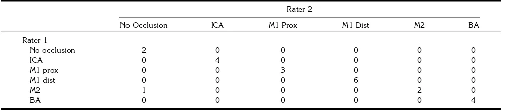
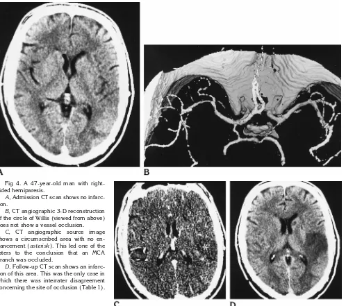
agreed in 21 of 22 assessments (95%,k5.78) (Table 1). They disagreed in one case of MCA branch occlusion (Fig 4).
In all patients with suspected occlusions in the anterior cerebral circulation, the two neuro-radiologists rated the LCBS with the use of a semiquantitative scale. They agreed in their as-sessment of 15 (88%) of 17 patients (Table 2). In two patients, LCBS was rated as “none” by one neuroradiologist and as “moderate” by the other.
The source CT angiographic images showed areas without parenchymal enhancement in eight (47%) of 17 patients with occlusion in the anterior cerebral circulation. In all cases, unen-hanced brain parenchyma became hypodense on the follow-up CT scan (Figs 4 and 5). The minimum size of the ischemic lesion was repre-sented consistently by the pattern of unen-hanced tissue. The hypodense area of brain tis-sue on the unenhanced follow-up CT scans was either the same size or larger than the unen-hanced tissue volume (Fig 5).
[image:3.612.60.564.601.711.2]On the basis of their assessment of occlusion site and collateral blood supply, the two neuro-radiologists tried to predict the size and pattern of hemispheric brain infarction in the 17 pa-tients with occlusion in the anterior circulation. When compared with CT performed at 24612 hours after the stroke, their ratings were correct in 21 (62%) of 34 assessments. In five patients with “none” or “moderate” LCBS, the infarct pattern and size were estimated correctly 90% of the time (nine of 10 predictions), whereas this rate dropped to 50% (12 correct predic-tions) in patients with “good” LCBS. In nine of the 12 false predictions, the infarcted area on the follow-up CT study was larger than pre-dicted.
TABLE 1: Assessment of occlusion site at CT angiography by two raters
Rater 2
No Occlusion ICA M1 Prox M1 Dist M2 BA
Rater 1
No occlusion 2 0 0 0 0 0
ICA 0 4 0 0 0 0
M1 prox 0 0 3 0 0 0
M1 dist 0 0 0 6 0 0
M2 1 0 0 0 2 0
BA 0 0 0 0 0 4
Discussion
Today, emergency CT is generally the first diagnostic step after physical examination in patients with acute focal neurologic deficit. CT is also currently used in major clinical trials to properly select patients for thrombolysis with remarkable results (12, 13). The European Co-operative Acute Stroke Study showed that the use of CT assessment to exclude patients with primary hemorrhage and larger ischemic le-sions (.33% of the MCA territory) within the first 6 hours after the onset of symptoms mark-edly influenced patients’ response to treatment (12).
Despite the enormous potential of magnetic resonance imaging in this regard (14), we pre-sume that, because of its greater availability and practicability for mostly uncooperative pa-tients, CT will continue to be the primary diag-nostic tool in stroke in the foreseeable future. All major controlled trials thus far have used CT to check patient inclusion and exclusion. It is therefore important to know whether a new CT technology with spiral scanning during bolus injection of a contrast agent can reliably give additional and important information about the site of arterial occlusion and, thus, the vascular territory at risk from low perfusion.
So far, CT angiography has been used to evaluate vascular lesions at the carotid bifurca-tion and to search for aneurysms at the circle of Willis (15–17). More recently, cerebral CT
venography has been used successfully to di-agnose dural sinus thrombosis (18).
CT angiography of the intracranial vascula-ture adds only a few minutes to standard CT protocols and thus does not significantly delay initiation of treatment. In our study, motion by uncooperative patients was a minor problem. No patient had an adverse reaction to the con-trast agent. The amount (130 mL) of nonionic contrast agent we injected is relatively high, but it does not preclude additional DSA if invasive angiography becomes necessary.
Pullicino and Kendall (19) reported a possible association between the use ofionic (and thus hyperosmolar) contrast agent and poor out-come in stroke patients. Although inconclusive, their findings resulted in a restrictive attitude toward the administration of contrast material in patients with cerebral ischemia. Surprisingly, no controlled study was performed to clarify a pos-sible toxic effect of contrast agents in acute cerebral ischemia. At this point, we want to stress that we exclusively usenonionic(almost isosmolar) contrast media for CT angiography. To obtain more information about potential risks of contrast infusion in acute stroke, we conducted an experimental study to investigate the effects of bolus injection of ionic and non-ionic contrast agents on infarction size and clin-ical course in a rat model of acute focal cerebral infarction. Our results suggest that bolus ad-ministration of nonionic contrast agents even in the double clinical dose does not significantly influence infarction volume and clinical course in acute ischemic stroke (Doerfler et al, unpub-lished data).
The two neuroradiologists assessed the oc-clusion sites of the intracranial internal carotid artery, the basilar artery, and the MCA trunk with a 100% sensitivity and specificity. Their evaluation was less certain in three patients with MCA branch occlusion. They disagreed in one patient (Fig 4), and neither rater could see one particular occlusion that was presumably re-TABLE 2: Assessment of collateral blood supply at CT
angiogra-phy by two raters
Rater 2
None Moderate Good
Rater 1
None 0 1 0
Moderate 1 3 0
Good 0 0 12
Fig 1. A 66-year-old man with brain stem symptoms and suspected basilar occlusion. AandB, Admission CT scans do not show early signs of cerebral infarction.
CT angiographic 3-D reconstruction of the basilar artery and the circle of Willis (C) and source image (D) show distal occlusion of the basilar artery extending into the P1 segments of the posterior cerebral arteries (straight arrows, CandD) as well as proximal MCA occlusion on the left (curved arrow, C).
E, DSA (injection of the right vertebral artery only) confirms the distal occlusion of the basilar artery but fails to show the length of the occlusion. Additional selective injection of the anterior circulation would have been required to illustrate the patency of the P2 segments of the posterior cerebral arteries.
[image:5.612.62.301.110.189.2]sponsible for small cortical infarctions (Fig 2). We counted this assessment as false-negative, although recanalization at the time of CT an-giography is another possibility. We did not re-peat the CT angiography to study recanaliza-tion.
For ethical reasons we were not able to com-pare CT angiographic findings with the DSA standard of reference in all patients. For exam-ple, if CT angiography showed MCA trunk oc-clusion and the patient turned out to have con-traindications for thrombolytic therapy, or if it was decided to perform systemic thrombolytic therapy, we could not perform DSA, which would have delayed therapy. In these cases we
compared CT angiographic findings with the pattern of infarction on the follow-up CT study. In all 11 patients who had both CT angiogra-phy and DSA, DSA confirmed the CT angio-graphic findings. Although the number of pa-tients is small, we have become convinced by this experience that CT angiography reliably shows the clinically relevant occlusions of ma-jor cerebral arteries. As mentioned earlier, CT angiography is less reliable in showing MCA
[image:6.612.62.565.81.536.2]branchocclusions. However, the natural course of MCA branch occlusion is relatively benign, whereas occlusions of the MCA trunk, intracra-nial internal carotid artery, and basilar artery are associated with high morbidity and mortality Fig 2. A 75-year-old man with
left-sided hemiparesis.
Admission CT scan (A) and CT angio-graphic 3-D reconstruction of the circle of Willis (viewed from above) (B) and source image (C) are nondiagnostic.
(20 –24) and are therefore considered the tar-gets of thrombolytic treatment.
The mismatch between the territory of an oc-cluded artery and the volume of ischemic lesion as shown by CT can be explained by collateral blood supply (25, 26). We found that CT an-giography reliably showed the effect of collater-als, that is, enhancement of MCA branches be-yond the site of occlusion. Although absence or reduction of this vascular enhancement was as-sociated with a large infarction, the presence of collaterals did not necessarily guarantee a small infarction (Fig 5).
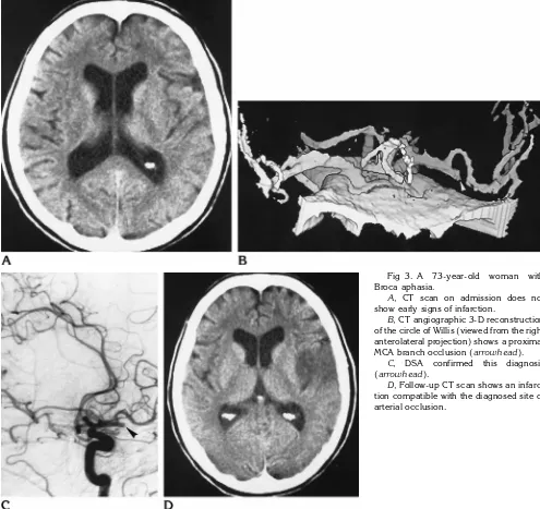
[image:7.612.60.555.88.555.2]If the LCBS was judged “none” or “moder-ate,” the extent of the infarcted area was cor-rectly predicted in 90% of the patients. By con-trast, if the LCBS was judged “good,” the rate of correct predictions dropped to 50%. We suggest that this variation may be due to the potential instability of collateral blood flow. The judgment of “good” at the time of CT angiography does not mean that the collaterals will remain viable over time. In these patients there is still brain tissue to be lost. This view is supported by the fact that in nine of the 12 incorrect predictions in patients with “good” collaterals, the infarcted Fig 3. A 73-year-old woman with Broca aphasia.
A, CT scan on admission does not show early signs of infarction.
B, CT angiographic 3-D reconstruction of the circle of Willis (viewed from the right anterolateral projection) shows a proximal MCA branch occlusion (arrowhead).
C, DSA confirmed this diagnosis (arrowhead).
area was underestimated. Perhaps it is this group of patients that is most likely to benefit from early thrombolytic recanalization.
We considered the brain parenchyma that ei-ther was hypodense on the admission CT scan or showed missing parenchymal enhancement at CT angiography to be the core of infarction, because without exception this area became hypodense on the follow-up CT scans (Figs 4 and 5).
With CT immediately followed by CT angiog-raphy, we may now have a tool that shows the core of infarction and the vascular territory at risk from arterial occlusion and hypoperfusion. In situations in which the territory of the
oc-cluded artery is large but the volume of hypo-dense or nonenhancing brain parenchyma is small, recanalization is more likely to be bene-ficial than in patients in whom the tissue volume that is hypodense or without parenchymal en-hancement matches the territory of the oc-cluded vessel.
[image:8.612.66.564.84.525.2]We conclude from our experience that CT angiography in cases of acute stroke is safe and can add important diagnostic information to that obtained from conventional CT; namely, the site of arterial occlusion, an estimate of the capacity of the collaterals, and the pattern of unenhanced (poorly perfused) brain tis-sue. The diagnostic information gained from Fig 4. A 47-year-old man with
right-sided hemiparesis.
A, Admission CT scan shows no infarc-tion.
B, CT angiographic 3-D reconstruction of the circle of Willis (viewed from above) does not show a vessel occlusion.
C, CT angiographic source image shows a circumscribed area with no en-hancement (asterisk). This led one of the raters to the conclusion that an MCA branch was occluded.
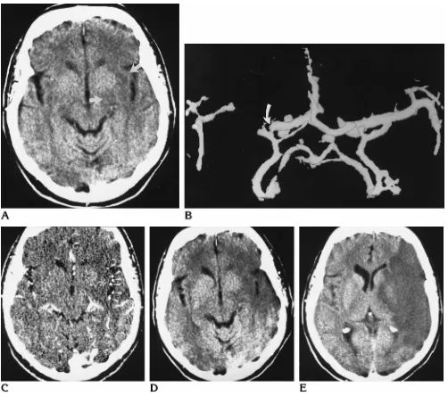
Fig 5. A 69-year-old woman with MCA trunk occlusion.
A, Baseline CT study 5 hours after symptom onset already shows hypodensity of the left insular cortex and the posterior part of the lentiform nucleus (arrows).
B, CT angiographic 3-D reconstruction shows a trunk occlusion of the left MCA (arrow).
C, CT angiographic source image shows a circumscribed area of no parenchymal enhancement (asterisk). The collateral blood supply in this case was rated as “good” (arrowheads).
D, Follow-up CT scan 1 day later shows an infarction exactly matching the area of missing parenchymal enhancement on CT angiogram.
conventional CT and CT angiography together may provide a rational basis by which to choose the optimal treatment for patients with acute stroke.
Acknowledgment
We thank Kathryn L. Allen for her careful review of the manuscript.
References
1. Tomura N, Uemura K, Inugami A, Fujita H, Higano S, Shishido F. Early CT finding in cerebral infarction.Radiology1988;168:463– 467
2. Truwit CL, Barkovich AJ, Gean-Marton A, Hibri N, Norman D. Loss of the insular ribbon: another early CT sign of acute middle cerebral artery infarction.Radiology1990;176:801– 806 3. Bozzao L, Bastianello S, Fantozzi LM, Angeloni U, Argentino C,
Fieschi C. Correlation of angiographic and sequential CT findings in patients with evolving cerebral infarction.AJNR Am J Neurora-diol1989;10:1215–1222
4. Horowitz SH, Zito JL, Donnarumma R, Patel M, Alvir J. Computed tomographic-angiographic findings within the first five hours of cerebral infarction.Stroke1991;22:1245–1253
5. von Kummer R, Meyding-Lamade´ U, Forsting M, et al. Sensitivity and prognostic value of early computed tomography in middle cerebral artery trunk occlusion.AJNR Am J Neuroradiol1994;15: 9 –15
6. von Kummer R, Nolte PN, Schnittger H, Thron A, Ringelstein EB. Detectability of hemispheric ischemic infarction by computed to-mography within 6 hours after stroke.Neuroradiology1996;38: 31–33
7. von Kummer R, Bozzao L, Manelfe C. Early CT Diagnosis of Hemispheric Brain Infarction.Heidelberg, Germany: Springer-Ver-lag; 1995
8. Bastianello S, Pierallini A, Colonnese C, et al. Hyperdense middle cerebral artery CT sign: comparison with angiography in the acute phase of ischemic supratentorial infarction.Neuroradiology1991; 33:207–211
9. Tomsick T, Brott T, Barsan W, Broderick J, Haley EC, Spilker J. Thrombus localization with emergency cerebral CT.AJNR Am J Neuroradiol1992;13:257–263
10. Leys D, Pruvo JP, Godefroy O, Rondepierre P, Leclerc X. Preva-lence and significance of hyperdense middle cerebral artery in acute stroke.Stroke1992;23:317–324
11. Landis JR, Koch GG. The measurement of observer agreement for categorical data.Biometrics1977;86:974 –977
12. Hacke W, Kaste M, Fieschi C, et al. Safety and efficacy of intra-venous thrombolysis with a recombinant tissue plasminogen ac-tivator in the treatment of acute hemispheric stroke.JAMA1995; 274:1017–1025
13. NINDS Stroke Study Group. Tissue plasminogen activator for acute ischemic stroke.N Engl J Med1995;333:1581–1587 14. Warach S, Dashe JF, Edelman RR. Clinical outcome in ischemic
stroke predicted by early diffusion-weighted and perfusion mag-netic resonance imaging: a preliminary analysis.J Cereb Blood Flow Metab1996;16:53–59
15. Napel S, Marks MP, Rubin GD, et al. CT angiography with spiral CT and maximum intensity projection.Radiology1992;185:607– 610
16. Dillon HE, VanLeeuwen MS, Fernandez MA, Mali WP. Spiral CT angiography.AJR Am J Roentgenol1993;160:1273–1278 17. Alberico RA, Patel M, Casey SO, Jacobs B, Maguire W, Decker R.
Evaluation of the circle of Willis with three-dimensional CT an-giography in patients with suspected intracranial aneurysms.
AJNR Am J Neuroradiol1995;16:1571–1578
18. Casey SO, Alberico RA, Patel M, et al. Cerebral CT venography.
Radiology1996;198:163–170
19. Pullicino P, Kendall BE. Contrast enhancement in ischaemic le-sions. I. Relationship to prognosis.Neuroradiology1980;19:235– 239
20. Saito I, Segawa H, Shiokawa Y, Taniguchi M, Tsutsumi K. Middle cerebral artery occlusion: correlation of computed tomography with clinical outcome.Stroke1987;18:863– 868
21. Furlan AJ. Natural history of atherothrombotic occlusion of cere-bral arteries: carotid versus vertebrobasilar territories. In: Hacke W, del Zoppo GJ, Hirschberg M, eds.Thrombolytic Therapy in Acute Ischemic Stroke. Heidelberg, Germany: Springer-Verlag; 1991:3– 8
22. von Kummer R, Holle R, Rosin L, Forsting M, Hacke W. Does arterial recanalization improve outcome in carotid territory stroke?Stroke1995;26:581–587
23. Jansen O, von Kummer R, Forsting M, Hacke W, Sartor K. Throm-bolytic therapy in acute occlusion of the intracranial carotid artery bifurcation.AJNR Am J Neuroradiol1995;16:1977–1986 24. Brandt T, von Kummer R, Mu¨ller-Ku¨ppers M, Hacke W.
Throm-bolytic therapy of acute basilar artery occlusion: variables affect-ing recanalization and outcome.Stroke1996;27:875– 881 25. Bozzao L, Fantozzi LM, Bastianello S, Bozzao A, Fieschi C. Early
collateral blood supply and late parenchymal brain damage in patients with middle cerebral artery occlusion.Stroke1989;20: 735–740
26. von Kummer R, Forsting M. Effects of recanalization and collat-eral blood supply on infarct extent and brain edema after middle cerebral artery occlusion.Cerebrovasc Dis1993;3:252–255