It is known that young Xenopus laevis tadpoles will swim in response to a sudden decrease in light intensity and that this response is mediated by the pineal eye (Roberts, 1978; Foster and Roberts, 1982). However, the normal role of this pineal dimming response in the behaviour and ecology of the tadpole is not yet clear. The first role suggested was as an escape reaction in which the tadpoles respond to shadows cast by predators. Such responses are seen in fish larvae, which respond to shadows by swimming downwards (Blaxter, 1968, 1969; Chamalbert et al., 1991; Burke et al., 1995; Forward et al., 1996), and similar responses are seen in crustacean larvae (Forward, 1974, 1976, 1977, 1986). Foster and Roberts (1982) considered a role in escape unlikely because the probability of tadpoles responding to light dimming was low. Other possible functions include an increase in spontaneous movement at low light levels (possibly leading to tadpoles congregating in more brightly lit regions), an increase in the strength of responses to other types of stimulus at low light levels, e.g. the tadpoles might swim more vigorously when touched in the dark, and an influence on the distribution of tadpoles that could vary with changes in light levels.
Unfortunately, there is little information on the distribution or responses of young tadpoles under natural conditions. However, in our breeding colony, tadpoles are raised from eggs in tanks of water and after hatching tend to be found attached
by their cement gland close to the water surface. A similar distribution was observed in deeper tanks by Bles (1905), who reported that tadpoles tended to swim in an upward direction, a response that he attributed to a weak positive heliotropism. Roberts et al. (2000) report similar observations, suggesting that nearly vertical upward swimming could be a stable form of swimming in the late embryo and hatchling tadpole. Thus, it seems important to determine whether light levels, detected by the pineal eye, could influence upward swimming behaviour.
This paper presents the results of a series of experiments designed to test the effects of changes in light levels on the swimming behaviour of young Xenopus laevis tadpoles. Physiological experiments link these behavioural responses to neuronal activity in the pineal eye and its influence on the central nervous circuits controlling swimming.
Materials and methods
Unless specified otherwise, all tadpoles of Xenopus laevis (Daudin) used were at stage 37/38 of development (Nieuwkoop and Faber, 1956) and were taken from a captive breeding colony. Only general methods are given here, and details of individual experiments are given briefly in the Results section. Some experiments involved pinealectomy or removal of the Printed in Great Britain © The Company of Biologists Limited 2000
JEB2628
When the light is dimmed, the pineal eye of hatchling Xenopus laevis tadpoles excites the central pattern generator for swimming, but the behavioural significance of pineal excitation is unclear. We show that tadpoles spend 99 % of their time hanging from the surface meniscus or solid objects using mucus secreted by a cement gland on the head. Attachment inhibits swimming, but unattached tadpoles swim spontaneously. Provided that their pineal eye is intact, they attach closer to the water surface in the dark than in the light and attach preferentially to the underside of floating objects that cast shadows. Dimming causes tadpoles swimming horizontally to turn upwards and is very effective in initiating upward swimming in unattached tadpoles. Similar pineal-dependent responses
during swimming are present up to stage 44. Pinealectomy blocks responses to dimming at all stages. Recordings from immobilised tadpoles reveal that light dimming induces faster fictive swimming and that pineal activity is increased for up to 20 min during sustained light dimming. We suggest that the increase in pineal discharge during dimming increases the probability of upward swimming and, in this way, increases the probability of tadpoles attaching to objects higher in the water column that cast shadows.
Key words: pineal eye, Xenopus laevis, photoreception, tadpole, swimming.
Summary
Introduction
RESPONSES OF YOUNG XENOPUS LAEVIS TADPOLES TO LIGHT DIMMING:
POSSIBLE ROLES FOR THE PINEAL EYE
D. JAMIESON ANDALAN ROBERTS*
School of Biological Sciences, University of Bristol, Bristol BS8 1UG, UK
*Author for correspondence (e-mail: A.Roberts@bristol. ac.uk)
lateral eyes. Dissections were carried out in saline (composition in mmol l−1): NaCl, 115; KCl, 3; CaCl
2, 2;
MgCl2, 1; NaHCO3, 2.4; Hepes, 10, at pH 7.4) on tadpoles
anaesthetized by exposure to 0.1 % MS-222. Tadpoles were pinned out on a rotatable Sylgard base, and the sense organs were removed after making an opening in the skin using fine mounted tungsten needles. After dissection, the tadpoles were placed in 50 % saline, which encourages healing. They were examined after at least 2 h to ensure that the skin was completely healed and that they still swam strongly in response to touch. For experiments in which the pineal or lateral eyes were removed, sham-operated tadpoles were used as controls; in these sham operations, the same dissections were performed but without removing the sense organs.
All behavioural experiments were performed using non-reflective black containers, with the exception of experiments involving video analysis and observations of upward movement, for which glass tanks were used. In behavioural experiments, standard 60 W incandescent light bulbs were used to provide even illumination from above the containers. In physiological experiments, an Intralux-Volpi fibre-optic light source with a manually operated iris was used to provide dimmings. The level of pre- and post-dimming illumination was set by the degree of opening of the iris and the distance from the tip of the fibre-optic cable to the tadpole. In all experiments, there was an interval of at least 2 min between trials. Photon irradiance was measured using a Cor Li-190SA quantum sensor (1µmol m−2s−1; approximately 54 lx).
Bright sunlight is approximately 2000µmol m−2s−1.
A rigidly mounted Sony handycam (CCD-TR303E) with a frame rate of 25 frames s−1was used for video analysis. The
video tape was either played back on a conventional television monitor for spontaneous activity experiments or analysed via a Sony Nicam video cassette player (EV-S9000E). The public domain NIH Image 1.60/ppc software (written by Wayne Rasband, US National Institute of Health) was used to map swimming paths. When mapping swimming routes, the camera was placed 1.93 m above the tank to reduce parallax. Mapping was performed by recording the x and y coordinates of the tadpole’s head every 15 frames for the overhead views. In the side-on view, the tadpole’s position was mapped every five frames.
Physiological experiments were carried out in a bath continuously perfused with saline. Impulse activity was recorded using glass suction electrodes filled with saline. Details of ventral root recording techniques are given by Perrins and Roberts (1995), and pineal recording techniques are described by Foster and Roberts (1982) and Jamieson (1997). Pineal activity or swimming episodes were recorded on video tape and digitised using a CED 1401 interface. Analysis was carried out using Signal Averager software (version 6.30, CED). The spike rate of pineal activity and the cycle period of ventral root activity were displayed using an analogue period meter constructed by Dr S. R. Soffe. All values given in the Results section use the standard error of the mean (S.E.M.). The temperature at which experiments was
performed was 18–22 °C. Data were analysed statistically using χ2-tests and Student’s t-tests.
Results Behaviour
Observations on swimming behaviour and the effects of daily light/dark cycles
To provide a general picture of the behaviour of the tadpoles, we made long-term video recordings of the tadpoles to determine how much time they spent attached by mucus or making spontaneous movements and whether these varied with cyclical changes in light levels. Twenty stage 35/36 tadpoles raised from eggs and kept in a 12 h:12 h light/dark cycle were transferred to a tray (23 cm×23.5 cm, 4 cm deep water) in a darkroom where the light cycle was maintained. The high light level (24µmol m−2s−1) was provided by fluorescent strip lights
and an overhead incandescent light; the low light level (0.1µmol m−2s−1) was provided by a red photographic safe
light. An overhead video camera recorded the movements of the tadpoles. A remote control connected to a timer was used to switch the camera on for 2 min in every 8 min. In this way, the tadpoles could be filmed for 12 h using one 3 h tape, providing ‘snapshots’ of activity without any interference from the experimenter. The tadpoles were filmed for 48 h, and they had reached stage 41 by the end of this period. This procedure was repeated on three groups of 20 tadpoles from different parents.
All tadpoles began each trial near the centre on the bottom of the container. Direct observations showed that after 30 min all tadpoles had swum spontaneously and attached by their cement glands either to the side of the container or to the water surface. Whilst attached, tadpoles exhibited no spontaneous movement. Only when the cement gland became detached, and the tadpoles sank, did they respond by swimming until reattachment occurred. Analysis of the video recordings showed that swimming was infrequent, averaging only 3.6±0.3 movements tadpole−1h−1over all three groups (N=60
tadpoles) for the first 12 h in the high-level light. Swimming usually lasted only a few seconds until the tadpole reattached. Movements were no more common in the dark, where the average for all groups was 3.5±0.4 movements tadpole−1h−1
(P>0.05, t-test) during the first 12 h dark period. Over the second 12 h light period, the numbers of movements were slightly higher, with an average of 4.4±0.3 movements tadpole−1h−1, but there was still no significant increase in the
second dark period (4.6±0.3 movements tadpole−1h−1, t-test,
P>0.05). No overall difference was observed in the frequency of swimming of individual tadpoles. However, attachment to the surface of the water was less secure than attachment to the solid side of the tray. Tadpoles attached to the surface of the water became detached more frequently (5.5±0.3 detachments tadpole−1h−1) than those attached to the
side (1.8±0.3 detachments tadpole−1h−1; t-test, P<0.001).
occurred, was measured in each of the three groups of tadpoles using the 10 unattached periods occurring closest to the middle of the first 12 h light and dark periods. The mean time unattached was 8.3±2.9 s (N=60). Swimming and reattachment normally occur quickly, but in six cases there was a substantial delay (the longest being 61 s) between the loss of cement gland attachment and reattachment, when the tadpole lay inactive on the bottom of the tray. Thus, with mean number of movements per hour ranging from 3.5 to 4.6 over the 48 h, and since the unattached period averaged 8.3 s, the tadpoles between stages 35/36 and 41 spent more than 99 % of their time attached (29–38 s h−1detached).
We conclude that tadpoles show virtually no spontaneous movement whilst attached by mucus from their cement glands. Swimming occurs only when the attachment breaks and usually leads to rapid reattachment, so tadpoles spend nearly all their time attached. Light/dark cycles make no difference to the probability of movement, which depends mainly on the probability of the mucus strand from which the tadpole hangs breaking. The lack of spontaneous movement after cement gland attachment raised the question of the possible effects of cement gland inhibition on any activity evoked by dimming.
Does cement gland attachment inhibit the dimming response?
Previous work has shown that stimulation of the cement gland reduces a tadpole’s responsiveness to touch stimuli (Roberts and Blight, 1975). We therefore tested whether tadpoles were less likely to swim in response to light dimming when attached by their cement gland than when they were unattached. Individual tadpoles were placed by pipette on the bottom of a tank so that the mucus from the cement gland was not attached, or they were allowed to attach naturally by swimming to the side or the surface of the tank. Each tadpole was tested once while attached and once while unattached to determine whether it swam in response to dimming (from 58–0.3µmol m−2s−1). There was a 2 min rest between trials,
and the order of the trials was randomized.
The results were clear. Swimming was evoked in only three out of 37 attached tadpoles but in 32 out of 37 unattached tadpoles (χ2=50.0, P<0.0001). We conclude that cement gland
attachment inhibits the swimming response to dimming. This makes it unlikely that the pineal eye is involved in the initiation of swimming when tadpoles are attached.
Is the vertical distribution of tadpoles influenced by light levels?
The observations described above indicate that tadpoles have to be unattached before light levels can influence them. Since tadpoles often swim upwards from the substratum either spontaneously or when stimulated (Bles, 1905; Roberts et al., 2000), one possible function of the pineal eye could be to affect the vertical distribution of unattached tadpoles. In preliminary observations on vertical distribution, groups of 20 tadpoles were released in the centre of a 25 cm diameter circular container with 10 cm deep water illuminated from above (46µmol m−2s−1). Tadpoles were allowed to adapt for 10 min,
and the water was then stirred to draw the tadpoles into the centre and onto the bottom so that they all started unattached. They were then left for 2 h to allow time for spontaneous swimming and redistribution to occur. The numbers of tadpoles in the bottom half (on the bottom or attached to the sides at depths of 5–10 cm) and in the top half (attached to the surface or sides at depths of 0–5 cm) of the container were then counted. Most tadpoles had moved into the top half of the water column (N=60 tadpoles, 46 in the top half after 2 h, significantly different from equal distribution, χ2=9.187,
P=0.005).
To determine whether the pineal eye played a role in this tendency to distribute upwards, groups of 20 sham-operated and 20 pinealectomized tadpoles were tested. Each group was placed in a 27 cm diameter circular container with 22 cm deep water illuminated from above (46µmol m−2s−1). A line around
the inside of the containers at 11 cm divided them into top and bottom halves. Tadpoles were allowed to adapt for 10 min, and the water was then stirred to draw the tadpoles into the centre and onto the bottom so that they all started unattached. After 15 min, most tadpoles will have swum spontaneously to redistribute themselves, so the number of tadpoles in the top half of the container was counted. The light was then dimmed (to 0.1µmol m−2s−1) and after a 10 min adaptation period the
water was stirred again to detach the tadpoles and return them to the bottom. The tadpoles were then left in dim light for another 15 min, after which the light level was raised and the tadpoles were immediately counted. Trials were performed on 120 sham-operated and 120 pinealectomized tadpoles.
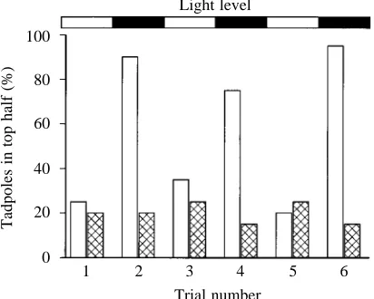
Fig. 1 shows results from one experiment in which the light/dark cycle was repeated three times to illustrate the differences in distribution between sham-operated and pinealectomized tadpoles. The group data show that more sham-operated tadpoles are found in the top half of the container in the dark (89 out of 120) than in the light (31 out of 120). This differs significantly from equal distribution (χ2=11.82, P=0.002). For the pinealectomized tadpoles, the
numbers in the top half in the dark (22 out of 120) did not differ from those in the light (19 out of 120; χ2=0.265, P=0.876).
There was no significant difference between the number of sham-operated tadpoles found in the top half in the light (31 of 120) and the number of pinealectomized tadpoles in the top half in the light (19 out of 120, χ2=3.638, P=0.162) or in the
dark (22 out of 120, χ2=1.961, P=0.375). This rules out the
possibility that the pinealectomized tadpoles might not be getting into the top half simply because they are poorer swimmers than the sham-operated tadpoles.
We conclude that reduced light levels led to unattached tadpoles attaching higher in the water column and that this depends on an intact pineal eye.
Do shadows cast by objects on the water’s surface affect the distribution of tadpoles?
object casting a shadow in the water could influence the distribution of tadpoles.
To test this, groups of 40 tadpoles were placed in 10 cm deep water in a tank 29 cm×29.5 cm (Fig. 2Ai,ii). Two flat pieces of plastic (12 cm×12 cm), one clear and one black opaque, were floated on the surface and held in place by thread attached to the side of the container. A single overhead light source (measured at 12µmol m−2s−1in the centre of the tank) was
positioned 40 cm above the surface to reduce parallax effects so that the black plastic cast a shadow onto the bottom of the container (1µmol m−2s−1beneath the centre of the plastic) that
did not spread beneath the clear plastic. The clear plastic cast minimal shadow (11µmol m−2s−1). In each trial, the water was
stirred to disturb the tadpoles and draw them into the centre. After 15 min, the numbers of tadpoles attached to the water surface, the tank and the underside of the clear plastic were counted so that the number attached to the underside of the black plastic could be deduced. In control trials, the black plastic was replaced by a second piece of clear plastic before stirring, and the numbers of tadpoles beneath both pieces of plastic were counted. Both the experiment and the control were performed on five groups of 40 tadpoles (N=200 tadpoles in total). The positions of the black and the clear plastic were switched randomly, and the direction of stirring was also randomized. Tadpoles attached to the side of any piece of plastic were not counted because they could have got there as a result of drift if attached to the water surface.
In all the tests, significantly more tadpoles attached to the underside of the black plastic (Fig. 2B). The number of tadpoles beneath the black plastic was 55 out of 200 compared
with eight out of 200 beneath the clear plastic (χ2=41.618,
PⰆ0.0001). There was no significant difference in the numbers attached to the two pieces of clear plastic in the control experiment (12 and 9 out of 200, χ2=0.429, P=0.807). The
fact that the shadow from the black plastic only covers approximately one-quarter of the bottom surface of the container means that many tadpoles would not encounter the shadow whilst swimming, which probably explains why most tadpoles are found attached to the sides of the tank or to the water surface.
We conclude that tadpoles attach preferentially to objects on the water’s surface that cast shadows.
How does dimming influence swimming pathways?
To determine what happens when tadpoles swim into shadow, they were video-recorded from the side as they swam horizontally in 23 cm deep water in a glass tank (10 cm×10 cm). They were illuminated by two light sources;
100
80
60
40
20
0
Tadpoles in top half (%)
1 2 3 4 5 6
[image:4.609.64.270.75.240.2]Trial number Light level
Fig. 1. Effects of pinealectomy on the vertical distribution of tadpoles at high and low ambient light levels. Results from the first six trials in a single experiment on 20 sham-operated and 20 pinealectomized tadpoles showing the percentage of tadpoles found in the top half of the container 15 min after they had been detached by stirring. Tadpoles were subjected to alternate trials at high and low light levels as indicated by the top bar. In this case, 75–95 % of the sham-operated tadpoles (open columns) were found in the top half in the low light level compared with 20–35 % at the high light level. Pinealectomy (hatched columns) abolishes the differences in distribution between high and low ambient light levels.
15
12
9
6
3
0
Number of tadpoles
Black Clear Clear Clear Experiment Control
Ai
Aii
B
Shadow cast by black plastic Black plastic
[image:4.609.321.551.78.397.2]Clear plastic
one was turned off to provide dimming from 31µmol m−2s−1
to 4 nmol m−2s−1. In every case (three tests each on 11
tadpoles), the tadpoles turned after dimming to swim upwards until they reached the surface, where they attached by their cement glands (Fig. 3). The orientation of the light source made no difference to this response: even with the light coming through the bottom of the tank, the tadpoles still swam upwards after dimming.
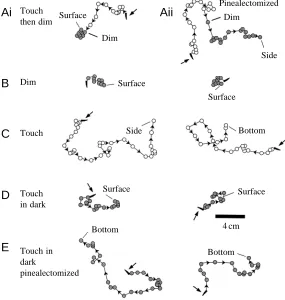
To determine whether this upward swimming depended on the pineal eye, the reactions of 10 sham-operated and 10 pinealectomized tadpoles to dimming and to touch were video-recorded from above (Fig. 4). Individual tadpoles were placed centrally on the bottom of a glass tank (35.5 cm×28.5 cm) in 23 cm deep water. They were then stimulated, and each test was repeated three times on each tadpole. To compare responses, we measured the maximum horizontal deviation during swimming from the tadpole’s initial position and the proportion of responses in which the tadpole reached the water’s surface rather than hitting the sides or the bottom of the tank. Tadpoles that are swimming more vertically would be expected to show less horizontal deviation and be more likely to reach the surface. Light sources similar to those used in the side-on video recordings were used to give dimmings from 24µmol m−2s−1to 5 nmol m−2s−1.
Examples of typical swimming paths seen from above are shown in Fig. 4. When swimming was initiated by touch with a fine hair, all sham-operated tadpoles turned to swim upwards when the light was dimmed during swimming (Fig. 4Ai), 4 cm
Dim Dim
Dim
Dim Water surface
Pinealectomized Dim
Side
Surface
Bottom
Surface
Bottom Surface
Dim
Dim
Surface
Touch Side
Surface
Bottom Touch
then dim
Touch in dark
Touch in dark
pinealectomized
4 cm
Ai
Aii
B
C
D
[image:5.609.52.295.74.309.2]E
Fig. 3. Responses of tadpoles to light dimming during horizontal swimming. Plots from video recordings made through the side of a glass tank showing swimming paths for four different tadpoles. Circles represent the position of the tadpole’s head every 0.2 s, the open circles before dimming and the filled circles after dimming. In each case, the tadpole turns to swim upwards after dimming and continues swimming upwards until it reaches the surface.
[image:5.609.280.565.441.741.2]and most reached the surface (26 out of 30 trials). After pinealectomy, dimming had no apparent effect on swimming (Fig. 4Aii) and fewer tadpoles reached the surface than after sham operations (4 out of 30; χ2=32.3, P<0.001). Together
with the previous observations on the effects of dimming during swimming (Fig. 3), this experiment suggests that the reliable upward swimming following dimming is a response mediated by the pineal eye. As expected, dimming evoked no upward turning responses in the pinealectomized tadpoles.
To extend these observations, we compared the swimming responses of sham-operated tadpoles to dimming (Fig. 4B) and touch stimuli (Fig. 4C). After dimming, more tadpoles reached the surface (30 compared with six, χ2=40, P<0.001)
and swimming was more vertical, so the horizontal deviation measurement was less (6.1±0.4 cm compared with 71.9±3.9 cm, t-test, P<0.001). The time taken to reach the
surface after dimming was 10.1±0.2 s (N=30). When tadpoles were touched in dim conditions after a 10 min adaptation period at the low light level (Fig. 4D), more reached the surface than in the high light level (17 compared with 6, χ2=8.5, P<0.01) and the horizontal deviation was reduced
(48.1±2.2 cm compared with 71.9±3.9 cm, t-test, P<0.05). After pinealectomy, the tadpoles’ response to touch was similar after adaptation to the light or dim condition (Fig. 4E). Few reached the surface (four in light and two in dim conditions), and the horizontal deviations were similar under both conditions (47.4±4.8 cm in light and 55.6±3.9 cm in dim conditions, t-test, P>0.05). However, in many cases, pinealectomized tadpoles hit the bottom of the tank soon after the start of swimming, which limited their horizontal deviation.
Since the pineal eye is excited by decreases in light levels (Roberts, 1978; Foster and Roberts, 1982), these experiments suggest (i) that pineal excitation can lead directly to reliable upward swimming, and (ii) that pineal excitation can also
increase the probability of upward swimming initiated by touch in tadpoles under low light conditions. We therefore used physiological recording to investigate whether these conclusions are compatible with the responses of pineal ganglion cells to dimming.
Physiology
Can pineal ganglion cell activity account for the behavioural responses to dimming?
Pineal ganglion cells exhibit continuous low-frequency background activity at constant light levels that can be recorded using suction electrodes applied to the caudal surface of the pineal eye (Fig. 5) (Foster and Roberts, 1982; Jamieson, 1997). Following dimming, the ganglion cells respond with a burst of impulse activity after a 40–50 ms delay. This ‘off-response’ grades with the level of the dimming, and with extended dimming the level of activity decreases as the pineal adapts. In immobilized tadpoles, the off-response is commonly followed by fictive swimming. Recordings of pineal ganglion cell activity were made in five tadpoles, and the light was dimmed by a similar amount as in the behavioural experiments (from 28µmol m−2s−1 to 2 nmol m−2s−1 for 20 min).
Fig. 5A–D shows that the adapted background discharge is slightly higher at the lower light level (Fig. 5C). Fig. 5E shows increased pineal activity following dimming compared with the previous background discharge for each of the five tadpoles. These increases are plotted as a proportion of the initial off-response after subtraction of the mean rate before the light was dimmed. This allows comparison of multiple unit recordings from different tadpoles with different overall levels of activity.
It was clear from pineal recordings (not illustrated) that there is a marked increase in pineal discharge for the 10 s that tadpoles needed to swim to the surface in the previous experiments. However, after 10 min of dark adaptation,
1
0.8
0.6
0.4
0.2
0
Pineal activity
0 5 10 15 20
250 ms
Time (min)
A
B
[image:6.609.295.558.71.309.2]E
C
D
Fig. 5. Sustained pineal ganglion cell activity in response to prolonged dimming (from 28µmol m−2s−1 to 2 nmol m−2s−1 at
tadpoles still tended to swim more vertically in response to touch than they did in the light (Fig. 4D). Is there a maintained pineal discharge to account for this? As Fig. 5 shows, 10–20 min after dimming, the pineal discharge was still higher than before dimming. From 10 to 15 min after dimming, the mean pineal discharge frequency was 170.5±18.9 % of the pre-dimming control level (N=5, t-test, P<0.001). From 15 to 20 min after dimming, the frequency was still increased at 173±32 % of the pre-dimming control value (N=5, t-test,
P>0.05).
These recordings suggest that for periods of many minutes following dimming the pineal eye could provide continuous increased excitatory input to the spinal cord circuitry controlling swimming.
Does dimming activate swimming more strongly than touch?
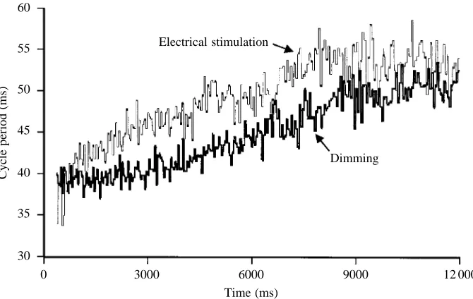
The behavioural experiments suggested that dimming leads to more reliable upward swimming than touch stimulation. We therefore recorded from trunk ventral roots supplying the swimming muscles in immobilised tadpoles and compared fictive swimming in response to dimming (from 28µmol m−2s−1to 4 nmol m−2s−1) and to a 1 ms current pulse
from an electrode on the tadpole’s skin to stimulate the touch-sensitive nerve endings in the skin (see Roberts, 1990). Fig. 6 shows an example from one tadpole in which the cycle period of ventral root activity for the first 10 s of a swimming episode is shorter following dimming than following skin stimulation. When the number of ventral root bursts over a 10 s period from 0.5 to 10.5 s after the start of fictive swimming was counted, skin-stimulation-evoked episodes averaged 197±3.4 bursts whereas dimming-evoked episodes averaged 210±3.3 (P<0.001, t-test, 30 trials in 15 tadpoles), an increase of approximately 7.5 %.
What are the effects of dimming during swimming in immobilized tadpoles?
For comparison with behavioural experiments in which
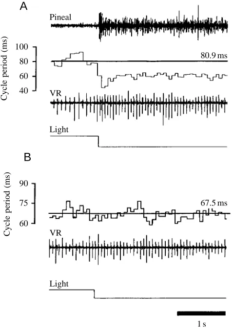
dimming was applied during swimming, we also looked at the effects of dimming during fictive swimming. Recordings were made simultaneously from the pineal eye and from the ventral roots of intact tadpoles and of pinealectomized preparations. Fictive swimming was evoked by electrical skin stimulation, and the light was dimmed after 40 s (Fig. 7A). A clear pineal off-response can be seen, and this is followed by a decrease in the cycle period of the ventral root recording. For 24 trials in 12 tadpoles, this decrease was measured by comparing the number of bursts over 1 s of swimming activity before dimming with that of a 1 s period starting 100 ms after the dimming. The mean number of ventral root bursts increased from 15.8±0.4 to 18.9±0.4 (t-test, P<0.0001). Pinealectomy prevented this acceleration (Fig. 7B), and in 18 trials on nine pinealectomized tadpoles the number of bursts before and after dimming remained unchanged (16.3±0.3 before and 16.4±0.4 after, P>0.05).
Responses of older tadpoles to dimming
Foster and Roberts (1982) demonstrated that the effectiveness of dimming in initiating swimming in detached tadpoles declined to less than 20 % by stage 41. Since the most significant effect of dimming may be to increase the frequency of swimming and evoke upward turns, we have re-examined the responses of older tadpoles from stages 41 to determine whether they turned upwards in response to dimming (from 28.72µmol m−2s−1to 5.13 nmol m−2s−1). At
stage 41, five sham-operated tadpoles and six tadpoles that had had both lateral eyes removed were each tested three times and exhibited a reliable upward turn in response to dimming applied during touch-evoked swimming. At stage 41, pinealectomy in six tadpoles abolished the upward turning response to dimming given during touch-initiated swimming, and similar results were obtained in six tadpoles that were both pinealectomized and had their lateral eyes removed. At stage 44, tadpoles swim continuously, so to separate ‘spontaneous’ upward movements from those caused 60
55
50
45
40
35
30
Cycle period (ms)
0 3000 6000 9000
Dimming Electrical stimulation
[image:7.609.229.564.73.285.2]12 000 Time (ms)
by dimming, tadpoles were observed for 10 s prior to dimming to ensure that no upward movement was occurring; the light was then dimmed, and we recorded whether the tadpole moved upwards during the next 10 s. Twenty tadpoles were tested three times each, and clear upward movement was found in 51 out of 60 dimming trials compared with only five out of 60 control trials without dimming (N=60, χ2=70.9,
P<0.001). None of the six tadpoles pinealectomized at stage
41 responded to dimming at stage 44, but six tadpoles sham-operated at stage 41 and six that had only their lateral eyes removed all responded to dimming by turning upwards when tested at stage 44. These observations suggest that pineal-mediated upward turning responses to light dimming may persist well into larval life.
Discussion
The present study is concerned with the behaviour of tadpoles of Xenopus laevis in the first day or two of their life after hatching. We have shown that during this period they spend 99 % of their time hanging from a mucus strand. In this state, they do not respond to changes in light level, but the mucus tends to break so, on average, they become unattached for very short periods 4–5 times per hour. When not attached, they sink in the water, can reach the bottom, tend to swim spontaneously, and respond to light dimming by swimming upwards. Tadpoles also turn to swim upwards as they swim horizontally into shadows cast by solid objects above them in the water. The tadpole’s vertical distribution is also dependent on light levels, and they tend to move upwards in the water column in the dark. During this period of development, all reactions to dimming depend on the pineal eye. Our physiological results demonstrate that the pineal discharge is increased for long periods when light levels are decreased and that the increase in pineal discharge is accompanied by an increase in swimming frequency. These observations do not explain how dimming and pineal excitation lead to upward swimming, but evidence is available in the companion paper (Roberts et al., 2000).
Roberts et al. (2000) have examined swimming responses in hatchling Xenopus laevis between stages 32 and 37/38. They show that during horizontal swimming the body is normally dorsal-side-up, but during upward swimming the body spirals about its long axis. Upward swimming is a common response and does not depend on vestibular reflexes mediated by the inner ears. The tadpole’s responses are compared with those of a simple mathematical model, and it is argued that during swimming dense yolk in the tadpole’s belly ballasts the body in a ventral-down position while two torques are generated. One acts to rotate the body about its long axis in the roll plane, while the other acts to raise the head in the pitch plane. During horizontal swimming, the ballast is sufficient to counter the torque acting in the roll plane, so the tadpole swims belly-down. However, if the pitch torque raises the head enough, this is no longer the case and the tadpole spirals as it swims upwards. Direct measurements of the tail position during swimming in tethered tadpoles showed that the body flexed dorsally when the light was dimmed and that dorsal flexion increased when swimming frequency increased. From these observations, it was concluded that the torque that raises the head could result from this dorsal body flexion at higher swimming frequencies. Since pineal excitation leads to increased swimming frequency, this mechanism could explain how tadpoles turn upwards when they swim into shadows (Fig. 3).
[image:8.609.52.292.72.412.2]Upward swimming into shadows does not seem appropriate as a predator escape reaction. It is in the opposite direction to the downward swimming response seen in fish larvae (Blaxter, 1968, 1969; Chamalbert et al., 1991; Burke et al., 1995; Forward et al., 1996). It is more likely that the pineal eye is contributing to the control of the vertical distribution of the tadpoles. Under natural conditions, young Xenopus laevis Fig. 7. Effects of dimming during fictive swimming in immobolised
tadpoles on cycle period. (A) In intact tadpoles, the cycle period during swimming (80.9±0.7 ms) decreased (to 58.5±0.2 ms) in response to dimming. Values are means ±S.E.M. (N=12). The top trace shows the off-response recorded from the pineal eye, below that is a plot showing the cycle period of ventral root activity. The next trace down shows ventral root activity (VR) and the bottom trace, in which downward deflection indicates dimming, indicates light level. The horizontal line in the cycle period plot shows the mean cycle period before dimming. (B) This decrease in cycle period does not occur in pinealectomized tadpoles, in which there is no change in mean cycle period after dimming (67.5±0.6 ms before and 67.7±0.6 ms after).
100 80
60 40
80.9 ms
Cycle period (ms)
Pineal
VR
Light
90
75
60
67.5 ms
1 s
Cycle period (ms) VR
Light
A
tadpoles could become unattached often enough for low light levels to affect their vertical distribution. We found that detached tadpoles generally reattached closer to the surface in the dark than in the light (Fig. 1). Perhaps this response is some form of vertical migration. Many aquatic organisms, including other amphibian larvae, move closer to the surface at night (diel vertical migration; Anderson and Graham, 1967; Anderson and Williamson, 1974; Holomuzki and Collins, 1983; Griffiths et al., 1988). The adaptive significance of vertical migration is thought to include predator avoidance (Zaret and Suffern, 1976; Stich and Lampart, 1981), maximisation of food intake (Haney, 1988; Lampert, 1989) or optimisation of some environmental variable such as temperature (McLaren, 1974; Enright, 1977; Dawidowicz and Loose, 1992; Williamson et al., 1996) or solar radiation (Huntsman, 1924; Williamson et al., 1996). The young tadpoles do not feed but they are in danger from predators, particularly cannibalistic adults (Leslie, 1890; McCoid and Fritts, 1980a,b; Schoonbee et al., 1992). Adult Xenopus laevis are nocturnal and feed largely on zooplankton and benthic invertebrates. Could moving closer to the surface reduce a tadpole’s chances of being eaten? Salamander larvae have been shown to alter their pattern of vertical distribution in response to predation by diving beetles (Holomuzki, 1986). Temperature has also been shown to play a part in vertical migrations of amphibian larvae (Beiswenger, 1977; Griffiths et al., 1988).
The effect of shadows cast by objects close to the water’s surface on the way tadpoles distribute suggests another possible role for the pineal eye. The upward turn in response to dimming applied while the tadpole is swimming (Fig. 3) mimics the tadpole moving underneath a shadow-casting object. Since attachment to the water’s surface is weaker than to a solid surface, the shadow response could increase the probability of tadpoles forming a secure attachment to solid objects such as plants. These may provide concealment from predators and protection from ultraviolet radiation. A similar function was proposed for the shadow response in larvae of the marine ascidian Botryllus schlosseri (Woodbridge, 1924; Grave and Woodbridge, 1924). The suggestion was that larvae swept underneath blades of eel grass would be stimulated to swim upwards by the shadow and attach to the underside of the blade. The larvae preferentially attached to the underside of eel grass blades, and this response was abolished by removing the shadow. Many ascidian larvae show upward swimming responses to shadows, and improved survival in shaded areas has also been demonstrated (Goodbody, 1963; Young and Chia, 1984), although the effect on patterns of distribution is debatable (Young and Chia, 1985).
Another way that the pineal eye may be excited is by the decrease in light intensity as tadpoles fall through turbid water after losing attachment. Algal blooms can lead to an approximately exponential attenuation in irradiance as a function of depth from the surface (Kirk, 1994). With a strong bloom, the vertical attenuation coefficient can be 3 m−1, which
leads to reductions in irradiance to 55 % at 22 cm from the
surface and to 5 % at 100 cm. Tadpoles sink 22 cm in approximately 20 s (see Roberts et al., 2000), so in this time would experience a 55 % reduction in light intensity at wavelengths around 500 nm at which the pineal has its maximum sensitivity (Foster and Roberts, 1982).
The discovery of pineal-mediated responses in stage 44 tadpoles is interesting since the pineal response was originally believed to disappear at around stage 40. This suggests that the pineal eye might still have a direct effect on behaviour after stage 45, which is when the lateral eyes become functional (Beazley et al., 1972).
Unfortunately, there is little information on the ecology of
Xenopus laevis. What little there is suggests that eggs are laid
on submerged vegetation (Balinsky, 1969; Tinsley et al., 1996). Plants such as grasses, sedges and other vegetation are common at the sides of ponds in South Africa where Xenopus
laevis tadpoles occur (A. Roberts, personal observation). Such
plants would cast shadows especially early and late in the day and could supply secure attachment points for the tadpoles.
The enhancement of touch-evoked vertical swimming responses in the dark (Fig. 4D) suggests a long-term increase in excitation in the spinal cord. Recordings from pineal ganglion cells confirmed that activity remains above pre-dimming levels for at least 20 min in the dark. The video experiments showed more direct vertical movement during touch-evoked episodes after 10 min in the dark. This is circumstantial evidence, but we can hypothesise that the higher rate of background pineal activity in the dark provides continuous input to the spinal cord, which strengthens touch-evoked vertical swimming. In contrast, tadpoles of the newt
Trituris vulgaris show no sustained elevation of pineal activity
in the dark (Roberts and Clarke, 1983). Recordings from the midbrain of Xenopus laevis tadpoles have shown that cells believed to connect the pineal ganglion cells to the hindbrain show activity similar to that of the ganglion cells (Jamieson and Roberts, 1999). Future studies may look at the neurons in the hindbrain or spinal cord to determine whether there is any evidence of continuous light-dependent input to the swim-generating systems.
ganglion cells for up to 20 min in the dark. This maintained activity could provide continuous excitatory input to the nervous system, thus enhancing the frequency of spontaneous or touch-evoked swimming responses and increasing upward movement.
We would like to thank the MRC for a studentship to D.J., Dr Steve Soffe for advice on experiments and drafts of the paper, Dillon Bright for advice on light measures, Dr Russell Foster and Dr Ray Perrins for advice on drafts and Linda Teagle and Derek Dunn for technical help.
References
Anderson, J. D. and Graham, R. E. (1967). Vertical migration and
stratification of larval Ambyostoma. Copeia 1967, 371–374.
Anderson, J. D. and Williams, G. K. (1974). Nocturnal stratification
in the larvae of the mole salamander Ambyostoma talpoideum. Herpetologica 30, 28–29.
Balinsky, B. L. (1969). The reproductive ecology of amphibians of
the Transvaal highveld. Zool. Africa 4, 37–93.
Beazley, L., Gaze, R. M. and Keating, M. J. (1972). The
appearance, during development, of responses in the optic tectum following visual stimulation of the ipsilateral eye in Xenopus laevis. Vision Res. 12, 407–410.
Beiswenger, R. E. (1977). Diel patterns of aggregative behaviour in
tadpoles of Bufo americanus in relation to light and temperature. Ecology 58, 98–108.
Blaxter, J. H. S. (1968). Visual threshold and spectral sensitivity of
herring larvae. J. Exp. Biol. 48, 39–53.
Blaxter, J. H. S. (1969). Visual thresholds and spectral sensitivity of
flatfish larvae. J. Exp. Biol. 51, 221–230.
Bles, E. J. (1905). The life-history of Xenopus laevis, Daud. Trans.
R. Soc. Edin. 41, 809–821.
Burke, J. S., Tanaka, M. and Seikai, T. (1995). Influence of light
and salinity on the behaviour of larval Japanese flounder (Paralichthys olivaceus) and implications for inshore migration. Neth. J. Sea Res. 34, 59–69.
Chamalbert, G., Macquart-Moulin, C., Patriti, G. and Chiki, D. (1991). Ontogeny of variation in phototaxis of larval and
juvenile sole (Solea solea L.). J. Exp. Mar. Biol. Ecol. 149, 207–225.
Dawidowicz, P. and Loose, C. J. (1992). Cost of swimming by
Daphnia during diel vertical migration. Limnol. Oceanogr. 37, 665–669.
Enright, J. T. (1977). Diurnal vertical migration: Adaptive
significance and timing. Part 1. Selective advantage: A metabolic model. Limnol. Oceanogr. 22, 856–880.
Forward, R. B. (1974). Negative phototaxis in crustacean larvae:
possible functional significance. J. Exp. Mar. Biol. 16, 11–17.
Forward, R. B. (1976). A shadow response in a larval crustacean.
Biol. Bull. 151, 126–140.
Forward, R. B. (1977). Occurrence of a shadow response in
brachyuran larvae. Mar. Biol. 39, 331–341.
Forward, R. B. (1986). A reconsideration of the shadow response of
a larval crustacean. Mar. Behav. Physiol. 12, 99–113.
Forward, R. B., Burke, J. S., Rittschof, D. and Welch, J. M.
(1996). Photoresponses of larval Atlantic menhaden (Brevoortia tyrannus Latrobe) in offshore and estuarine waters: implications for transport. J. Exp. Mar. Ecol. Biol. 199, 123–135.
Foster, R. G. and Roberts, A. (1982). The pineal eye in Xenopus
laevis embryos and larvae: A photoreceptor with a direct excitatory effect on behaviour. J. Comp. Physiol. 145, 413–419.
Goodbody, I. (1963). The biology of Ascidia nigra (Savigny). II. The
development and survival of young ascidians. Biol. Bull. 124, 31–44.
Grave, C. and Woodbridge, H. (1924). Botryllus schlosseri (Pallas):
The behaviour and morphology of the free-swimming larva. J. Morph. 39, 207–247.
Griffiths, R. A., Getliff, J. M. and Mylotte, V. J. (1988). Diel
patterns of activity and vertical migration in tadpoles of the common toad, Bufo bufo. Herpetol. J. 1, 223–226.
Haney, J. F. (1988). Diel patterns of zooplankton behaviour. Bull.
Mar. Sci. 43, 583–603.
Holomuzki, J. R. (1986). Predator avoidance and diel patterns of
microhabitat use by tiger salamanders. Ecology 67, 737–748.
Holomuzki, J. R. and Collins, J. P. (1983). Diel movement of larvae
of tiger salamanders, Ambyostoa tigrinum nebulosum. J. Herpetol.
17, 276–278.
Huntsman, A. G. (1924). Limiting factors for marine animals. 1. The
lethal effect of sunlight. Contrib. Can. Biol. 2, 83–87.
Jamieson, D. (1997). Synaptic transmission in the pineal eye of
young Xenopus laevis tadpoles: a role for NMDA and non-NMDA glutamate and non-glutaminergic receptors. J. Comp. Physiol. A
181, 177–186.
Jamieson, D. and Roberts, A. (1999). A possible pathway
connecting the photosensitive pineal eye to the swimming central pattern generator in young Xenopus tadpoles. Brain Behav. Evol.
54, 323–337.
Kirk, J. T. O. (1994). Light and Photosynthesis in Aquatic
Ecosystems. Cambridge: Cambridge University Press.
Lampert, W. (1989). The adaptive significance of diel vertical
migration of zooplankton. Funct. Ecol. 3, 21–27.
Leslie, J. M. (1890). Notes on the habits and oviposition of Xenopus
laevis. Proc. Zool. Soc. Lond. 1890, 69–71.
McCoid, M. J. and Fritts, T. H. (1980a). Observations of feral
populations of Xenopus laevis (Pipidae) in Southern California. Bull. S. Calif. Acad. Sci. 79, 82–86.
McCoid, M. J. and Fritts, T. H. (1980b). Notes on the diet of a feral
population of Xenopus laevis (Pipidae) in California. SW Nat. 25, 257–282.
McLaren, I. A. (1974). Demographic strategy of vertical migration
by a marine copepod. Am. Nat. 108, 91–102.
Nieuwkoop, P. D. and Faber, J. (1956). Normal Table of Xenopus
laevis (Daudin). Amsterdam: North-Holland.
Perrins, R. and Roberts, A. (1995). Cholinergic and electrical
motoneuron-to-motoneuron synapses contribute to on-cycle excitation during swimming in Xenopus embryos. J. Neurophysiol.
73, 1005–1012.
Roberts, A. (1978). Pineal eye and behaviour in Xenopus tadpoles.
Nature 273, 774–775.
Roberts, A. (1990). How does a nervous system produce behaviour?
A case study in neurobiology. Sci. Prog. 74, 31–51.
Roberts, A. and Blight, A. R. (1975). Anatomy, physiology
and behavioural role of sensory nerve endings in the cement gland of embryonic Xenopus. Proc. R. Soc. Lond. B 192, 111–127.
Roberts, A. and Clarke, J. D. W. (1983). The sensory systems of
embryos of the newt: Trituris vulgaris. J. Comp. Physiol. 152, 529–534.
organise orientation of escape swimming in embryos and hatchling tadpoles of Xenopus laevis. J. Exp. Biol. 203, 1869–1885.
Schoonbee, H. J., Prinsloo, J. F. and Nxiweni, J. G. (1992).
Observations on the feeding habits of larvae, juvenile and adult stages of the African clawed frog, Xenopus laevis, in impoundments in Transkei. Water SA 18, 227–236.
Stich, H. B. and Lampert, W. (1981). Predator evasion as an
explanation of diurnal vertical migration by zooplankton. Nature
293, 396–398.
Tinsley, R. C., Loumont, C. and Kobel, H. R. (1996). Geographical
distribution and ecology. In The Biology of Xenopus (ed. R. C. Tinsley and H. R. Kobel), pp. 35–39. Oxford: Oxford University Press.
Williamson, C. E., Sanders, R. W., Moeller, R. E. and Stutzman, P. L. (1996). Utilization of subsurface food resources for
zooplankton production – implications for diel vertical migration theory. Limnol. Oceanogr. 41, 224–233.
Woodbridge, H. (1924). Botryllus schlosseri (Pallas): The behaviour
of the larva with special references to the habitat. Biol. Bull. 47, 223–230.
Young, C. M. and Chia, F. S. (1984). Microhabitat-associated
variability in survival and growth of subtidal solitary ascidians during the first 21 days after settlement. Mar. Biol. 81, 61–68.
Young, C. M. and Chia, F. S. (1985). An experimental test of the
shadow response fucntion in ascidian tadpoles. J. Exp. Mar. Biol. Ecol. 85, 165–175.
Zaret, T. M. and Suffern, J. S. (1976). Vertical migration in