Preembolization Functional Evaluation in Brain Arteriovenous Malformations:
The Ability of Superselective Amytal Test to Predict Neurologic Dysfunction
before Embolization
Ronald A. Rauch,1.2 Fernando Vinuela,1 Jacques Dion,1 Gary Duckwiler,1
Edwin C. Amos,3
Sheldon E. Jordan,3
Neil Martin,4 Mary E. Jensen, 1·5 and John Bentson 1
Purpose: To describe the incidence of neurologic dysfunction following embolization of supraten-torial A V Ms, and to correlate findings with results of preembolization Amytal tests. Materials and Methods: Data from 147 embolizations of supratentorial AVMs following Amytal tests in 30 awake patients were analyzed retrospectively. Results: Of five embolizations done after a positive Amytal test, two were followed by neurologic complications. Eighty-two embolizations done as single embolizations immediately after a negative Amytal test were associated with no neurologic complications. The remaining embolizations were parts of multiple series of embolizations, each beginning with an Amytal test and followed by a number of embolizations without catheter movement or repeat Amytal testing. Since any prior embolization in the series might reduce the sump effect of the AV M, embolic agent delivered later in the series could potentially reach functional brain tissue not fully tested by the Amytal test. Therefore, repeat embolizations (not immediately preceded by an Amytal test) were considered separately. In 60 repeat embolizations, six (10%) were associated with some neurologic complication. Conclusions: Repeat Amytal testing might detect the loss of sump effect as the AV M is embolized. We conclude that use of data from superselective Amytal tests adds to the safety of A V M embolizations and that repeat Amytal testing potentially could be valuable when serial embolization of a vessel is planned.
Index terms: Arteriovenous malformations, cerebral; Embolism, therapeutic blockade; lnterven-tional neuroradiology, provocative testing
AJNR 13:309-314, January/February 1992
Intracranial arteriovenous malformations (A V Ms) may hemorrhage, producing potentially devastat-ing neurologic symptoms ( 1 ). Therapy of these lesions requires total ablation of the nidus of these lesions if the risk of hemorrhage is to be
elimi-Received January 30, 1991; revision requested May 9; revision re-ceived July 3; final acceptance August 12.
Presented at the 28th Annual Meeting of the ASNR, Los Angeles, CA, March 19-23, 1990.
1
Department of Radiology, University of California, Los Angeles Med-ical Center, Los Angeles, CA 90024.
2
Address reprint requests to R.A. Rauch at Department of Radiology, The University of Texas Health Science Center at San Antonio, 7703 Floyd Curl Drive, San Antonio, TX, 78284-7800.
3
Department of Neurology, University of California, Los Angeles Medical Center, Los Angeles, CA 90024.
4
Department of Neurosurgery, University of California, Los Angeles Medical Center, Los Angeles, CA 90024.
5
Present address: Diagnostic and lnterventional Radiology, Medical College of Virginia, Richmond, VA.
AJNR 13:309-314, Jan/Feb 1992 0195-6108/92/1301-0309
© American Society of Neuroradiology
309
nated (2). While small A V Ms may be totally obliterated by embolization alone, larger lesions with multiple feeding vessels not infrequently require surgical ablation. To make surgical
resec-tion easier, some of these A V M patients are
referred to interventional neuroradiologists for
presurgical embolization. In these cases, the goal of the interventional neuroradiologist is to block as much of the A V M nidus as possible and to
reduce blood flow to the A V M without reducing
blood flow to the nearby functional cerebral tis-sue. This is done by meticulous evaluation of the preembolization superselective angiogram to rule
out the presence of arterial branches to normal
brain tissue. In addition, careful patient
monitor-ing during embolization will help detect any neu-rologic complication during embolization that
could be due to decreased blood flow to func-tional areas of the brain. A modification of the
Wada test (3) has been used at our institution
brain tissue within an arterial distribution prior to embolization. The results of our experience with the superselective Amytal (amobarbital, a short acting barbiturate) test was described in an earlier paper (4). In this paper, we describe the incidence
of neurologic dysfunction following embolization
correlated with the preembolization Amytal test.
Materials and Methods
The data from patients with A V Ms of the brain who underwent embolization were analyzed retrospectively to evaluate the efficacy of preembolization injection of Amytal
into the feeding vessel of the A V M as a predictor of safety
for embolization. As was discussed in our earlier paper (4) on the Amytal test, all patients had cerebral A V Ms and all patients were awake during the entire embolization. A neurologist was present throughout the procedure, to mon-itor both the clinical examination as well as the electroen-cephalogram (EEG). The neurointerventional embolization procedure was carried out through a coaxial catheter
sys-tem that was placed through a sheath in the femoral artery,
with a microcatheter (either a Tracker catheter, Target
Therapeutics Inc., San Jose, CA, or a Bait catheter, Bait,
Montmorency, France) positioned in the brain A V M feeding
vessel. A digital superselective angiogram was done to show that the microcatheter was properly positioned near the A V M and to confirm that there was no filling of normal-appearing cerebral vessels. Intraarterial injection of 30 mg of Amytal was then performed through the microcatheter. Positive Amytal tests consisted of focal slowing of the EEG (decrease in alpha activity or an increase in delta activity) or development of new focal neurologic deficits. If no change in the baseline clinical neurologic examination or EEG was seen, the Amytal test was considered negative and embolization of the A V M was done.
Over the last 36 months, 33 patients with supratentorial brain A V Ms were evaluated with Amytal tests for possible embolization. Thirty of these patients were judged to be suitable candidates for embolization. The embolization was performed through microcatheters using either particles (polyvinyl alcohol particles suspended in a water-soluble iodine contrast agent) or a liquid embolic mixture (isobutyl-2-cyano acrylate mixed with pantopaque and tantalum powder), using techniques previously described (5, 6).
Results
Patient Population
Thirty-three patients between ages 16 and 73
years were evaluated by Amytal test. Of these,
30 patients' A V Ms were embolized. The
remain-ing three patients had positive Amytal tests on all vessels injected and were never embolized.
Vessels Studied
The vascular distributions of the vessels
stud-ied by Amytal tests were as follows: anterior
cerebral artery, 13 patients; middle cerebral ar-tery, 32 patients; posterior cerebral artery, 18 patients. Note that the number of vessels ex-ceeded the number of patients because many
patients had A V Ms fed by more than one artery.
Am ytal Tests
Each of the vessels embolized was first injected
with Amytal. If multiple embolizations were
per-formed on a single vessel (without movement of
the catheter), the Amytal injection was not
re-peated between embolizations in many cases. Because of this, the total number of Amytal tests, 109, was less than the number of embolizations completed. The Amytal test was considered pos-itive if either the EEG or clinical exam or both were changed following Amytal injection. There were 23 positive Amytal tests. The neurologic changes that were seen were evident within 1 minute of Amytal injection and all changes had resolved within 10 minutes.
In general, embolization was not done if the
Amytal test was positive. This was true in 18 of
23 positive Amytal tests. However, in five cases, embolization was performed despite a positive
Amytal test. This was done only when the
loca-tion of the A V M suggested that any neurologic deficit that might be produced would not be of
great clinical significance. Of the 86 negative
Amytal tests, 82 were followed by embolization. The remaining four were not embolized for tech-nical reasons. Generally this was due to partial occlusion of the microcatheter.
Embolizations Performed
There were 147 embolizations performed. This number exceeded the number of major trunks embolized. There were often two or more small vessels of a main trunk that were embolized (for instance, two different anterior temporal arteries feeding the A V M). In addition, a single vessel often required more than a single embolization to achieve satisfactory occlusion, and each of these fractionated embolizations was counted as a
sep-arate embolization. (A single embolization will be
considered to consist of an injection of sufficient
AJNR: 13, January/February 1992
of liquid embolic agent. This is described in the
discussion section below.)
If the five embolizations done following a
pos-itive Amytal test are excluded, there were a total
of 142 embolizations performed after a negative
Amytal test. Of these, 82 were done immediately
following a negative Amytal test. The remaining
60 embolizations had one or more embolizations
done in the interval between the last Amytal test
and the embolizations. These 60 embolizations will be designated "repeat" embolizations to
dif-ferentiate them from embolizations done
imme-diately after a negative Amytal test. We feel these
embolizations deserve separate consideration be-cause the intervening embolizations had the po-tential to alter the blood flow to the A V M. With progressive embolization, the sump effect of the A V M could be diminished and a larger fraction of blood in the feeding vessel might reach functional brain tissue not previously detected. This change
in blood flow could direct embolic agent to
func-tional brain tissue. This same change in blood flow might also have had the potential to direct
more Amytal to the functional brain tissue, if the
Amytal test had been repeated.
Complications of Embolization
Eighty-two embolizations were done immedi-ately following a negative Amytal test. There were no neurologic sequelae of these embolizations
identifiable by clinical exam or EEG. Of the 60
repeat embolizations done without an immedi-ately preceding Amytal test, five embolizations were followed by changes in the neurologic ex-amination, plus one additional embolization was
followed by development of focal slowing on
EEG, without change in the clinical examination. Of the five patients embolized after a positive Amytal test, two patients had changes on neu-rologic exam following embolization. Both of these occurred in patients whose Amytal test showed only slowing on EEG, with no change in the clinical neurologic exam.
This data is summarized in Table 1.
Discussion
There were no technical difficulties or
perma-nent neurologic deficits associated with the
Amy-tal test. This is more completely described in our
previous paper on the superselective Amytal test
technique (4). The preembolization angiogram
and Amytal test were done to help determine
311
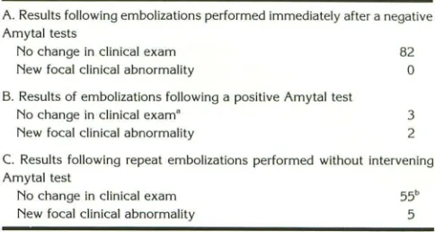
TABLE 1: Summary of data of results following embolizations
A. Results following embolizations performed immediately after a negative Amytal tests
No change in clinical exam
New focal clinical abnormality
B. Results of embolizations following a positive Amytal test No change in clinical exam•
New focal clinical abnormality
82 0
3 2
C. Results following repeat embolizations performed without intervening Amytal test
No change in clinical exam New focal clinical abnormality
55b 5
• One of these three patients had the catheter moved prior to emboli
-zation and a second patient was (correctly) believed to be protected by
this flow pattern even though the Amytal test was positive (see Discu s-sion), giving a total of only one patient who had a simple positive Amytal test in whom embolization was done and there were no postembolization
findings on neurologic exam.
b In one of these 55 embolizations there was focal slowing seen on the
EEG without any change on neurologic examination.
whether it was safe to embolize the A V M. The
embolizations were performed using a
fraction-ated embolization technique.
Table 1 summarizes our data for a single
em-bolization of an A V M feeding vessel immediately
following a negative superselective Amytal test.
None of these 82 embolizations produced any
change in neurologic exam or the EEG. Thus, a
negative Amytal test appeared to predict that it
was safe to embolize an A V M using a single embolization.
Since none of the patients were embolized
without a preceding Amytal test, no true control
group exists for comparison. The Amytal tests
done with the microcatheter in place for a
possi-ble embolization did show a 20% positive rate
(23 of 109 Amytal tests). How many of these
cases would have had neurologic sequelae if the
vessels had been embolized is difficult to
deter-mine. Most of these positive Amytal tests were
not followed by embolization. However, the five
cases that were embolized despite a positive
Amytal test showed a 40% rate of neurologic
sequelae (Table 1). This is significantly greater
than the 0% rate of neurologic sequela of
em-bolization immediately following a negative
Amy-tal test (P
<
.001 using ax
2test with continuity
correction). Furthermore, of the five
emboliza-tions performed after a positive Amytal test, two
were performed using the data of the angiogram
and the Amytal test to improve the safety of the
embolization (see below), and these two had no
neurologic complications. Of the remaining three
[image:3.612.313.555.99.228.2]Amytal test without some change in technique,
two patients developed neurologic deficits. This
equaled a complication rate of 67% for
emboli-zation after a positive Amytal test and a false
positive rate of 33% for the Amytal test. This
suggests that doing embolization after a positive
Amytal test ·is associated with a high rate of
neurologic complication.
Embolization after a positive Amytal test was
performed in only a few cases. These were cases
where the likely neurologic sequela of damage to nearby neuronal tissue was judged to be relatively minor. This was based on the known functional
organization of the brain (7, 8) (ie, production of
a quadrantanopia, if the region of the optic
radia-tions in Meyer's loop was embolized). The
possi-bility of such a deficit was discussed with the
patient prior to the time of embolization. As noted
above, the positive Amytal test led to minor
changes in the embolization technique in two
cases (moving a catheter beyond the take-off of
a small normal vessel or selection of particulate embolic material to avoid a small normal vessel arising at a right angle from the main vessel) making the embolization potentially safer. Of the two cases developing neurologic symptoms
fol-lowing embolization, one is particularly
interest-ing. The embolization involved a left
temporal-parietal A V M following an Amytal test in which there were very subtle changes on the EEG but
no changes on the patients clinical examination.
The subtle changes were not detected on the computer-analyzed EEG but were evident on the standard paper EEG record, both of which were
available in the angiographic suite. The EEG
changes were so subtle that they were felt to be
insignificant during the procedure. However,
these changes were recognized at one of the
weekly EEG review sessions. Even if these EEG
changes had been appreciated at the time of
embolization, the significance of such minor
changes had not been previously shown and the embolization likely would have been performed.
Unfortunately, following a single embolization,
this patient developed a fairly dense receptive
aphasia. Luckily, this deficit largely resolved over
a period of 1 month, but the case does illustrate
the potential significance of even minor EEG
changes during the Amytal test. Based on this
case, we now feel that even subtle EEG changes
should be searched for following the Amytal test
injection. If these EEG changes are seen, one
should consider avoiding embolization of this
ves-sel, especially if an eloquent region of the brain
might be involved.
In many instances, the A V M was not optimally
occluded by embolic material after a single em-bolization. If the microcatheter was moved after
initial embolization, the entire procedure was
re-peated with angiography, Amytal testing, and
embolization, if the Amytal test was negative.
However, in many cases the microcatheter did
not move during the initial embolization. In these cases, if angiography confirms that the A V M needed additional embolization, a decision was made to either embolize the A V M again (called
repeat embolization and listed in Table 1 C) or the
Amytal test was repeated prior to embolization (such an embolization would then be called a single embolization post-Amytal test and listed in
Table 1A). In general, a repeat Amytal test was
done when the feeding vessel to the A V M was
likely also to supply an eloquent region of brain
(an area involved with speech such as Broca's or
Wernicke's area or the motor-sensory strip near
the Rolandic fissure). The reasons for limiting the
repeat Amytal testing to only eloquent regions
were 1) Amytal has a cumulative effect,
produc-ing drowsiness in the patient that could poten-tially interfere with both the clinical examination and EEG, and 2) a complication involving a noneloquent region of the brain was less likely to be of clinical significance.
Of the 60 repeat embolizations done without
an immediately preceding Amytal test, there were
five episodes of neurologic dysfunction (8% ). This
is significantly more than the 0% rate associated
with embolization immediately after a negative
Amytal test (P
<
.05 using ax
2 test withconti-nuity correction). This difference is even more
significant if an additional case is included in
which repeat embolization was followed by focal EEG slowing only (without change on the clinical
examination). There were no similar cases seen
in the group embolized immediately after a
neg-ative Amytal test. In total, there were six cases
of some type of change in neurologic function (examination or EEG) in the 60 repeat
emboliza-tions (10%).
The meaning of this comparison of emboliza-tions performed immediately after a negative Amytal test to repeat embolizations is somewhat
unclear. A possible explanation for the increased
incidence of neurologic sequelae seen in the
AJNR: 13, January /February 1992
low pressure and resultant sump effect that ex-isted in the A V M may have made these normal
vessels difficult to detect on angiography. These
vessels supplying normal brain may have been so small as to be difficult to detect even under optimal angiographic conditions. With emboliza-tion of the A V M, the sump effect would gradually disappear and more of the embolic material could
then flow to other vessels (9). Thus, the repeat
embolization group, which only included A V Ms that were embolized multiple times, might be expected to have a higher complication rate than the embolization group with immediately preced-ing negative Amytal testpreced-ing (which contained two
types of cases: 1) those in which embolization
was discontinued after a single embolization, and 2) those in which multiple embolizations were performed, each preceded by its own Amytal test).
Another explanation of this data should be considered. The same sump action of the A V M that initially acted to direct the embolic agents to the A V M and which was later lost with progres-sive embolization, might also be expected to initially direct a large fraction of the Amytal to the A V M. The small fraction of Amytal that did reach normal brain at the time of the initial Amytal test might not have been sufficient to produce detectable changes in neurologic func-tion. If repeat Amytal testing had been done after partial A V M embolization, it is possible that more
of the Amytal would have reached the normally
functioning brain that was later adversely affected by the embolization. If this were the case, one might expect the Amytal test to have become positive, although it had been negative prior to embolization. Because Amytal tests were not re-peated before all repeat embolizations and
be-cause the neurologic complication rate was
rela-tively low, there are few such cases of conversion of Amytal test from negative to positive. How-ever, there was one patient in whom an initially negative Amytal test was followed by an embo-lization and who subsequently underwent an ad-ditional Amytal test (without catheter movement) who then had a positive Amytal test (with tran-sient focal neurologic dysfunction). Because of this, additional embolization was not performed.
However, if this patient had had repeat
emboli-zation, it is likely that a neurologic complication
would have been produced. This case suggests
that the Amytal test can indeed change from
negative to positive following embolization.
313
If one accepts the above arguments, one should consider doing repeat Amytal tests follow-ing embolizations to determine whether the risk
of additional embolization has increased. It must
be realized that all these patients had A V Ms that
were at risk of hemorrhage, or had hemorrhaged
in the past, and who were to undergo surgical
excision of the A V M. Either hemorrhage or
sur-gery could result in neurologic complication.
Fur-thermore, the embolizations may make the
sur-gery less complicated. The risk of neurologic
complication, especially relatively minor ones,
must be considered in this context.
Conclusion
This article reviews our experience with the
superselective Amytal test, consisting of the
in-jection of 30 mg of Amytal through a
microcath-eter prior to embolization of an A V M, which was
performed to identify the existence of blood
ves-sels supplying functional brain tissue. Our
find-ings are:
1. A positive Amytal test suggested there was
a high likelihood of neurologic complication if the
patient was embolized without change in catheter
placement. This was true even if the Amytal test
produced changes only in the EEG, without a change in the clinical examination.
2. Single embolizations immediately following
negative Amytal tests were performed without
neurologic complication.
3. Using the fractionated embolization
meth-od, the incidence of neurologic complication was
significantly higher in cases where a series of
embolizations was performed after a single Amy
-tal test as compared to a single embolization
performed after an Amytal test. Repeat Amytal
tests should be considered during a series of
multiple step embolizations (also called
fraction-ated embolizations), especially when an eloquent
region of the brain could be involved.
Acknowledgments
I would like to extend a special thank you to UCLA's electroencephalographic technologists, Christopher Barn-hart, James Jackson, Walt Banoczi, Sharon Locke, and Mark Garson, for their diligent work in annotating the EEG throughout the embolizations, which made the retrospec
References
1. Vinuela F, Fox AJ. lnterventional neuroradiology and the
manage-ment of arteriovenous malformations and fistulas. Neural C/in
1983;1:131-153
2. Wolpert SM. Silastic sphere embolization of intracranial arteriovenous
malformations. In: Wilson CB, Stein BM, eds. Intracranial arteri
ove-nous malformations. Baltimore: Williams & Wilkins, 1984:274-294
3. Wada J, Rasmussen T. lntracarotid injection of sodium amytal for
the lateralization of cerebral speech dominance: experimental and
clinical observations. J Neurosurg 1960; 17:266-282
4. Rauch R, Vinuela F, Dion J, et al. Preembolization functional evalu
a-tion in brain A V Ms: the superselective amytal test. AJNR
1992; 13:303-308
5. Debrun G, Vinuela F, Fox AJ, Drake G. Embolization of cerebral
arteriovenous malformations with bucrylate: experience in 46 cases.
J Neurosurg 1982;56:615-627
6. Luessenhop AJ. Natural history of cerebral arteriovenous
malforma-tions. In: Wilson CB, Stein BM, eds. Intracranial arteriovenous
mal-formations. Baltimore: Williams & Wilkins, 1984:14-16
7. Geshwin N. The organization of language and the brain. Science
1970; 170:940-944
8. Vinken PJ, Bryun GW, eds. Handbook of clinical neurology: locali
za-tion in clinical neurology. Amsterdam: North-Holland Publishing, 1969;2:640-783
9. Batjer HH, Purdy PD, Giller CA, Samson DS. Evidence of redistribution
of cerebral blood flow during treatment for an intracranial
arteriove-nous malformation. Neurosurgery 1989;25:599-605
10. Vinuela F, Fox AJ, Debrun G, Pelz D. Preembolization superselective
angiography: role in the treatment of brain arteriovenous