Birth represents a dramatic change of environment that poses both physiological and behavioural challenges to a newborn mammal. Physiological systems have to readjust in a host of ways to extrauterine life: breathing, circulation, metabolism, digestion, muscular action, electrolyte balance, thermoregulation, etc. all require rearrangements within a short time frame (Guyton, 1991). Mastering behavioural challenges is also crucial to survival. Many species of marine mammals give birth in the water (all whales and sirenians), and swimming skills for these newborns are essential to avoid drowning. Seals give birth on solid surfaces, land or ice, but some species use floe ice or intertidal rocks that are quite unstable temporally, and some species face high risks of predation in the terrestrial environment (Lydersen and Kovacs, 1999). These circumstances require the early development of swimming and diving skills.
Although it is clear that the development of swimming and diving skills requires both physiological and behavioural components, a basic asymmetry between the two exists: physiology can, in principle, develop faster than is reflected in the behaviour of the animal, whereas the behavioural repertoire is effectively limited by the physiological scope. Investigations of mammals for which the period from birth to independence
is short may reveal the degree to which behaviour or physiology constrains neonatal development.
The period from birth to weaning is extremely short among phocid seals, and for the phocid pup the end of lactation also means the end of parental care (Kovacs and Lavigne, 1986; Oftedal et al., 1987). Within the phocid group, bearded seals Erignathus barbatus and harbour seals
Phoca vitulina are exceptional in that they normally enter
water on the day they are born (Bigg, 1981; Kovacs et al., 1996), and they stay highly active and aquatic throughout the nursing period (Lydersen et al., 1994; Bowen et al., 1999). Physiology has been suggested to set the limits for behavioural development in several seal species (Burns, 1999; Baker and Donohue, 2000; Donohue et al., 2000), while neonatal behavioural development in other pinnipeds is slow enough that it is unlikely to determine the rate of physiological development (Horning and Trillmich, 1997a,b).
This study explores the physiological and behavioural ontogeny of harbour seal pups during the first weeks of life. Harbour seals give birth on rocks and beaches that are often flooded at high tide, and pups normally enter the water within hours of being born (Knudtson, 1977). Studies of pups JEB3603
This study investigated physiological and behavioural aspects of diving development in pups of the harbour seal Phoca vitulina. Behavioural data (4280 h, 6027 dives) from time/depth recorders (N=13) deployed on pups aged 0–19 days are presented concomitantly with physiological measurements (N=8, sampled both early and late in the nursing period) of blood oxygen stores and body composition. Pups grew from 12.6±1.8 kg (mean age 2 days, total body fat 16±4 %) to 22.2±2.5 kg (mean age 16 days, total body fat 35±5 %; means ± S.D.) over the duration of the experiment. Pups less than 5 days of age had an elevated haematocrit and reduced plasma volume compared with older pups. Although plasma volume and blood volume increased, mass-specific blood oxygen stores (total haemoglobin) fell during the study period. Simultaneously, the following behavioural indicators of
diving ability increased: the proportion of time spent in the water, dive depth, dive duration, bottom time and maximum daily swimming velocity. In addition, the proportion of dives that were identified by cluster analyses as being U-shaped increased significantly with age. On the basis of the measured blood oxygen stores, less than 1 % of the recorded dives exceeded the calculated aerobic dive limit. Thus, development in blood oxygen stores or rates of oxygen consumption did not seem to restrain the rate of neonatal dive development in harbour seals. It appears that behavioural modifications (experience and learning) may be the primary rate-limiting factors for ontogeny of diving skills in neonates of this species.
Key words: harbour seal, Phoca vitulina, postnatal development, diving, oxygen stores, haematocrit, body composition.
Summary
Introduction
Diving development in nursing harbour seal pups
Christian Jørgensen
1,2, Christian Lydersen
1, Ole Brix
2and Kit M. Kovacs
1,*
1Norwegian Polar Institute, Polar Environmental Center, N-9296 Tromsø, Norway and 2Department of Zoology,
University of Bergen, Allégaten 41, N-5007 Bergen, Norway
*Author for correspondence (e-mail: Kit.Kovacs@npolar.no)
equipped with time/depth recorders (TDRs), which allow undisturbed collection of diving behaviour in free-living animals, have revealed that suckling harbour seal pups spend as much as 40 % of their time in the water (Bowen et al., 1999). They swim and dive with their mothers, although they dive for short periods compared with their mothers when the latter are performing bouts of relatively deep dives (Bowen et al., 1999). The lactation period in this species lasts approximately 25 days, but pups may not be able to catch enough prey to balance their energy consumption for several weeks thereafter (Muelbert and Bowen, 1993).
This experiment was designed to investigate whether behaviour or physiology sets the rate of neonatal development of diving in harbour seal pups. Time/depth recorders were deployed on young, free-living harbour seal pups to record their diving and activity patterns. The behavioural data were then compared with development in blood oxygen stores and changes in body composition, which are physiological variables that have a pronounced influence on diving (Butler and Jones, 1997; Donohue et al., 2000).
Materials and methods
This study was conducted from 16 June to 10 July in both 1999 and 2000 on the west coast of Prins Karls Forland (78°20′N, 11°30′E), the westernmost island of the high Arctic Archipelago Svalbard (Fig. 1). This is the breeding site for the world’s most northerly harbour seal population (Prestrud and Gjertz, 1990; Gjertz and Børset, 1992). As a consequence of the high latitude, the study area experienced 24 h of daylight throughout the study period.
In this study, 117 harbour seals were live-captured, 10 in 1999 and 107 in 2000, using set-nets deployed close to the shore, hoop-nets from zodiacs or direct capture on land by hand. Adults and subadults were secured in restraint-nets and treated with Zoletil (1 mg kg−1body mass, Virbac S.A., France). Pups were manually restrained when necessary during sampling and left free to move in the proximity of their mothers at other times during the experimental treatments. All seals were tagged (Rototags in the hind flippers and colour marks on the head) and weighed (Salter spring scales), and blood samples were taken from the extradural intravertebral vein.
Seal captures commenced on June 16 each year, which coincided with the onset of the pupping season (Gjertz and Børset, 1992). Ten very young pups were selected in 1999 and again in 2000 from the captured mother/pup pairs for the investigation of dive development. In this study, newborn status was determined by the presence of placental remains or a fresh (bleeding) umbilical cord (age 0). Age up to 3 days (1, 2 or 3 days) was determined from the condition of the umbilical cord in conjunction with a subjective assessment of the overall body condition and coordination abilities of the pup. Thirty-three pups were young enough to have their age determined in this way; 16 of these were also recaptured and weighed at a later stage. A linear regression of age (A, days) versus body
mass (M, kg) for these pups resulted in the relationship that was used to estimate the age of older pups:
where 11.6 kg is newborn body mass and 0.73 kg day−1is daily mass gain (K. M. Kovacs, C. Lydersen and C. Jørgensen, unpublished data). The coefficient of determination for this relationship was r2=0.91. Only one of the pups sampled for physiological ontogeny (see below) was aged on the basis of body mass; the others were newborn or were captured and aged in the first 3 days of life.
TDRs (model Mk6, Wildlife Computers Inc., Redmond, Washington, USA) were deployed on 20 pups to document undisturbed diving behaviour. They were attached dorsally using quick-setting epoxy resin. A VHF tag (Televilt Positioning AB, Lindesberg, Sweden) was also glued next to the TDR to facilitate recapture. Recapture attempts were initiated from 1 July onwards in both years. Dive recordings from pups that had been abandoned by their mothers were discarded.
(1)
M−11.6 0.73
[image:2.612.312.562.326.670.2]A = ,
Fig. 1. Map of Svalbard (inset) with Prins Karls Forland enlarged. Pups with time/depth recorders were captured and recaptured in the hatched area around Forlandsøyane, except for pup 1999-09, which was captured and recaptured in the hatched area towards the southern tip of the island. All other animals included in the study were caught along the western coast of Prins Karls Forland.
10m 10m
Prins Karls Forland
F orlands
-øy an
e
10 km
50 km 50 km
76° 77° 78°
79° 80°
20° 25° 15°
The TDRs were set to time dry periods and to sample depth (±2 m) and velocity (±0.05 or ±0.02 m s−1) every 10 s while the pups were in the water. The dive recordings were corrected for baseline drift with Zero Offset Correction (ZOC, Wildlife Computers Inc.). All dives performed within 300 s after a haul-out period of 350 s or more were excluded from the analysis because the pressure transducers on the TDRs are unstable following prolonged haul-out periods (Lesage et al., 1999). Furthermore, TDR data from the day of capture were excluded to avoid potential effects of disturbance on the activity budgets or diving behaviour of the pups. The shortest dive recording contained 10 full 24 h periods of sampling, so 10 days were chosen at random from longer recordings to achieve a balanced design when age-related trends (assessed by least-squares linear regression) were analysed.
Depth registrations had to be at least twice the resolution of the TDR (i.e. ⭓4 m) to be considered a dive. For all dives of 4 m or greater, five variables were computed using Dive Analysis (DA, Version 4.0, Wildlife Computers Inc.): (i) duration (s); (ii) maximum depth (m); (iii) bottom time (s) (the time spent at ⭓80 % of the maximum depth of the dive); (iv) average vertical descent rate (m s−1); and (v) average vertical ascent rate (m s−1). In addition to the first three variables, bottom time divided by dive duration, bottom time divided by maximum depth, maximum depth divided by dive duration, average ascent rate divided by average descent rate and average descent rate divided by average ascent rate were selected for cluster analyses (Schreer and Testa, 1996; Schreer et al., 1998; Lesage et al., 1999). Clustering was performed on all dives of 4 m or greater, and was again performed on all dives of 6 m or greater using the procedures outlined by Krafft et al. (2000). Briefly described, multicolinearity was reduced
via principal components analysis on logarithmically transformed values (PRINCOMP, SAS Institute Inc.); the resultant uncorrelated variables with an eigenvalue greater than 1 that explained more than 5 % of the variance were retained. Variables for 1000 randomly selected dives were introduced to a hierarchical cluster analysis (CLUSTER, SAS Institute Inc.). The appropriate number of clusters was identified by the agglomeration coefficient (TREE, SAS Institute Inc.), and their variable means were introduced as seed points in a non-hierarchical k-means-clustering of all the dives (FASTCLUS, SAS Institute Inc.).
The baseline-corrected TDR records were transformed to ASCII files (BINEX, Wildlife Computers Inc.) and merged with velocity data extracted from the TDR records (3M, Wildlife Computers Inc.). Information was retrieved from the merged files on (i) duration and (ii) temporal distribution of haul-out periods, (iii) maximum recorded velocity and (iv) total distance swum.
The following relationship was used to transform all velocity readings (x) to true swimming speeds (y):
y = 1.343x−0.167x2 + 0.049x3−0.072 , (2) (Boyd et al., 1995). Negative true swimming speeds for low-velocity readings were set equal to zero.
The pups were considered ‘hauled out’ whenever the salt-water switch of the TDR had been dry for 3 min or more and to be ‘in the water’ the rest of the time. Tide data were taken from ‘Tide Tables for the Norwegian Coast and Svalbard’ calculated by the Norwegian Mapping Authority.
In the year 2000, blood from the 10 pups carrying TDRs was analysed for haematocrit (Hct) and whole-blood haemoglobin (Hb) concentration, and blood volume and body composition were determined both at the time of capture early in the nursing period and upon recapture late in the nursing period. Haematocrit was measured using microhaematocrit centrifugation of six replicates (Compur M1100 Minicentrifuge; Compur-electronic GmbH, München, Germany). Because seals can sequester red blood cells in the spleen and temporarily lower their Hct (Zapol, 1987; Hurford et al., 1993), the highest value was used wherever several Hct measurements were made from the same animal (Ponganis et al., 1993; Castellini et al., 1996). The first blood sample taken had the highest Hct in all instances. Thus, comparisons with animals that were sampled only once were deemed acceptable. Haemoglobin concentration was measured using the cyan-methaemoglobin photometric method (Sigma Diagnostics, Product no. 525A). Both Hct and Hb were also measured for additional pups (N=37), subadults (N=28) and adults (N=32) for comparative purposes. Calculations were also made of the mean corpuscular haemoglobin concentration (MCHC; g Hb dl−1red blood cells):
where Hb is the measured whole-blood Hb concentration (in g dl−1) and Hct is the measured venous Hct.
Plasma volume was determined using Evans Blue dye. A solution of Evans Blue (Sigma, product no. E2129; 8 mg ml−1 dissolved in 0.9 % NaCl solution) was injected into the extradural intravertebral vein at a dosage of 0.5 mg kg−1body mass using finely graded 2 ml syringes calibrated by weighing. Blood was sampled just prior to injection and 10, 20 and 30 min after injection of the dye. Samples were centrifuged for 5 min at 3000 revs min−1, and the plasma was then removed and kept frozen until analysis. Because haemolysis and lipaemia impeded direct measurement on the plasma (Farjanel et al., 1996), the dye was extracted as described by Campbell et al. (1958). Briefly, the plasma containing the dye was dissolved in a solution of Teepol detergent and KH2PO4and run through a column made up of syringes (2 ml) with 20 layers of circular fitted blotting paper (grade 3MM Chr, Whatman Intl. Ltd, UK). After the column had been washed with KH2PO4solution, the dye was eluted into a known volume of acetone and water. Photometric absorption was then read at 619 nm (Perkin-Elmer Lambda Bio spectrophotometer). Additional eluate did not show any absorption. The standard was prepared from dye at a known concentration in distilled water and run through the columns as described above. Recovery from distilled water was 93–97 %, the same as that for dye added to fresh plasma. No
(3) Hb ×100
traces of Evans Blue were found in plasma or standards that had been run through the columns.
Absorbance values were logarithmically transformed, and linear regression was used to determine the dye concentration at the time of injection since the dye is gradually washed out (Lawson, 1962; El-Sayed et al., 1995). In instances where the slope of log(absorbance) versus time was positive, the absorbance values were averaged, and the mean observed dye loss was calculated from all the samples in which concentrations fell (7 % in 20 min). This relationship was used to find the original concentration of the injected dye. Plasma volume (VP) was calculated as the distributional volume of the injected dye and this, in turn, was used to find the blood volume (VB):
where Hct is the measured venous Hct.
The aerobic dive limit (ADL) was calculated as by Kooyman et al. (1983), except that values for blood oxygen stores were taken from the present study and we used the muscle myoglobin content (1.4 g myoglobin per 100 g muscle) from another study of harbour seal pups (Burns et al., 1999).
Body water content was assessed by hydrogen isotope dilution using tritiated water (HTO). Blood was sampled by venipuncture for background values before 1.0 ml of HTO (specific activity 2.8 MBq ml−1) was injected into the extradural intravertebral vein using finely graded 2 ml syringes calibrated by weighing. Following injection, blood was drawn into the syringe to verify contact with the vein and to wash all the HTO from the syringe. One hour was allowed for mixing. Mothers were handled simultaneously, and the pair was kept in close proximity during the sampling. The pre- and post-injection blood samples were stored frozen until analyses. The water from vacuum-distillation of 1.0 ml of whole blood was then added to 10.0 ml of Opti-fluorine scintillation cocktail (Packard), and 3H activity was measured with a liquid scintillation counter (Packard Tri-Carb 4530). Because HTO also exchanges loosely bound hydrogen ions, the measured distributional volume for tritiated water overestimates total body water (TBW). This was corrected for using:
TBW = 0.003 + 0.968(HTO space) , (5) (Bowen and Iverson, 1998).
Total body fat was calculated according to values provided by Oftedal et al. (1993) for hooded seal Cystophora cristata pups rather than using the more commonly cited relationship obtained from grey seals Halichoerus grypus (Reilly and Fedak, 1990) to facilitate comparison with results for the harbour seal pups studied by Bowen et al. (1992).
Age-related trends were examined using least-squares linear regression. To have a balanced design for the exploration of age-related diving trends, all dive recordings were reduced to the shortest recorded period for an individual (10 days) via
random sampling of days in the 12 longer data sets. All values are given as means ±1 S.D.
The experimental protocol was approved by the Norwegian Animal Research Authority and permission to conduct the fieldwork and to perform all experimental manipulations was granted by the Environmental branch of the Office of the Governor of Svalbard.
Results
All 20 pups carrying TDRs were recaptured. In 1999, two TDRs failed to download. Five pups (three in 1999, two in 2000) were not fed by their mothers, and their dive recordings were therefore discarded. Thus, 13 dive recordings (average duration 14.0±1.4 days) were analysed. Eight of the pups for which dive recordings were obtained were also sequentially sampled (twice) for blood oxygen stores and body composition (Fig. 2). During the study period, the body mass of these pups increased from 12.6±1.8 kg (mean age 1.9 days; fat content 16±4 % of body mass) to 22.2±2.5 kg (mean age 15.9 days; fat content 35±5 % of body mass) (Fig. 2).
Behaviour
In total, 4280 h of activity and 6027 dives were contained in the dive recordings from the 13 pups. Descriptive details from the TDRs are shown in Table 1, and the distributions of depth and duration for the dives are presented in Fig. 3. The pups spent half their time in the water (51±21 %) and the other half hauled out (49±21 %). Diving took place 1.1±0.8 % of the total time (range 0.1–2.8 %). Age-related diving trends are explored in a data set containing information from 10 days from the diving record of each pup, which included 3120 h of activity during which 4154 dives were performed.
Pups spent a large proportion of their time in the water
(4)
VP×100
100 −Hct
[image:4.612.314.561.499.689.2]VB= ,
Fig. 2. Body composition of harbour seal pups captured serially (twice) on Svalbard (N=8). ‘Fat’ (open columns) is total body fat, ‘Blood’ (shaded columns) is total blood volume and ‘Lean body mass’ (hatched+shaded columns) is fat-free body mass.
Body mass (kg)
0 5 10 15 20
+316%
+21%
+37%
Fat
Blood
Lean body mass
2 16
Age (days) 12.6 kg
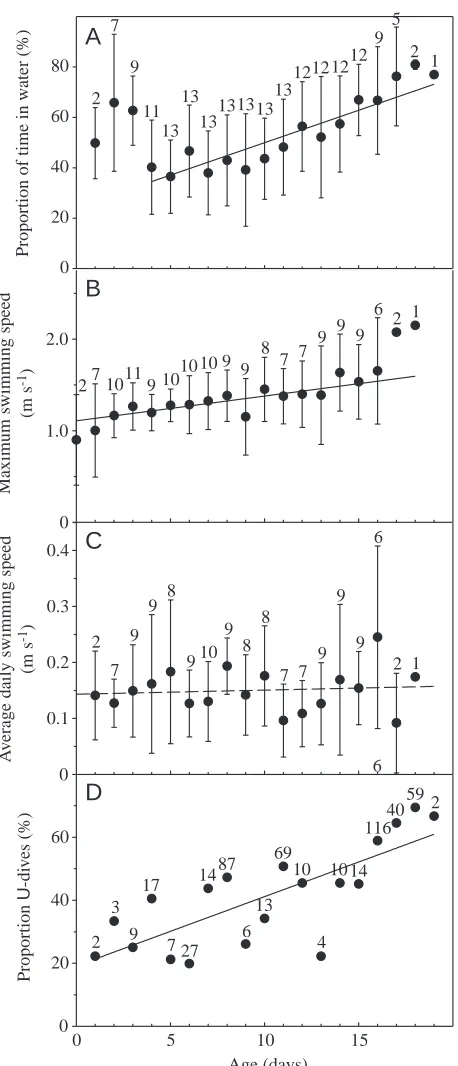
during the first few days of each recording (Fig. 4A). This behaviour declined in prevalence and then subsequently increased once again from approximately 35 % at 4 days of age to approximately 70 % towards the end of the study period (r2=0.20, F
1,115=28.9, P<0.0001) (Fig. 4A). Pups hauled out at the same frequency throughout the study period. The increased proportion of time spent in the water was therefore the result of a doubling of the average duration of aquatic periods from 1.5 h at 4 days of age to more than 3 h in 18-day-old pups (r2=0.01, F
1,1535=18.1, P=0.0001).
Considerable variation existed among the pups with respect to their aquatic behaviour. The pup that spent most time in the water (68.8 %) was twice as aquatically active as the individual that spent the least time in the water (32.8 %) (Table 1). Five pups spent periods in the water that exceeded 20 h, with a maximum of approximately 26 h. The longest recorded haul-out period was 16.75 h. All pups hauled haul-out for more than 5 h at some point during the study.
Analysis of covariance (GLM, SAS Institute Inc.) revealed that year, individual, age, body mass and tidal state, as well as interactions between age and tidal state and between time of day and tidal state, were all significant factors contributing to the overall activity pattern. Pups spent significantly more time in the water during the night and were hauled out 40 % of their time between 23:00 and 08:00 h compared with 55 % during the day (two-tailed Student’s t-test, P<0.0001). Apart from a general increase in time spent in the water, the diurnal pattern did not change from early to late in the nursing period. A tidal pattern was also discernible. Pups spent 60 % of their time hauled out around low tide, compared with approximately 40 % when the tide was high (two-tailed Student’s t-test,
P<0.0001). This pattern was most pronounced early in the
[image:5.612.50.566.98.321.2]nursing period.
Table 1. Descriptive details of dive records for 13 harbour seal pups equipped with time/depth recorders on Svalbard in 1999
and 2000
TDR data
Overall % Year Capture Age Duration No. of Dive duration (s) Dive depth (m) of time and identity Sex date (days) (days) dives Mean Maximum Mean Maximum in water
1999-01 乆 17 June 1–15 14.1 153 22±16 80 4.0±0.3 6 55
1999-02 么 17 June 1–15 14.3 151 24±20 150 4.3±0.8 8 46
1999-09 么 18 June 5–19 13.8 858 34±25 150 6.2±3.9 30 69
1999-13 么 19 June 0–16 16.3 579 34±27 160 5.2±3.9 30 33
1999-29 么 22 June 3–16 12.9 319 31±22 100 4.2±0.5 6 39
2000-01 么 17 June 1–15 13.9 55 16±11 60 4.0±0.3 6 33
2000-05 么 17 June 1–17 15.8 530 23±16 90 4.3±1.0 16 60
2000-07 么 17 June 1–16 15.0 539 27±23 150 4.7±1.8 22 48
2000-14 么 18 June 2–17 14.7 308 26±21 120 5.3±1.8 14 46
2000-18 么 18 June 3–17 13.7 381 22±22 180 4.2±0.7 10 47
2000-20 乆 19 June 2–16 14.2 1105 25±22 220 4.2±0.6 8 62
2000-30 乆 21 June 0–11 11.0 607 26±26 180 4.4±1.0 14 58
2000-34 乆 21 June 5–18 12.7 442 27±20 100 4.4±0.8 8 65
TDR, time/depth recorder. Values are means ±S.D.
Dive duration (s)
10 20 30 40 50 60 70 80 90 100ù110
P
ro
p
ortion o
f di
v
es (
%
)
0 10 20 30 40 50
Dive depth (m)
4 6 8 10 12 14 16 18 ù20
0 10 20 80 90
A
B
[image:5.612.58.293.355.701.2]The number of dives recorded per pup ranged from 55 to 1105, with an average of 464±292 dives (Table 1). The number of dives per day varied considerably among individuals and among days for the same individual. There was no significant increase in the number of dives performed per day with age. Ten of the 13 pups had days in the middle of the sampling period when they did not perform a single dive; the maximum number of dives performed by a pup on a single day was 216 (8 days old). The average dive duration was 27±23 s, and average dive depth was 4.7±2.2 m. Three pups made dives of 3 min or longer; the longest dive recorded was 3 min 40 s. The depth of the dives varied: two pups dived as deep as 30 m, while three never exceeded 6 m, and three more never went below 8 m.
Average dive duration increased significantly with age (r2=0.07, F
1,108=8.36, P<0.005), as did average dive depth (r2=0.04, F
1,108=4.48, P=0.037) and maximum daily dive depth (r2=0.04, F
1,108=4.56, P=0.04). The pups also increased their average bottom time with age (r2=0.06, F
1,108=6.72, P=0.01). Maximum dive duration also increased significantly with age when complete dive recordings for pups were analysed (r2=0.04, F
1,149=5.85, P=0.017), but this trend was not significant in the case of the randomly selected 10-day records. There was a significant increase in the maximum daily swimming speed as pups grew older (r2=0.10, F
1,90=10.11, P=0.002), but when velocity readings over a whole day were
averaged, the average daily swimming speed did not exhibit any relationship with age (Fig. 4B,C).
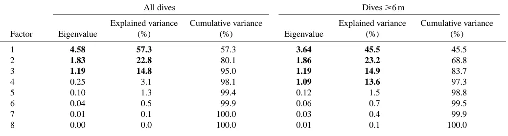
Three principal components were retained (Table 2), and two clusters were identified when all recorded dives were subjected to clustering. One of the clusters contained all the dives that were at the minimum resolution of the TDR, i.e. one minimal depth reading, which means a dive to 4 m depth and of 10 s duration, while the rest of the dives constituted a second grouping. Since this had limited value when it came to interpreting possible dive functions, the 1118 dives that were to depths of 6 m and deeper were clustered independently. Four principal components were retained (Table 2) and three clusters were identified (Table 3). Two of the dive types were V-shaped with skews in opposite directions, while the third cluster group consisted of U-shaped dives. The proportion of dives of 6 m or deeper performed by all pups of a given age that were U-shaped increased with age from approximately 20 % early in the experimental period to 60 % towards the end of the experiment (Fig. 4D) (r2=0.59, F
1,17=24.63, P=0.0001). Repeated periods of diving to depths greater than 10 m were performed by only four pups. Three of these pups were 14–16 days old when these isolated bouts of ‘deep’ diving took place. A 16-day-old pup performed the most extreme dives, reaching depths of more than 10 m 35 times over a period of 2.5 h. Eighteen of these dives were to depths between 20 and 30 m. The fourth pup spent 4 days diving repeatedly to depths that exceeded 10 m at 6, 8, 11 and 18 days of age.
Physiology
In total, 107 animals were sampled for Hct. Clotting prevented Hb analyses for 28 of these, so whole-blood Hb Age (days)
0 5 10 15
Proportion U -d iv es ( % ) 0 20 40 60 A v er age da il y s w imm in g s p eed (m s -1) 0 0.1 0.2 0.3 0.4 M ax im u m s w imm in g s p eed (m s -1) 0 1.0 2.0 Proportion o f ti m e in w at er ( % ) 0 20 40 60 80 2 7 9 7 7 9 8 9 9 910 9 6 8 8 9 2 1 1011 10
10 10 9
9 9 8 7 7 9 9 27 9 6 2 1 2 7 9 11 13 13 13131313 13 121212 12 9 5 2 1
A
B
C
D
2 3 9 7 10 17 27 4 10 6 1487 6 13 69 14 40 116 592 [image:6.612.54.283.68.601.2]concentration was determined for 79 animals, which then allowed MCHC to be calculated (Table 4). Pups had significantly lower Hct levels than juveniles or adults (Tukey, P<0.05) and significantly lower whole-blood Hb concentrations than juveniles or adult males (Tukey, P<0.05). The MCHC of pups was significantly lower than that of adult males, but not significantly different from those of juveniles or adult females (Tukey, P<0.05) (Table 4). No correlations between Hct and age or body mass were found over the entire range of pup age (0–26 days), but when pups less than 5 days old were evaluated separately they had a significantly higher Hct (58.0±5.0 compared with 52.3±3.0; two-tailed Student’s t-test, P<0.01) (Fig. 5) and lower MCHC (36.0±0.9 compared with 38.2±1.2 g dl−1; two-tailed Student’s t-test, P<0.0001) than older pups. The Hct also increased significantly with age for pups 5 days of age and older (r2=0.20, F
1,35=8.87, P=0.005) (Fig. 5). The Hb concentration showed no
relationship with age, although most young pups had quite high values.
Plasma volumes increased sharply in very young pups (Fig. 6A,B) (Table 5). Blood volume also increased rapidly, changing by 17 % between the two sampling periods (from 2480±300 to 2900±250 ml; paired one-tailed Student’s t-test;
P=0.006) (Fig. 6A). Mass-specific plasma and blood volumes
increased for pups less than 5 days but decreased for older pups. Simultaneous changes in Hct resulted in the maintenance of circulating Hb levels (from 515±52 to 560±66 g).
Discussion
[image:7.612.51.570.98.237.2]Harbour seal pups are extremely precocial at birth and are already equipped in many ways for an aquatic lifestyle (Bigg, 1981). They are born with a subcutaneous blubber layer that is a good insulator, the white lanugo has already been moulted in favour of the more streamlined adult pelage (Oftedal et al., 1991), their eyes are open and they can vocalise sufficiently for individual recognition (Renouf, 1985) and their locomotor muscle strength and control are sufficient for them to survive being born directly into the water (Ronald and Thomson, 1981). However, they must undergo physiological adjustments
Table 2. Eigenvalues and percentages of explained variance for the eight components used to classify dives from nursing harbour
seal pups on Svalbard
All dives Dives ⭓6 m
Explained variance Cumulative variance Explained variance Cumulative variance
Factor Eigenvalue (%) (%) Eigenvalue (%) (%)
1 4.58 57.3 57.3 3.64 45.5 45.5
2 1.83 22.8 80.1 1.86 23.2 68.8
3 1.19 14.8 95.0 1.19 14.9 83.7
4 0.25 3.1 98.1 1.09 13.6 97.3
5 0.10 1.3 99.4 0.12 1.5 98.8
6 0.04 0.5 99.9 0.06 0.7 99.5
7 0.01 0.1 100.0 0.03 0.4 99.9
8 0.00 0.0 100.0 0.01 0.1 100.0
[image:7.612.49.299.566.673.2]Values in bold type indicate the components having an eigenvalue >1 and explaining >5 % of the variance.
Table 3. Characteristics of the groups identified by
k-means-clustering of all (1118) dives 6 m or deeper collected by time/depth recorders from harbour seal pups on Svalbard
Cluster 1 2 3
Dive type V-shaped V-shaped U-shaped*
Skew Left Right
Number of dives 390 219 509
Depth (m) 6.9±2.3 6.8±1.7 8.9±5.1 Duration (s) 31±17 45±24 66±26 Bottom time (s) 3.0±4.6 3.7±6.4 33±19 Vertical descent rate (m s−1) 1.0±0.4 0.3±0.1 0.8±0.5 Vertical ascent rate (m s−1) 0.6±0.4 0.9±0.5 0.7±0.4
*Three pups performed only one or two dives that were deep enough to be included in the clustering process; none of these three pups performed any U-dives. The remaining five pups performed 16–366 clustered dives, of which 29–61 % were U-dives.
Values are means ±S.D.
Table 4. Haematocrit, whole-blood haemoglobin
concentration and mean corpuscular haemoglobin concentration for harbour seals at Svalbard
Haemoglobin MCHC Haematocrit N (g dl−1) N (g dl−1) N
Pups 53.6±4.2 47 20.3±1.2c 35 37.7±1.5e 34 Juveniles 58.1±3.2a 28 22.1±1.2d 21 38.2±0.9e,f 21 Adult 乆 55.5±4.1b 16 21.4±1.1c,d 13 38.7±0.5e,f 13 Adult 么 57.8±1.9a,b 16 22.4±0.7d 10 39.2±0.4f 10
Identical superscripts indicate statistically non-significant differences (Tukey, P<0.05).
Some blood samples clotted, which prevented subsequent determination of haemoglobin concentration.
Values are means ±S.D. (N).
to extrauterine life and must face the additional challenge of dealing with both terrestrial locomotion and aquatic swimming and diving from the day of birth.
The diving performed by harbour seal pups is not extraordinary by phocid standards, except for its early onset. Harbour seals are generally shallow divers throughout their lives. TDRs deployed on adult seals from the Svalbard population revealed that half the dives were shallower than 40 m and only 5 % exceeded 200 m (Gjertz et al., 2001).
The maximum depths measured in the present study (30 m) are comparable with maximum depths recorded in nursing harbour seal pups elsewhere (35 m) (Bowen et al., 1999), but the pups in this study dived for remarkably shorter periods (maximum 3 min 40 s) than other investigators have found for pups of a similar age: 8 min, age 1 week, experimental dive
(Harrison and Tomlinson, 1960); approximately 9 min, age less than 12 days (Bowen et al., 1999); 8.5 min, age less than 25 days (Bekkby and Bjørge, 2000). It has been suggested that the dive durations of harbour seal pup are correlated with depth in various localities (Bekkby and Bjørge, 2000). The observed differences between this study and others are in all probability due to the shallowness (1–6 m) of the water surrounding the breeding grounds at Prins Karls Forland. The only pup captured and recaptured in an area where the bathymetry is more diverse (Fig. 1) had the longest mean dive duration and the greatest mean dive depth measured in the present study.
The weeks following birth were characterised by gradually increasing diving skills. Not only did pups spend a greater proportion of their time in the water, but concomitantly all measures of dive performance also showed improvement. Dives became longer and deeper, bottom time increased, the pups were capable of swimming faster and U-dives, which are strongly associated with feeding in adult harbour seals (Lesage et al., 1999), became more common. At the same time, significant physiological changes took place. The pups nearly doubled their body mass during the study period. Most of the accumulated mass was blubber, but muscle mass also increased. Blood oxygen stores, however, which are crucial to any air-breathing diver, did not increase significantly over the study period. In fact, the available oxygen in the circulatory system decreased relative to body mass and even relative to mass raised to the power 0.67.
[image:8.612.44.563.85.251.2]Phocid seals store approximately 65 % of their on-board oxygen in the blood; the remaining stores are divided between muscle myoglobin (28 %) and the lungs (7 %) (Kooyman, 1989). It is a shortcoming of the present study that the second largest oxygen store, muscle myoglobin, was not assessed. However, myoglobin levels vary between muscles (Dolar et al., 1999) and within the same muscle (Polasek and Davis, 2001), so it is difficult to obtain a sample that is representative of total muscle oxygen stores. Although developmental trends can be detected from muscle biopsies from live animals,
Table 5. Blood oxygen stores and body composition of eight harbour seal pups that were sequentially sampled twice
First capture Second capture
Year Age Mass [Hb] Plasma Fat Age Mass [Hb] Plasma Fat
and identity (days) (kg) Hct (g dl−1) (ml) (kg) (days) (kg) Hct (g dl−1) (ml) (kg)
2000-01 1 12.0 59.0 21.2 985 − 15 20.7 49.8 18.8 1318 7.4
2000-05 1 11.5 65.3 22.6 803 2.3 17 19.4 50.2 19.4 − 8.1
2000-07 1 10.6 59.0 20.6 1106 1.0 16 23.4 50.0 18.3 1419 8.0 2000-14 2 14.1 52.0 19.4 1163 2.3 17 25.8 50.0 18.7 1495 9.2 2000-18 3 14.5 55.2 20.2 1314 1.4 17 23.6 57.5 20.2 1444 6.9 2000-20 2 12.2 58.2 21.1 1106 1.6 16 22.7 51.8 19.1 1341 8.4 2000-30 0 10.5 65.0 23.1 669 2.0 11 18.4 54.3 21.1 1305 6.6 2000-34 5 15.4 49.0 18.7 1273 2.3 18 23.7 50.5 18.9 1383 7.1
Mean 1.9 12.6 57.8 20.9 1052 1.8 15.9 22.2 51.8 19.3 1386 7.7
S.D. 1.6 1.8 5.7 1.5 223 0.5 2.2 2.5 2.8 0.9 70 0.9
Age, estimated age; mass, total body mass; Hct, measured haematocrit; [Hb], whole-blood haemoglobin concentration; plasma, plasma volume; fat, total body fat.
Fig. 5. Haematocrit versus pup age for harbour seals on Svalbard (linear regression for pups more than 5 days old: y=49.2+0.23x, r2=0.20). The eight pups that were sampled twice are represented by individual symbols; open circles represent additional pups.
Age (days)
0 5 10 15 20 25
H
ae
m
atocrit
[image:8.612.50.289.506.691.2]reliable estimates for the calculation of total muscle oxygen stores require complete carcass analysis. Studies suggest that, although muscle myoglobin levels limit behaviour for young that have started independent feeding, this may not be the case for younger divers that are provided with milk (Thorson and LeBoeuf, 1994; Ponganis et al., 1999; Noren et al., 2001). Previous investigations of myoglobin levels in nursing harbour seal pups concluded that early modification of blood oxygen stores is more important than changes within the muscle (Burns et al., 1999). It has also been shown that hooded seal pups store a larger fraction of their oxygen in blood than do adults (Burns et al., 2000).
Blood oxygen stores that do not scale in proportion to body mass are not unique to harbour seal pups. Blood volume, measured by bleeding southern elephant seal Mirounga
leonina pups, was correlated to fat-free body mass. However,
Hct levels in this species fall during the first 3 weeks of life, resulting in a relative decrease in red blood cell volume (Bryden and Lim, 1969).
The development of blood oxygen stores can be followed more closely by focusing on the total volume of red blood cells rather than from a consideration of total blood volume. For pups that are 3 weeks of age and older, increasing volumes of red blood cells have been measured in both harbour seals (Kodama et al., 1977) and southern elephant seals (Bryden and Lim, 1969). For the latter species, red blood cell volume started to increase simultaneously with the onset of diving and was therefore assumed to be related to increased physiological demand. In the present study, the volume of red blood cells did not increase significantly over the study period, which included a considerable amount of diving experience. However, total Hb content was only measured for pups that were approximately 2 and approximately 16 days old. This could mask changes that occur on a short time scale. Changes in Hct take place on a very short time scale through redistribution of fluids and blood cells. Alterations to Hb content, however, involve the production or destruction of large quantities of proteins and cells and are therefore likely to occur more slowly. Thus, the absence of a significant increase in total Hb content might indicate that additional constraints act to restrict the rate at which new red blood cells can be put into circulation shortly after birth.
The rate of erythropoiesis is controlled by the hormone erythropoietin, which stimulates the production and maturation of red blood cells (Hadley, 1992). Foetal erythropoietin is produced first in the yolk sac then in the liver. After birth, the kidneys take over erythropoietin secretion (Hadley, 1992). In general, hypoxia causes an increase in erythropoietin levels, whereas hyperoxia has the opposite effect (Hadley, 1992). Foetal life is lived in conditions of relatively low oxygen supply (Guyton, 1991; Palis and Segel, 1998). The comparatively plentiful supply of oxygen available as a result of breathing thus decreases erythropoiesis at birth (Matoth et al., 1971; Guyton, 1991). Although newborns are more sensitive to erythropoietin than adults (Weinberg et al., 1992), a 10-fold decrease in the rate of erythropoiesis has been found in humans during the week following birth (Palis and Segel, 1998). By approximately the sixth week of life in humans, the rate of erythropoiesis reaches a level that balances the loss of erythrocytes (Matoth et al., 1971). A similar phenomenon might restrict the expansion of red blood cell volume in young harbour seal pups.
In most mammals, neonates are special in that foetal red blood cells, which normally contain a distinct foetal Hb, are gradually replaced by cells that have the adult form (Gilles, 1985). This is the case for human neonates, but foetal Hb has not been found in phocids: Weddell seal Leptonychotes
weddellii (Qvist et al., 1981); harp seal Phoca groenlandica
Fig. 6. (A) Plasma volume and the corresponding blood volume calculated from haematocrit expressed as absolute volumes (ml). (B) Plasma volume and the corresponding blood volume calculated from haematocrit expressed as a percentage of body mass. (C) Total body fat (kg) measured by hydrogen isotope dilution. Symbols represent individual pups, and all lines are for illustrative purposes only.
Age (days)
0 5 10 15
To
ta
l
bo
dy
fa
t (
k
g)
0 2 4 6 8
V
ol
um
e (
m
l)
0 1000 2000 3000
V
ol
um
e (
%
bo
dy
m
ass)
0 5 10 15 20
Plasma Blood
Plasma Blood
A
B
[image:9.612.55.295.226.662.2](Nævdal, 1965); hooded seal (Nævdal, 1966); harbour seal (C. Jørgensen, K. M. Kovacs, C. Lydersen, and O. Brix, unpublished data). However, it has not been established whether phocid neonates also lack the foetal type of red blood cell. Replacing short-lived foetal red blood cells prior to parturition would be a mechanism by which erythrocyte loss shortly after birth could be reduced. Whether this strategy is possible, or is actually employed, by phocids requires further investigation; the absence of foetal Hb from phocid neonates suggests that such a strategy is a possibility.
The lack of a significant increase in blood oxygen stores may therefore be inherent in the mechanisms by which new red blood cells are formed. It seems likely, therefore, that any demand an organism has for red blood cells during the first few weeks of life must be met by stores that are present at birth. At first glance, the slow rate of development may seem inadequate, but the red blood cell volume could still be tailored to suit the requirements of pups that are 2–3 weeks old by having volumes that exceed actual requirements at birth.
Haematocrit and whole-blood Hb concentration are difficult to compare directly between studies for several reasons. Phocid haematological variables are known to differ with age, sex, stress level, dive history, season, birth history, anaesthesia and methodology (Chaplin and Mollison, 1952; McConnell and Vaughan, 1983; Kuiken, 1985; Castellini et al., 1996). Comparisons across studies must therefore be viewed with caution. In the present study, the first few days after birth were characterised by an elevated Hct and a low plasma volume. A 40 % increase in plasma volume subsequently restored Hct levels to within the normal range for pups. Previous investigations of Hct and Hb concentrations in pinniped pups have reported low levels (Qvist et al., 1981; Hall, 1998) or levels that gradually increased towards juvenile and adult values (Horning and Trillmich, 1997a).
The results from the present study are consistent with these findings. When Hct was analysed for all pups grouped as a single sample, the average pup value was lower than that of adults and juveniles, and there was no increase with age. However, when very young pups, most of which had above-average Hct levels, were excluded, pups 5 days of age and older exhibited a statistically significant increase in Hct with age. The different trends that arose among different age groups of pups stresses the importance of knowing and reporting accurately the ages of pups when measuring blood variables.
An elevated Hct, and thus Hb content, in very young neonates is a common mammalian phenomenon. It has been reported in grey seals (Hall, 1998), Weddell seals (Lenfant et al., 1969) (but not in Qvist et al., 1981), Steller sea lions
Eumetopias jubatus (Lenfant et al., 1970), Juan Fernández fur
seals Arctocephalus philippii (Sepúlveda et al., 1999) and humans (Matoth et al., 1971) and for the harbour seals in the present study.
Haematocrit and Hb have been shown to increase during the first few hours in newborn humans (Gairdner et al., 1958). Gairdner et al. (1958) argued that, since there was no source of extra red blood cells, the observed increase in Hct values in
their study had to result from a decreasing plasma volume resulting from a loss of plasma water. This explanation is consistent with the low plasma volume found in very young pups in the present study, but results from other studies from phocids suggest that water movement in the blood is more complicated (Castellini et al., 1990). Taking into account the complex shifts of body fluids between different compartments in young neonates (Cheek et al., 1984), our inadequate understanding of their regulatory mechanisms (Simpson and Stephenson, 1993), the extent of spleen sequestering in phocids (Hurford et al., 1993) and differing degrees of precocity between species, it is clear that we know too little to explain how water moves in the blood.
Some support for the suggestion that changes in water balance may play a role can be derived from the analyses of body composition in the present study. The method of calculating body fat based on isotope dilution assumes that fat-free body mass has a constant degree of hydration. This assumption can be violated, e.g. if the animal is dehydrated. Under these conditions, the equations applied would yield an artificially elevated fat content. Thus, the apparently declining fat content, despite simultaneous fat-rich milk intake in 0- to 3-day-old pups in this study (Fig. 6C), could indicate that the pups were actually somewhat dehydrated when they were newborn. However, this pattern could be an artefact of small sample size or of other assumptions of the methods involved since hydration levels were not measured directly in this study. For comparison, hydration has been shown to increase by 1.72 % during the first 3 days in newborn humans (Rodriguez et al., 2000), but was 4 % higher in pups than in adult Antarctic fur seals Arctocephalus gazella by carcass analysis (Arnould et al., 1996).
Total body fat values both early and late in nursing in this study (16 % and 35 % at 2 and 16 days of age, respectively) are comparable with studies from Sable Island that reported a fat content of 11 % in newborns and approximately 35 % in older pups (Bowen et al., 1992; Muelbert and Bowen, 1993).
The behavioural data and the physiological data in this study appear to be paradoxical in that dive performance increases during the study period despite the fact that relatively less blood oxygen is available to the diving pups as they grow older. There are two ways in which this ‘paradox’ might be explained. Either oxygen is limiting but the rates of consumption decrease, presumably through the acquisition of a more adult-like dive response, or oxygen is simply never a limiting factor during the study period but behavioural limitations prevent full utilisation of the physiological potential.
ADL of 1 min 40 s using a diving metabolic rate of four times BMR. Less than 1 % of the recorded dives and only eight of the 13 pups exceeded even the shortest calculated ADL. Moreover, the proportion of dives that exceeded the shortest ADL did not increase significantly with age over the study period. If decreasing rates of oxygen consumption restricted the rate of dive development, dive durations would be expected to parallel ADL more closely. Since this is not the case in the present study, it is suggested that neither oxygen stores nor oxygen consumption restrain the rate of dive development.
In summary, nursing harbour seal pups increased their activity levels and dive performance between 0 and 19 days of age. Pups spent a larger proportion of their time in the water but hauled out at the same frequency. They dived deeper and for longer periods of time. A greater proportion of the dives were U-shaped, and the pups were capable of swimming faster, although their average swimming speed did not increase. Over the same period, body mass increased by 75 %, mainly as a result of the deposition of blubber. Although plasma and blood volume rose sharply in pups less than 5 days old, a concomitant fall in haematocrit led to a reduction in blood oxygen stores relative to body mass. Dives that exceeded the calculated aerobic dive limit, on the basis of the measured oxygen stores, occurred with a low frequency and were independent of age. These findings suggest that neither available oxygen nor the rate of oxygen consumption restrains the rate of dive development. Thus, it appears that behavioural modifications, presumably experience and learning, play a major role in determining the rate of dive development in harbour seal pups. We thank Magnus Andersen, Bjørn Krafft, Hans Lund and Sonja Reder for valuable help in the field. Claus Beck kindly loaned us his microhaematocrit centrifuge. Bjørn Krafft also helped with the dive clustering analyses. We also thank Sofie Van Parijs and Øystein Wiig for reading and commenting on the manuscript and Anne Estoppey for designing the map. The Norwegian Polar Institute, the Norwegian Research Council (NFR) and the University of Bergen financed this research.
References
Arnould, J. P. Y., Boyd, I. L. and Speakman, J. R. (1996). Measuring the
body composition of Antarctic fur seals (Arctocephalus gazella): validation of hydrogen isotope dilution. Physiol. Zool. 69, 93–116.
Baker, J. D. and Donohue, M. J. (2000). Ontogeny of swimming and diving
in northern fur seal (Callorhinus ursinus) pups. Can. J. Zool. 78, 100–109.
Bekkby, T. and Bjørge, A. (2000). Diving behaviour of harbour seal Phoca vitulina pups from nursing to independent feeding. J. Sea Res. 44, 267–275. Bigg, M. A. (1981). Harbour seal. In Handbook of Marine Mammals, vol. 2, Seals (ed. S. H. Ridgway and R. J. Harrison), pp. 1–27. London: Academic Press.
Bowen, W. D., Boness, D. J. and Iverson, S. J. (1999). Diving behaviour of
lactating harbour seals and their pups during maternal foraging trips. Can. J. Zool. 77, 978–988.
Bowen, W. D. and Iverson, S. J. (1998). Estimation of total body water in
pinnipeds using hydrogen-isotope dilution. Physiol. Zool. 71, 329–332.
Bowen, W. D., Oftedal, O. T. and Boness, D. J. (1992). Mass and energy
transfer during lactation in a small phocid, the harbor seal (Phoca vitulina). Physiol. Zool. 65, 844–866.
Boyd, I. L., Reid, K. and Bevan, R. M. (1995). Swimming speed and
allocation of time during the dive cycle in Antarctic fur seals. Anim. Behav.
50, 769–784.
Bryden, M. M. and Lim, G. H. K. (1969). Blood parameters of the southern
elephant seal (Mirounga leonina, Linn.) in relation to diving. Comp. Biochem. Physiol. 28, 139–148.
Burns, J. M. (1999). The development of diving behavior in juvenile Weddell
seals: pushing physiological limits in order to survive. Can. J. Zool. 77, 737–747.
Burns, J. M., Blix, A. S. and Folkow, L. P. (2000). Physiological constraint
and diving ability: a test in hooded seals, Cystophora cristata. FASEB J. 14, A440.
Burns, J. M., Costa, D. P., Harvey, J. T. and Frost, K. (1999). Physiological
development in juvenile harbor seals. In 13th Biennial Conference on the Biology of Marine Mammals, Maui, Hawaii, USA, 28 November – 3 December 1999, pp. 26–27.
Butler, P. J. and Jones, D. R. (1997). Physiology of diving of birds and
mammals. Physiol. Rev. 77, 837–899.
Campbell, T. J., Frohman, B. and Reeve, E. B. (1958). A simple, rapid and
accurate method of extracting T-1824 from plasma, adapted to the routine measurement of blood volume. J. Lab. Clin. Med. 52, 768–777.
Castellini, J. M., Castellini, M. A. and Kretzmann, M. B. (1990).
Circulatory water concentration in suckling and fasting northern elephant seal pups. J. Comp. Physiol. B 160, 537–542.
Castellini, J. M., Meiselman, H. J. and Castellini, M. A. (1996).
Understanding and interpreting hematocrit measurements in pinnipeds. Mar. Mammal. Sci. 12, 251–264.
Chaplin, H., Jr and Mollison, P. L. (1952). Correction for plasma trapped in
the red cell column of the hematocrit. Blood 7, 1227–1238.
Cheek, D. B., Wishart, J., MacLennan, A. H. and Haslam, R. (1984). Cell
hydration in the normally grown, the premature and the low weight for gestational age infant. Early Human Dev. 10, 75–84.
Dolar, M. L. L., Suarez, P., Ponganis, P. J. and Kooyman, G. L. (1999).
Myoglobin in pelagic small cetaceans. J. Exp. Biol. 202, 227–236.
Donohue, M. J., Costa, D. P., Goebel, M. E. and Baker, J. D. (2000). The
ontogeny of metabolic rate and thermoregulatory capabilities of northern fur seal, Callorhinus ursinus, pups in air and water. J. Exp. Biol. 203, 1003–1016.
El-Sayed, H., Goodall, S. R. and Hainsworth, R. (1995). Re-evaluation of
Evans Blue dye dilution method of plasma volume measurement. Clin. Lab. Haematol. 17, 189–194.
Farjanel, J., Denis, C., Chatard, J. C. and Geyssant, A. (1996). An accurate
method of plasma volume measurement by direct analysis of Evans blue spectra in plasma without dye extraction: origins of albumin space variations during maximal exercise. Eur. J. Appl. Physiol. 75, 75–82.
Gairdner, D., Marks, J., Roscoe, J. and Brettell, R. (1958). The fluid shift
from the vascular compartment immediately after birth. Arch. Dis. Child.
33, 489–498.
Gilles, R. (1985). Circulation, Respiration and Metabolism. Berlin: Springer. Gjertz, I. and Børset, A. (1992). Pupping in the most northerly harbor seal
(Phoca vitulina). Mar. Mammal. Sci. 8, 103–109.
Gjertz, I., Lydersen, C. and Wiig, Ø. (2001). Distribution and diving of
harbour seals (Phoca vitulina) in Svalbard. Polar Biol. 24, 209–214.
Guyton, A. C. (1991). Textbook of Medical Physiology. Eighth edition.
Philadelphia: W. B. Saunders Co.
Hadley, M. E. (1992). Endocrinology. Third edition. London: Prentice-Hall
Inc.
Hall, A. J. (1998). Blood chemistry and hematology of gray seal (Halichoerus grypus) pups from birth to postweaning. J. Zoo Wildlife Med. 29, 401–407. Harrison, R. J. and Tomlinson, J. D. W. (1960). Normal and experimental
diving in the common seal (Phoca vitulina). Mammalia 24, 386–399.
Horning, M. and Trillmich, F. (1997a). Development of hemoglobin,
hematocrit and erythrocyte values in Galápagos fur seals. Mar. Mammal. Sci. 13, 100–113.
Horning, M. and Trillmich, F. (1997b). Ontogeny of diving behaviour in the
Galápagos fur seal. Behaviour 134, 1211–1257.
Hurford, W. E., Guyton, G. P., Zapol, W. M., Schneider, R. C., Stanek, K., Zapol, D. G., Hochachka, P. W. and Liggins, G. C. (1993). Splenic
contraction, blood volume and seal muscle saturation of free-diving Weddell seals. Antarctic J. 28, 152–155.
Kleiber, M. (1961). The Fire of Life: An Introduction to Animal Energetics.
New York: Wiley.
Knudtson, P. M. (1977). Observations on the breeding behavior of the harbor
Kodama, A. M., Elsner, R. and Pace, N. (1977). Effects of growth, diving
history and high altitude on blood oxygen capacity in harbor seals. J. Appl. Physiol. 42, R852–R858.
Kooyman, G. L. (1989). Diverse Divers. Berlin: Springer-Verlag.
Kooyman, G. L., Castellini, M. A., Davis, R. W. and Maue, R. A. (1983).
Aerobic diving limits of immature Weddell seals. J. Comp. Physiol. B 151, 171–174.
Kovacs, K. M. and Lavigne, D. M. (1986). Maternal investment and neonatal
growth in phocid seals. J. Anim. Ecol. 55, 1035–1051.
Kovacs, K. M., Lydersen, C. and Gjertz, I. (1996). Birth-site characteristics
and prenatal molting in bearded seals (Erignathus barbatus). J. Mammal.
77, 1085–1091.
Krafft, B. A., Lydersen, C., Kovacs, K. M., Gjertz, I. and Haug, T. (2000).
Diving behaviour of lactating bearded seals (Erignathus barbatus) in the Svalbard area. Can. J. Zool. 78, 1408–1418.
Kuiken, T. (1985). Influences of diet, gestation and age on haematology and
plasma chemistry of the harbour seal, Phoca vitulina. Aquat. Mammals 11, 40.
Lawson, H. D. (1962). The volume of blood −a critical examination of methods for its measurement. In Handbook in Physiology, section 2, Circulation, vol. I (ed. W. F. Hamilton), pp. 23–49. Washington, DC: American Physiological Society.
Lenfant, C., Elsner, R., Kooyman, G. L. and Drabek, C. M. (1969).
Respiratory function of blood of the adult and fetus Weddell seal Leptonychotes weddelli. Am. J. Physiol. 216, 1595–1597.
Lenfant, C., Johansen, K. and Torrance, J. D. (1970). Gas transport and
oxygen storage capacity in some pinnipeds and the sea otter. Respir. Physiol.
9, 277–286.
Lesage, V., Hammill, M. O. and Kovacs, K. M. (1999). Functional
classification of harbor seal (Phoca vitulina) dives using depth profiles, swimming velocity and an index of foraging success. Can. J. Zool. 77, 74–87.
Lydersen, C., Hammill, M. O. and Kovacs, K. M. (1994). Diving activity
in nursing bearded seal (Erignathus barbatus) pups. Can. J. Zool. 72, 96–103.
Lydersen, C. and Kovacs, K. M. (1999). Behaviour and energetics of
ice-breeding, north Atlantic phocid seals during the lactation period. Mar. Ecol. Prog. Ser. 187, 265–281.
Matoth, Y., Zaizov, R. and Varsano, I. (1971). Postnatal changes in some
red cell parameters. Acta Paediat. Scand. 60, 317–323.
McConnell, L. C. and Vaughan, R. W. (1983). Some blood values in
captive and free-living common seals (Phoca vitulina). Aquat. Mammal.
10, 9–13.
Muelbert, M. M. C. and Bowen, W. D. (1993). Duration of lactation and
postweaning changes in mass and body composition of harbor seal, Phoca vitulina, pups. Can. J. Zool. 71, 1405–1414.
Nævdal, G. (1965). Protein polymorphism used for identification of harp seal
populations. Årbok Univ. Bergen. Mat.-Naturv. Ser. 9, 1–20.
Nævdal, G. (1966). Hemoglobins and serum preoteins in four North Atlantic
seals, studied by electrophoresis. FiskDir. Skr. Ser. HavUnders. 14, 37–50.
Noren, S. R., Williams, T. M., Pabst, D. A., McLellan, W. A. and Dearolf, J. L. (2001). The development of diving in marine endotherms: preparing
the skeletal muscles of dolphins, penguins and seals for activity during submergence. J. Comp. Physiol. B 171, 127–134.
Oftedal, O. T., Boness, D. J. and Tedman, R. A. (1987). The behavior,
physiology and anatomy of lactation in the Pinnipedia. In Current
Mammalogy, vol. 1 (ed. H. H. Genoways), pp. 175–245. New York: Plenum Press.
Oftedal, O. T., Bowen, W. D. and Boness, D. J. (1993). Energy transfer by
lactating hooded seals and nutrient deposition in their pups during the four days from birth to weaning. Physiol. Zool. 66, 412–436.
Oftedal, O. T., Bowen, W. D., Widdowson, E. M. and Boness, D. J. (1991).
The prenatal molt and its ecological significance in hooded and harbor seals. Can. J. Zool. 69, 2489–2493.
Palis, J. and Segel, G. (1998). Developmental biology of erythropoiesis. Blood Rev. 12, 106–114.
Polasek, L. K. and Davis, R. W. (2001). Heterogeneity of myoglobin
distribution in the locomotory muscles of five cetacean species. J. Exp. Biol.
204, 209–215.
Ponganis, P. J., Kooyman, G. L. and Castellini, M. A. (1993). Determinants
of the aerobic dive limit of Weddell seals−analysis of diving metabolic rates, postdive end-tidal pO∑s and blood and muscle oxygen stores. Physiol. Zool. 66, 732–749.
Ponganis, P. J., Starke, L. N., Horning, M. and Kooyman, G. L. (1999).
Development of diving capacity in emperor penguins. J. Exp. Biol. 202, 781–786.
Prestrud, P. and Gjertz, I. (1990). The most northerly harbor seal, Phoca vitulina, at Prins Karls Forland, Svalbard. Mar. Mammal. Sci. 6, 215–220. Qvist, J., Weber, R. E. and Zapol, W. M. (1981). Oxygen equilibrium
properties of blood and hemoglobin of fetal and adult Weddell seals. J. Appl. Physiol. 50, R999–R1005.
Reilly, J. J. and Fedak, M. A. (1990). Measurement of the body composition
of living gray seals by hydrogen isotope dilution. J. Appl. Physiol. 69, 885–891.
Renouf, D. (1985). A demonstration of the ability of the harbour seal Phoca vitulina (L.) to discriminate among pup vocalizations. J. Exp. Mar. Biol. Ecol. 87, 41–46.
Rodriguez, G., Ventura, P., Samper, M. P., Moreno, L., Sarria, A. and Perez-Gonzalez, J. M. (2000). Changes in body composition during the
initial hours of life in breast-fed healthy term newborns. Biol. Neonate 77, 12–16.
Ronald, K. and Thomson, C. A. (1981). Parturition and postpartum
behaviour of a captive harbour seal, Phoca vitulina. Aquat. Mammals 8, 79–90.
Schreer, J. F., O’Hara Hines, R. J. and Kovacs, K. M. (1998). Classification
of dive profiles: A comparison of statistical clustering techniques and unsupervised artificial neural networks. J. Agric. Biol. Envir. Stat. 3, 383–404.
Schreer, J. F. and Testa, J. W. (1996). Classification of Weddell seal diving
behavior. Mar. Mammal. Sci. 12, 227–250.
Sepúlveda, M. S., Ochoa-Acuña, H. and Homer, B. L. (1999). Age-related
changes in hematocrit, hemoglobin and plasma protein in Juan Fernandez fur seals (Arctocephalus philippii). Mar. Mammal. Sci. 15, 575–581.
Simpson, J. and Stephenson, T. (1993). Regulation of extracellular fluid
volume in neonates. Early Human Dev. 34, 179–190.
Thorson, P. H. and LeBoeuf, B. J. (1994). Developmental aspects of diving
in northern elephant seal pups. In Elephant Seals: Population Biology, Behavior and Physiology (ed. B. J. LeBoeuf and R. M. Laws), pp. 271–289. Berkeley, CA: University of California Press.
Weinberg, R. S., He, L. and Alter, B. P. (1992). Erythropoiesis is distinct at
each stage of ontogeny. Pediatr. Res. 31, 170–175.
Zapol, W. M. (1987). Diving adaptions of the Weddell seal. Scient. Am. 256,