Ira F. Braun1 James C. Hoffman, Jr.1
Deborah Reede2 William Grist3
Received October 24, 1983; accepted after re-vision January 17, 1984.
1 Department of Radiology, Section of Neurora-diology, Emory University School of Medicine, 1365 Clifton Rd., N.E., Atlanta, GA 30322. Address re-print requests to I. F. Braun.
2 Department of Radiology, New York University School of Medicine, New York, NY 10016.
3 Department of Otolaryngology, Emory Univer-sity School of Medicine, Atlanta, GA 30322.
AJNR 5:611-616, September/October 1984
0195-6108/84/0505-0611 © American Roentgen Ray Society
Computed Tomography of
the Buccomasseteric
Region:
2. Pathology
611
Part 2 of this article concerns itself with alterations in the normal anatomy (described in part 1) by various disease processes. Ten patients are described with various facial masses. The role of computed tomography in the clinical workup of these patients is stressed.
Part 2 of this article concerns itself with alterations in the normal anatomy (presented in part 1 [1]) by various neoplastiC and nonneoplastic disease entities seen in a series of patients with extraparotid cheek masses. In addition, we emphasize the utility of computed tomography (CT) as the imaging method of choice for investigating patients with buccomasseteric region masses and the impact CT makes on the patient's clinical management.
Materials and Methods
Scanning was performed on the G.E. CTrr 8800, Siemens DR-3, and Philips Tomoscan-60 scanners. The 10 studies chosen for review were from a group of about 50 cases with facial region masses. Axial scans were obtained with the patient supine and head slightly extended, so that nonangled slices were obtained about parallel to the hard palate. Several patients were also scanned in the coronal position. This position was used when corroboration of suspected findings on axial studies was indicated. Contrast material was administered intravenously in all cases. This was helpful in one of our cases for defining the limits of the hemangioma. Contrast material was also administered to enhance carotid sheath structures in cases where retropharyngeal and parapharyngeal adenopathy could have been confused with normal vascular structures. Several studies were performed after administration of sialographic contrast material. The gamut of pathologic entities studied is listed in table 1 .
Results and Discussion
Benign Masseteric Hypertrophy
Benign masseteric hypertrophy, a rare disorder, is important because it must be included in the differential diagnosis of parotid-masseteric region masses [2-6]. About one-half of the patients are seen with unilateral enlargement, while the rest are seen with bilateral swelling [7].
612 BRAUN ET AL AJNR:5, Sept/Oct 1984
TABLE 1: Buccomasseteric Region: Pathologic Entities Studied with CT
Pathology (No. of Cases)
Neoplastic (benign):
Capillary hemangioma (1)
Capillary and venous hemangioma (1) Lipoma (1)
Pleomorphic adenoma in accessory parotid (1) Neoplastic (malignant):
Adenocarcinoma Stensen duct (1)
Squamous cell carcinoma of tonsil with masseteric extension (1) Recurrent osteosarcoma of mandible involving masseter (1) Inflammatory (infectious):
Masseteric space abscess (2) Miscellaneous:
Benign masseteric hypertrophy (1)
Guggenheim and Cohen [7] suggest that a severe emotional disturbance is seen in some cases and believe it to be
contributory, while Tempest [13] believes a deranged
tem-poromandibular joint is an underlying factor.
The first mention of an area of mandibular bony
hyperos-tosis associated with this condition (and also seen in our
patient) was made by Masters et al. [12]. They describe a spinel ike, rough, bony projection of cortical bone along the
anterior surface and free margin of the mandible at the
insertion of the hypertrophied masseter. This finding is, how-ever, inconstant, and it is not known why it is present in some cases and not in others.
Histologic changes of questionable significance have been reported in this condition, while many authors report no abnormalities in either resected muscle or bone [6, 10-12, 14], again consistent with our findings. Interstitial edema with poorly staining muscle fibers [10], muscle fiber enlargement with loss of striations [6], as well as histologic changes
consistent with true work hypertrophy [15] have all been
reported.
Previous radiologic findings and descriptions in the litera-ture are all from the pre-CT era. Prominence and flaring of the mandible have been reported [6, 8]. Several authors
specifically mention the absence of any bony radiographic
abnormality [13, 15].
Our first patient was referred for evaluation of a parotid mass. CT clearly revealed an enlarged masseter (fig. 1) as the cause for facial swelling associated with an area of cortical bony irregularity. The absence of any clinical signs to suggest infection, as well as the CT finding of normally preserved
fascial and soft-tissue planes, discounted a masseteric
ab-scess with associated osteomyelitis. However, the area of
cortical irregularity on CT made a localized neoplasm causing bony destruction a diagnostic possibility. At surgery a normal
but enlarged masseter was encountered, along with several
areas of bony hyperostosis which were excised. No evidence
of malignancy was noted on histologic evaluation.
Intramasseteric Hemangiomata
Intramuscular hemangiomata are probably congenital and
are distinctly uncommon in the head and neck region. In a 1957 review of 393 cases, Ott [16] reported 28 in the head
A
B
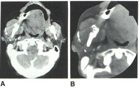
Fig. 1.-73-year-old woman with 4 month history of right facial mass. She was referred for CT evaluation of right parotid mass. She had mild pain over right parotid region. A, Axial CT scan through midmasseteric region after administration of sialographic contrast material into Stensen duct on right. Right masseter is noticeably larger than its counterpart on left. Parotid gland appears normal, as do surrounding fascial spaces and planes. Cortical irregularity of mandible is noted. B, Magnification view at wide window for visualization of bony detail. Cortical irregularity in lateral aspect of mandible (arrows). Inflam-matory disease was not clinically or radiologically suspected; however, neo-plasm causing bony erosion could not be excluded. At surgery, an enlarged, histologically normal masseter was encountered along with bony exostosis.
and 15 in the neck. Sixty percent occurred in trapezius and masseter muscles and 19 involved the masseter exclusively. These lesions are rarely diagnosed preoperatively [17] and most often present as a localized swelling [18]. Pulsations, thrills, and bruits normally are absent [16, 18]. Pain is a presenting complaint in over one-half of the patients and is thought to be from local pressure caused by the enlarging mass.
The appearance of phleboliths has been reported as a radiologic finding in some cases [16]. These presumably contained a venous component, as in one of our cases. The CT appearance, however, of this lesion has not to our knowl-edge been reported before. Angiography has been reported to reveal the vascular nature of this neoplasm [16].
Surgical therapy has been used with varying success in most reported cases. We believe that intraarterial treatment using microembolization techniques provides a valuable ad-junct to surgery and in inoperable cases it may be the only therapy that can be used [19, 20].
Our second case (fig. 2) presented clinically with a parotid mass. CT clearly revealed the true extraparotid, intramasse -teric location of this lesion. In addition, the associated bony deformity suggested a benign chronic process diagnosed as an intramuscular hemangioma by subselective external ca-rotid angiography. The cause of the facial swelling in another case was obvious clinically (fig. 3). CT followed by angiogra-phay was invaluable, however, in defining the true extent of this mixed capillary and venous hemangioma as well as in defining the relative contributions of each component to the entire lesion. Since each of the two components of the lesion is treated by a different interventional radiologic method (direct puncture with subsequent instillation of absolute alcohol into the venous lesion and transarterial microembolization of the
capillary lesion), the importance of this determination cannot
[image:2.612.317.560.74.230.2] [image:2.612.56.300.98.237.2]AJNR:5, Sept/Oct 1984 CT OF BUCCOMASSETERIC REGION PATHOLOGY 613
A
B
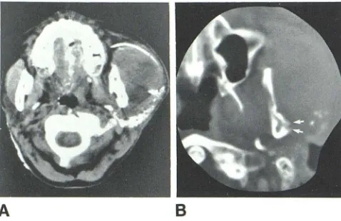
Fig. 2.-lntramasseteric hemangioma. A, Axial CT scan through hard palate/ floor of maxillary antral level. Mass clearly involves entire right masseter muscle without parotid involvement. Right masseter is greatly enlarged in comparison with left-side counterpart. Fascial planes and spaces are maintained and uninvolved. No difference in enhancement in this muscle is appreciated when compared with normal counterpart. Diagnosis of hemangioma causing swelling would be difficult to make. B, Axial CT scan through mid-maxillary antral level demonstrates pressure deformity of right posterolateral wall of maxillary antrum secondary to enlarged masseter (arrows). This finding attests to chronicity of this process. After subselective angiography, embolization of this lesion, sup-plied primarily by transverse facial artery, was performed, reducing much swellinq and alleviating pain.
A
B
Fig. 3.-8-year-old boy with life-long left facial swelling. His lesion had enlarged recently; the lesion was soft and compressible, and firm nodules were felt within its depths. A diagnosis of hemangioma was made from previous surgical biopsy; the hemangioma was believed to be confined to the parotid. Contiguous axial CT scans after intravenous administration of contrast material clearly demonstrate lesion to be bicompartmental. Peripheral mass (V) within subcutaneous soft tissues was venographically demonstrated to represent pure venous hemangioma. Scattered high-density flecks within hemangioma represent phleboliths, which were clinically palpable (arrows). Fairly well defined fatty soft-tissue plane is appreciated between lateral aspect of masseter and medial aspect of venous hemangioma. Left masseter is enlarged compared with right, and it enhances in homogeneously. This was angiographically proven to represent capillary (arterial) hemangioma (C). This CT study clearly demon-strates parotid to be uninvolved. Both lesions were treated via interventional radiologic techniques: venous part by instillation of absolute alcohol; capillary leSion (C) by transarterial microembolization of transverse facial artery.
Salivary G/and Tumor in Accessory Parotid Tissue
Salivary gland tumors constitute less than 3% of all head and neck neoplasms [21]. Between 75% and 80% of these lesions arise in the parotid gland. Mixed tumor or pleomorphic adenoma is found in the parotid 10 times more often than in
Fig. 4.-48-year-old woman with right-sided cheek mass. Physical ex-amination revealed a mobile, firm, non tender mass. Axial CT scan through maxillary antra reveals well circumscribed homogeneous mass (M) just lateral to right masseter. Well defined soft-tissue plane between mass, adjacent masseter muscle, and parotid gland. Mass was excised and pathologically proven to represent pleomorphic adenoma in accessory parotid tissue along Stensen duct.
the submandibular gland. It is also rarely found in the sublin-gual glands [22]. Eighty percent of tumors of the parotid are found in the superficial and caudal part of the gland. The mixed tumor is slow growing, well demarcated, and usually found in the tail. It accounts for 65% of all parotid neoplasms [2], is more common in females, and presents as a painless mass that on physical examination is firm, smooth, and mo-bile. Histologically, both epithelial and mesenchymal compo-nents are necessary for the diagnosis.
In our case, CT revealed a well defined mass of higher attenuation than normal parotid tissue, distinctly separate from the main parotid gland and underlying masseter (fig. 4). The mass appeared radiographically benign because of its sharp outline, but a more precise histologic diagnosis was impossible on the basis of the images. Connective or neural tissue neoplasm such as fibroma or neuroma should be included in the differential diagnosis.
Infectious Processes of the Masticator and Buccal Spaces
The superficial layer of the deep cervical fascia investing the muscles and organs of the buccomasseteric region
seg-regates these structures into clinically important potential spaces. Infections may be confined early in the process. However, avenues of communication exist between all of them, and an infection may spread along fascial and muscle planes, blood vessels, nerves, salivary ducts, and fat pads and thus can involve multiple contiguous spaces relatively quickly [23]. Infectious processes that spread by direct ex-tension do so by following the path of least resistance. The superficial layer of the deep cervical fascia within the face varies in structure from coarse areolar tissue to dense, fibrous sheets of connective tissue with obviously varying abilities to contain infectious processes [23].
[image:3.612.321.558.77.217.2] [image:3.612.56.299.78.233.2] [image:3.612.57.299.358.512.2]614 BRAUN ET AL. AJNR:5, Sept/Oct 1984
A
B
Fig. 5.-40-year-old man with left-sided erythematous facial mass and fever 2 weeks after extraction of several upper molars. A, Axial CT scan through superior alveolar ridge revealed marked soft-tissue swelling on left in region of subcutaneous tissues, buccinator region (b), masseter and masseteric space (m), and buccal fat pad (f). Parapharyngeal space medial to mandible is uninvolved. B, Axial CT scan through floor of maxillary antra reveals similar findings, but superior aspect of buccal fat pad is less involved at this level. Bone window images (not shown) revealed no evidence of osseous destruction. Masticator space was incised posteriorly, and 25 ml of foul-smelling pus was drained from this region Treatment for osteomyelitis was not instituted.
A
B
Fig. 6.-35-year-old woman developed swelling of left anterior cheek region after upper respiratory tract infection. No recent history of dental treatment or trauma. Physical examination revealed a tender, nonfluctuant mass in the left parotid region, and a parotid-space abscess was diagnosed clinically. A, Axial CT slice through superior alveolar ridge and level of Stensen duct after administration of sialographic contrast material. Masseter of decreased density compared with normal counterpart and greatly enlarged. Lucent area in pos-terior medial aspect of masseter. Stensen duct and parotid gland are com-pressed but uninvolved, as are pterygoid region and parapharyngeal space. B, Magnification view of bone-window image through mandible reveals evidence of bony irregularity laterally (arrows), consistent with osteomyelitis. At surgery 20 ml of purulent and foul-smelling material was drained from medial masseteric space. Osteomyelitis of mandible was confirmed.
The buccal space is limited medially by the buccinator. In most adults the roots of the molars are inferior to the attach-ment of the buccinator, so root abscesses are usually con-fined to the oral vestibule medial to the cheek [23]. In adults with long roots, however, or in children with incompletely
erupted maxillary or mandibular teeth whose roots extend
beyond the insertion of the buccinator, infection can more
easily involve the buccal space [26].
A
B
Fig. 7.-22-year-old man with left-sided facial mass. On physical examina-tion the mass was firm, mobile, and non tender. A, Axial CT scan through midmasseter region after injection of sialographic contrast material into Stensen duct. Lucent mass (m) just lateral to masseter and anterior to parotid gland. Soft tissue and fascial planes are well preserved. Mass is clearly extraparotid. B, Magnification view of left buccomasseteric region with cursor placed over mass reveals clearly negative attenuation value consistent with lipoma. Contrast within Stensen duct (arrows). Lipoma was removed at surgery.
Fig. 8.-44-year-old man with right-sided, firm cheek mass. On physical examination mass was slightly tender and relatively mobile. Axial CT slice through superior alveo-lar ridge reveals well circumscribed mass just lateral to buccinator muscle (b). Mass is confined by anterior ex-tension of masseteric fascia (white arrow) and relatively clear soft-tissue plane is between mass and buccina-tor. Mass is within anterior reaches of buccal fat pad (black arrow), without involving masseter itself. Adenocar-cinoma of Stensen duct was removed surgically.
Clinically, infections of the masticator space are manifested by cheek swelling, pain, and trismus. Treatment consists of surgical drainage and appropriate antibiotic therapy. Gaugh-ran [27] considers it important that a preoperative determi-nation be made as to whether the fat pad or the muscle is involved within the masticator space. The particular compo-nent of the involved masticator space should be approached directly to effect proper drainage with a minimum of tissue destruction. It is apparent that this preoperative determination can be aided by CT.
[image:4.612.57.303.83.236.2] [image:4.612.65.560.84.238.2] [image:4.612.57.296.349.504.2]AJNR:5, Sept/Oct 1984 CT OF BUCCOMASSETERIC REGION PATHOLOGY 615
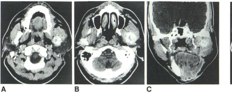
Fig. 9.-41-year-old man 8 months after resection of osteosarcoma of left mandible. Swelling was present in left cheek region. A, Axial CT slice through superior alveolar ridge reveals recurrence of osteosarcoma within posterior aspect of masseter on left (arrows). Mass does not extend to parapharyngeal space medially nor does it extend into parotid posteriorly. Tissue planes are relatively well preserved. B, Axial CT slice through mid aspect of maxillary antra. Most superiorly, mass involves lateral pterygoid muscle as well. Parapharyngeal space and parotid are radiographically uninvolved. C, Coronal scan through central aspect of mass. Both masseter and pterygOid muscles are involved at this level. Lateral pterygoid plate is intact, as is skull base. Radical removal of recurrent osteosarcoma was performed.
Fig. 10.-69-year-old man with left tonsillar region and left cheek mass.
Axial CT scan through inferior aspect of
maxillary antra reveals irregular necrotic mass in left tonsillar region that extends
to masseteric space (arrows), involving
medial surface of masseter and produc-ing facial swelling. Biopsy proved this to be squamous carcinoma of tonsil.
Miscellaneous Masses
Lipomas of the head and neck are benign lesions usually found within the subcutaneous tissues. They are usually well encapsulated and of little clinical significance. Over one-half involve the buccal mucosa [2]. While they are rarely large enough to be detectable as a mass, they can do so, as shown by the patient illustrated in figure 7, who presented with a cheek mass. CT was able to accurately define the extent of and determine the histological diagnosis of this lesion because of the negative attenuation value of fat.
Carcinoma of Stensen duct is a rare lesion that may be either mucoepidermoid, squamous, or adenomatous [2]. Pa-tients usually present with a cheek mass. Our representative patient had a well defined mass in the course of the duct,
simulating a benign neoplasm (fig. 8). A diagnosis of malig-nancy is difficult to arrive at, given this CT picture. Parotidec-tomy, along with a very wide excision of duct and adjacent soft tissues, is the treatment of choice [28].
Patients with osteosarcoma of the jaws usually present at a later age than patients harboring this neoplasm elsewhere in the skeleton [2]. This entity is usually seen in males, with the mandible being the most likely site of occurrence.
Radiographically the lesion may be predominantly blastic or lytic. A local recurrence of a blastic bone-producing lesion is illustrated (fig. 9). In this patient with a recurrent cheek mass, the tumor was noted to involve the substance of the masseter as well as neighboring structures. CT in both the axial and coronal planes accurately depicted the extent of the neoplasm and directed further radical surgery.
Squamous cell carcinomas of the tonsil are second in frequency to carcinoma of the larynx among malignancies of the upper respiratory tract [29]. Men are afflicted more often
then women by this disease seen in older age groups. Since the tonsillar region is relatively insensitive from a clinical point of view, patients often present with a sore throat, neck mass,
or, as in our example, a cheek mass (fig. 10). Local extension of tonsillar carcinomas is common and occurs early in the
disease. Radiation therapy is the treatment of choice [2].
Conclusions
CT is the imaging method of choice for patients with buccomasseteric region masses. In cases of neoplasm, the
extent of involvement of neighboring vital structures is usually seen easily. The assessment of malignancy versus benign disease, however, may be difficult (fig. 8). In cases of inf
ec-tious disease, CT can accurately reveal which fascial spaces
and planes are involved as well as if underlying bone is
affected and will direct the best surgical approach based on the altered anatomy. CT provides important anatomic detail not previously available to the head and neck surgeon, and we believe has made, and will continue to make, a great
impact on the treatment of patients with facial masses.
REFERENCES
1. Braun IF, Hoffman JC Jr. Computed tomography of the bu
cco-masseteric region: 1. Anatomy. AJNR 1984;5:605-611
2. Batsakis JG. Tumors of the head and neCk, 2d ed. Baltimore:
Williams & Wilkins, 1979
3. Waldhart E, Lynch JB. Benign hypertrophy of the masseter
muscles and mandibular angles. Arch Surg 1971;102: 115-118
4. Hersch JG. Hypertrophy of the masseter muscle. Arch Otolar
-yngo/1946;43:593-596
5. Drummond JA, Mcintosh CA. Unilateral hypertrophy of the m
as-seter muscle. Am J Surg 1954;87:711-714
6. Maxwell JH, Waggoner RW. Hypertrophy of the masseter mu
s-cles. Ann Otol Rhinal Laryngo/1951;60:538-548
7. Guggenheim P, Cohen L. The nature of masseteric hypertrophy.
Arch Otolaryngo/1961;73:15-28
8. Barton RT. Benign masseteric hypertrophy. JAMA 1957;164:
1646-1657
[image:5.613.54.442.82.237.2]616 BRAUN ET AL. AJNR:5, Sept/Oct 1984
mandible. J Oral Maxillofac Surg 1957;15:329-331
10. Gurney CEo Chronic bilateral benign hypertrophy of the masseter
muscle. Am J Surg 1947;73:137-139
11. Eubanks R. Surgical correction of masseter muscle hypertrophy associated with unilateral prognathism. J Oral Maxillofac Surg 1957;15:66-69
12. Masters F, Georgiade N, Pickrell K. Surgical treatment of benign
masseteric hypertrophy. Plast Reconstr Surg 1955; 15: 215-221
13. Tempest MN. Simple unilateral hypertrophy of the masseter
muscle. Br J Plast Surg 1951;4:136-139
14. Bloem JJAM, Van Hoff RF. Hypertrophy of the masseter
mus-cles. Plast Reconstr Surg 1971;47: 138-144
15. Coffey RJ. Unilateral hypertrophy of the masseter muscle.
Sur-gery 1941 ;11 : 815-818
16. Ott JES. Hemangiomata in skeletal muscle. Br J Surg
1957;44: 496-501
17. Clem is JD, Briggs DR. Changus GW. Intramuscular hemangioma
in the head and neck. J Otolaryngo/1975;4:339-347
18. Conley JJ, Clairmont AA. Intramuscular hemangioma of the
masseter muscle. Plast Reconstr Surg 1977;60: 121-124
19. Berenstein A, Kricheff II. Microembolization techniques of
vas-cular occlusion: radiologic, pathologic and clinical correlation.
AJNR 1981;2:261-267
20. Kendall B, Moseley I. Therapeutic embolization of the external
carotid arterial tree. J Neural Neurosurg Psychiatry 1977;40: 937-950
21. Leegaard T, Lindeman H. Salivary gland tumors: clinical picture
and treatment. Acta Otolaryngol [Suppl] (Stockh)
1979;263: 155-159
22. Leading article. Salivary gland tumors. Lancet 1969;1 :655-657
23. Paonessa OF, Goldstein JC. Anatomy and physiology of head
and neck infections with emphasis on the fascia of the face and neck. Otolaryngol Clin North Am 1976;9:561-580
24. Hall C, Morris F. Infections of the masticator space. Ann Otol
Rhinal Laryngo/1941;50: 1123-1140
25. Collier FA, Yglesias L. Infections of the lip and face. Surg Gynecol Obstet 1935;60:277-290
26. Archer WHo Oral and maxillofacial surgery, 5th ed. Philadelphia:
Saunders, 1975:447-473
27. Gaughran GRL. Fasciae of the masticator space. Anat Rec
1957;129:383-400
28. Gaisford JC. Hanna DC, Sotereanos GC. Primary cancer of
Stensen's duct. Arch Otolaryngo/1965;82:45-48
29. Whicker JM, DeSanto LW, Devine KD. Surgical treatment of