printed in Great Britain © The Company of Biologists Limited 1984
THE IONIC BASIS OF THE RESTING POTENTIAL IN
A CROSS-STRIATED MUSCLE OF THE AQUATIC
SNAIL LYMNAEA STAGNALIS
BY B. L. BREZDEN AND D. R. GARDNER
Carleton University, Department of Biology, Ottawa, Ontario KJS 5B6, Canada
Accepted 27 June 1983
SUMMARY
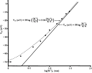
The mean resting potential in the heart ventricle muscle cells of the freshwater snail Lymnaea stagnalis was found to be —61-2 ± 3*5 (..) mV (ranging from — 56mV to — 68mV). The average intracellular potassium concentration was estimated to be 51 -5 ± 14-6 (..) m (ranging from 27-8 m to 77-3 m). The membrane of the heart ventricle muscle cells appears to be permeable to both potassium and chloride, as changes in the extracellular concentration of either of these ions resulted in a change in the membrane potential. A ten-fold change in the extracellular potassium concentration was associated with a 50-4 ± 3-8 (..) mV slope when the potassium con-centration was above about 6 m. Deviations from the straight-line relation predicted for a potassium electrode could be accounted for by introducing a term for sodium permeability. The ionic basis of the membrane potential in these cells can be described by a modified form of the Goldman-Hodgkin-Katz equation.
INTRODUCTION
The ionic basis of the membrane potential in molluscan muscle cells has received little detailed attention. In the few cases examined so far, the membrane potential appears to be a function, to a greater or lesser extent, of the extracellular potassium concentration (Hill, Greenberg, Irisawa & Nomura, 1970; Burnstock, Greenberg, Kirby & Willis, 1967; Twarog, 1967). Chloride ions may also be involved (Shigeto, 1970).
Resting potentials of between — 27 mV and —65 mV have been reported for dif-ferent molluscan muscles (Kater, Heyer & Hegmann, 1971; Kidokoro, Hagiwara & Henkart, 1974; Florey & Kriebel, 1969; Kobayashi & Shigenaka, 1978; Twarog, 1967; Shigeto, 1970). These resting potentials are low compared to those reported for vertebrate muscles (cf. —92 mV for frog sartorius muscle; Adrian, 1956). This could reflect either damage to the sarcolemma during impalement, or a genuine difference. In some cases the resting potential declined rapidly following impalement (e.g. Kidokoro et al. 1974) which would suggest that the membrane had been damaged. The values for resting potentials determined by high resistance gap voltage clamp
techniques, on the other hand, incorporate other errors. For example, Sugi 8? Yamaguchi (1976), using an oil gap voltage clamp, reported a resting potential of -12mV to - 2 5 mV for the ABRM of Mytilus edulis (cf. - 6 5 mV; Twarog, 1967). However, the resting potentials of the muscles of different invertebrates, e.g. Ascaris
lumbricoides, may be genuinely low ( — 30 mV to —40 mV; Brading & Caldwell, 1964;
del Castillo, de Mello & Morales, 1964a,6; Toida, Kuriyama, Tashiro & Ito, 1975). The apparently low resting potentials of some molluscan muscles, therefore, does not represent a unique situation. This could reflect the comparatively low values of the potassium equilibrium potential calculated for some molluscan muscles. For exam-ple, the K+ equilibrium potential of myocardial muscle fibres of Crassostrea gigas was calculated from measurements of intracellular K+ concentration to be —68 mV (Shigeto, 1970), which is considerably less negative than the K+ equilibrium potential of about —100mV in vertebrate skeletal muscles (Adrian, 1956; Hodgkin & Horowicz, 1959).
If muscle cell membranes are freely permeable to K+ and Cl~, the Donnan equilibrium theory holds that under conditions of a constant [K+]e X [Cl~]e product
(Boyle & Conway, 1941), the membrane potential is given by:
S (1)
[K Jiwhere a decade change in the extracellular K+ concentration should be associated with a 58 mV change in the membrane potential on the condition that the membrane is not significantly permeable to ion species other than K+ or Cl~ (cf. Hodgkin & Horowicz, 1959).
In the present investigation we have examined the ionic basis of the resting potential in heart ventricle muscle cells of the aquatic snail, Lymnaea stagnalis.
MATERIALS AND METHODS
The experimental animals
Two subspecies of the freshwater snail Lymnaea stagnalis were used in these investigations. L. stagnalis appressa were collected locally whileL. stagnalis stagnalis were imported from England (Griffin & George Ltd., Gerrard Biological Centre, East Preston, West Sussex, U.K.). The animals were kept in large aquaria containing standing, aerated water and were fed lettuce leaves ad libitum. During the course of the experiments no intersubspecific physiological differences were observed, and the two subspecies were used interchangeably.
The preparation and apparatus
Electrophysiology of molluscan heart muscle 307
Table 1. Composition of the constant [K+]e X [Cl~] e product salines ([K+] X [Cl~] = 60mm) K+ 1-0 2 0 4-0 6-0 8-0 10-0 150 20-0 25-0 30-0 40-0 Na+ 41-65 40-8 391 37-4 35-7 34-0 29-75 25-5 21-25 17-0 8-5 Ca2+ 0-5 0-5 0-5 0-5 0-S 0-5 0-5 0-5 0-5 0-5 0-5
Mg2*
100 10-0 10-0 10-0 10-0 10-0 100 10-0 10-0 10-0 10-0 Ac" 0-2 0-4 0-8 1-2 1-6 2-0 3 0 4-0 5-0 6-0 8-0 Glu" 1-0 2-0 4-0 6 0 8-0 100 150 20-0 25-0 30-0 40-0
cr
60-0 30-0 15-0 10-0 7-5 6 0 4 0 3-0 2-4 2-0 1-5 HEPES 5-0 5-0 5-0 50 5 0 5-0 5-0 5-0 5-0 5-0 5-0Concentrations given in w .
Glu = glutamate, Ac = acetate (note elevated Mg2* and reduced Caz+ concentrations).
HEPES was adjusted to pH 7-6 with NaOH. 5 mm HEPES added 2-35 mM-Na+ to the salines.
face. The Sylgard block with attached ventricle was placed in a preparation chamber (volume = 1 ml) in which the superfusion fluid could be readily exchanged via a nine-way non-return valve (see Brezden & Gardner, 1983).
The membrane potential of the heart muscle cells was measured using 3 M-KC1-filled intracellular glass microelectrodes and standard electrophysiological tech-niques, employing a WPI model KS 700 microelectrode amplifier (WPI Instruments Co., Ridgewood, New Jersey). Permanent recordings of membrane potential were made on a Linear Instruments model 395 chart recorder (Linear Instruments Corp., Irvine, California).
The saline solutions
Salines with K+ concentrations of 1 mM to 40 mM were prepared such that the product of the K+ and Cl~ concentrations was constant at 60mM (Table \). These salines contained elevated Mg2* and reduced Ca2+ concentrations; the reason for this is discussed below.
In addition to the above salines, normal Lymnaea saline (l-6mM-K+), low Cl~ (5 mM) saline, Na+-free saline and Na+-free saline with low Ca2+ and high Mg2"1" concentrations were used (Table 2).
The experimental regime
I n these experiments, the response of the membrane potential of individual Lymnaea heart ventricle muscle cells to changes in the extracellular K+ and Cl~ concentrations was measured. The K+ concentration was varied in a random sequence to reduce systematic experimental error. After each change in the K+ concentration the prepara-tion was returned to 1 mM-K+ saline (baseline membrane potential). The experiment was considered successful only if a complete recovery of the baseline membrane
Table 2. Composition of normal and substituted Lymnaea salines
Salt
NaCl
KC1
CaCl2
MgCfe HEPES-Na HEPES-K BDAC
Normal
50-0
1-6 3-5 20
5-0 0 0
Na+-free
0 0 3 5 2 0 0 5-0 50-0
Na+-free, low Ca2+, high Mg*+
0 0 0-5 10-0
0 5-0
42-5
L o w C P
5C1, 45 G 1-6G 3-5A 2-OA
5-0 0 0
Concentrations given in mM.
BDAC = bis(2-hydroxyethyl) dimethyl ammonium chloride.
HEPES was adjusted to pH 7-6 with 10 M-NaOH or 10 M - K O H (the latter was used in Na+-free saline). 5 mM-HEPES-Na added 2-35mM-Na+ to the saline while SmM-HEPES-K added 2-72mM-K+ (i.e., the Na+-free saline contained 2-72mM-K+).
In the low Cl~ saline, Cl = chloride salt, G = glutamate salt and A = acetate salt.
ventricle muscle cells. As a result, it was not possible to maintain successful microelectrode insertion. Various mechanical means (e.g. 'floating' microelectrodes, more rigid preparation mounting, etc.) were not successful in eliminating this problem, probably because of the extremely fragile nature of this tissue. Therefore, the Mg2"1" concentration was elevated to lOmM and the Ca2+ concentration was reduced to 0-5 mM to inhibit contraction. Following this procedure, successful im-palement could be maintained through many solution changes.
A further criterion for an acceptable experiment was that the membrane potential in 1 mM-K+ be more negative than, or equal to, —55 mV; cells with more positive resting potentials were judged to be damaged.
In the following text, '±x' denotes the standard deviation of the given value.
RESULTS
The resting potential
The mean resting potential oiLymnaea heart ventricle muscle cells in normal saline was found to be - 6 1 - 2 ± 3-5mV (N= 30) (ranging from -56mV to —68mV).
The potassium and chloride dependence of the resting potential
A reduction of the extracellular Cl~ concentration from 62-6 mM to 5 mM resulted in a transient depolarization of the membrane potential. This depolarization was of the order of 6 mV in magnitude and the membrane potential reverted to a level which was 2 mV less negative than the original resting potential after about 3 min in the reduced Cl~ saline. Restoration of the original Cl~ concentration induced a hyper-polarization of about 7mV, followed by a rehyper-polarization of the membrane potential to a stable level about 1 mV more negative than the original resting potential (Fig. 1).
Electrophysiology of molluscan heart muscle
309
- 5 4 '
- 5 6
' - S 8
0 2 4 0 0-5 1 1 - 5 2 2-5 3 3-5 0 0-5 1 1-5 2 2-5 3 3-5 4
- 6 0
-62
[image:5.451.57.374.360.611.2]- 6 4
Fig. 1. Effect of reduced chloride concentration on the membrane potential of a Lymnaea ventricle muscle cell. Note change in time scale. N 8. Data from a single cell; one of eight experiments.
- 1 0
- 2 0
£ - 3 0
-40
-50
-60
-70
V. (mV, = 58 log ( Q + 0-04^) „
-Vm (mV) = 581og
41-4
0-5 1 0
log[K+] c (maj)
1-5 2 0
In an attempt to account for the observed deviation from an ideal potassium electrode1; a modified form of the Goldman-Hodgkin-Katz equation (Hodgkin & Katz, 1949) as applied by Moreton (1968) was used. This equation is given by:
V F / R T _ [ K+]C PN.[Na+]e
[iTTT P K [ K
+] , ^ 'and predicts a linear relationship between eV F'R T and [K+]e if [K+]i and the ratio
P N8/ P K remain unchanged (cf. Moreton, 1968). The inverse of the slope of the line
gives an estimate of [K+]i, and the value of the term PN«/PK can be calculated from
the Y-intercept. The data from Fig. 2 were plotted using equation (2) giving a value for [K+]i of 41-4mM, and a PN^/PK ratio of 0-04 (Fig. 3). Substituting these values
in equation (2) and rearranging to solve for V (where [Na+]e was known) gave the
values for the dashed line in Fig. 2.
The above calculations were performed for each of the 20 successful experiments. The mean estimated intracellular K+ concentration was 51-5 ± 14-6 mM (ranging from 27-8 mM to 77-3 mM) and the mean ratio of PN«/PK was 0-07 ± 0-003 (ranging from 0-01 to 0-14). The experimental points were very close to those predicted by equation (2) (Table 3).
TTie effect of sodium-free saline on the membrane potential
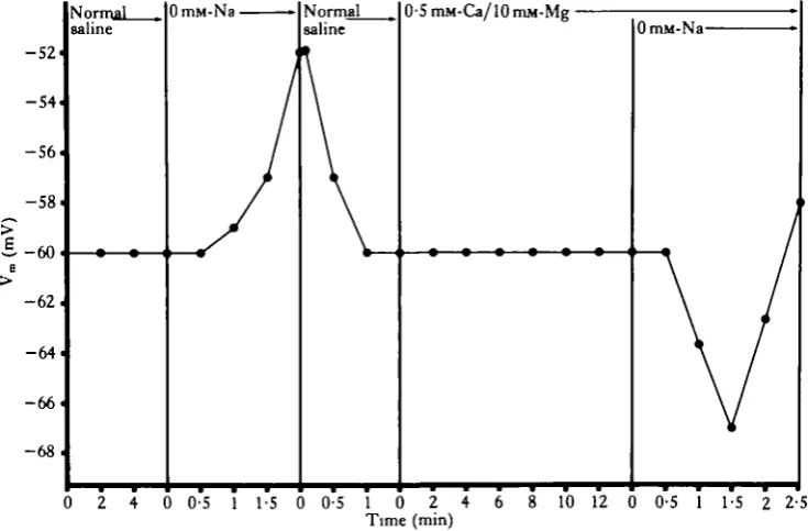
The exposure of heart ventricle muscle cells to Na+-free saline resulted in a gradual depolarization of the membrane potential and eventual contraction of the muscle cells.
0-8
0-7
0-6
StO-5
~0-4
S
0-3 •
0-2 i
0 1
OH
6 8 10 12 14 16 18 20 22 24 26 28 30 32 [ K+]
t (mM)
Fig. 3. Relationship between eVF'RT and the extracellular potassium concentration in
Electrophysiology of molluscan heart muscle
311
Table 3. Deviation between predicted and observed values for the membrane
poten-tial of Lymnaea heart ventricle muscle cells at various extracellular potassium con-centrations
l 2 4 6
8
10 15 20 25 30 40
The predicted values are from equation (2). JV = 20.
This depolarization could be reversed and the contraction prevented if the cells were exposed to normal saline shortly after the onset of depolarization (Fig. 4). However, if the cells were pre-treated with 0-5 mM-Caz+/l0mM-Mg2+ saline, the subsequent exposure to 0mM-Na+/0-5mM-Ca2+/10mM-Mg2+ saline resulted in a transient hyperpolarization of the membrane potential (Fig. 4). Thus it would appear that the
Observed Vm( ± s . D . )
-62-2 (5-2) -58-7 (S-4) -52-5 (5-4) -45-1 (5-5) -38-4(5-8) -31-6(6-0) -24-8 (6-6) -19-5(6-3) -15-5(6-3) -11-0(6-3) -5-5 (5-9)
Expected Vo ( ± !
-63-0 (9-8) -55-3 (140) -51-1 (7-9) -44-4 (7-0) -40-3 (6-7) -34-2 (6-4) -27-0 (5-9) -22-7 (5-6) -17-5 (6-2) -12-9 (6-0) - 5 - 3 (6-3)
0 2 4 0 0-5 1 1 - 5 0 0-5 1 0 2 4 6 8 10 12 0 0-5 1 1-5 2 2-5
[image:7.451.42.410.367.608.2]removal of extracellular Na results in a hyperpolarization only if the influx of Ca " is prevented, although even 0-5mM-Ca2+/l0mM-Mg2+ saline did not prevent the contraction induced by the removal of extracellular Na+.
DISCUSSION
The chloride dependence of the resting potential
The membrane potential of Lymnaea heart ventricle muscle cells did not change by the predicted amount during the reduction or subsequent restoration of the original extracellular Cl~ concentration. In the experiment presented in Fig. 1, for example, the membrane potential should have changed from —58 mV to —26 mV when the Cl~ concentration was reduced from 62-6 miw to 5 rriM (cf. Hodgkin & Horowicz, 1959), whereas only a 6 mV depolarization was observed. However, the full magnitude of the expected change can only be measured when the extracellular Cl~ concentration is changed very rapidly (Hodgkin & Horowicz, 1959). In the present experiments about 1 min was required to exchange the bath contents completely (faster flow rates dis-lodged the microelectrodes).
Hodgkin & Horowicz (1959) reported that the resting potential in frog skeletal muscles was quickly restored following changes induced by lowering or restoring the extracellular Cl~ concentration, and attributed the restoration of the resting potential to an insignificant change in the intracellular K+ concentration during variations in the extracellular Cl~ concentration. However, a hysteresis in the value of the mem-brane potential following a change in the extracellular Cl~ concentration was observed in all of the Lymnaea muscle cells examined, i.e., within the 2—3 min time frame of the experiment the membrane potential always stabilized at a level about 2 mV less negative (following a reduction in the extracellular Cl~ concentration) and about 1 mV more positive (following the restoration of the original Cl~ concentration) than the original resting potential. These results suggest the possibility that the intracellular K+ concentration may have changed by as much as 4 HIM in these cells during alterations in the extracellular Cl~ concentration. If the intracellular K+ con-centration is low (as may be the case in Lymnaea ventricle muscle cells) the Donnan equilibrium theory [and using equation (2)] predicts that this change in the intracellular K+ concentration may be sufficient to account for the observed changes in the resting potential.
The potassium dependence of the resting potential
The membrane potential of Lymnaea heart ventricle muscle cells was found to be relatively insensitive to changes in extracellular K+ concentrations below about 6 mM. An average slope of 50-4 ± 3-8 mV per decade change in the extracellular K+ con-centration was obtained between 6mM and 40 mM. This is about 8mV lower than predicted by equation (1), suggesting that these cells are permeable to some ion(s) other than K+ and Cl~. It is unlikely that changes in the intracellular concentrations of K+ or Cl~ contributed to the reduction in the K-slope, since constant [K+]e 1
Electrophysiology ofmolluscan heart muscle 313
The sodium dependence of the resting potential
In all the muscle cells examined, the resting potential was considerably lower than that predicted by equation (1). This type of deviation could be explained if the cell membrane has a resting permeability to Na+. However, upon removal of extracellular Na+, the expected hyperpolarization (cf. Sattelle, 1974) was only observed if the magnesium concentration was elevated and the calcium concentration reduced. It would seem, therefore, that under normal conditions, the depolarization was a direct or indirect result of Ca2+ entry into the cell, and that this entry could be prevented by elevating the extracellular Mg2"1" concentration and lowering the extracellular Ca2+ concentration. Removal of extracellular Na+ inhibits, or possibly reverses the Na+-Ca2+ exchange mechanism (Baker, 1972), and increases the influx of Ca2+ into the cell (Coraboeuf, Gautier & Guiraudon, 1981). Whether or not an elevation of free intracellular Ca2+ levels alone could be responsible for the observed depolarization cannot be determined on the basis of the available evidence. Nevertheless, under appropriate conditions the removal of extracellular Na+ does cause a hyperpolariza-tion of the heart ventricle muscle cell membrane, an observahyperpolariza-tion which is consistent with the proposal that this membrane has a resting permeability to Na+.
A quantitative description of the membrane potential
The mean resting potential of —61-2 ± 3 5 mV in Lymnaea heart ventricle muscle cells is substantially less negative than that in vertebrate cardiac muscle (—90 mV to —100mV; Weidmann, 1956). Also, the mean estimated intracellular K+ concentra-tion of 51-5 ± 14-6 mM is considerably lower than that of vertebrate cardiac muscles (>100mM; Kunze & Russell, 1981; Walker, 1981). Apart from the relatively low level of resting potential and of intracellular K+ concentration, the ionic basis of the resting potential in Lymnaea heart ventricle muscle cells was found to be similar to that of frog skeletal muscle (cf. Hodgkin & Horowicz, 1959): the heart ventricle muscle cell membrane appears to be permeable to both K+ and Cl~ since changes in the extracellular concentration of either of these ions resulted in a change in the membrane potential. They also appear to have a resting permeability to Na+.
In conclusion, the membrane potential (assuming PNI/PK does not change) can be defined by equation (2). This equation produced values for the membrane potential which approximated those observed at extracellular potassium concentrations be-tween 1 mM and 40 mM in Lymnaea heart ventricle muscle cells.
One of us (DRG) thanks the Natural Sciences and Engineering Research Council of Canada for an operating grant (A-4290).
R E F E R E N C E S
ADRIAN, R. H. (1956). The effect of internal and external potassium concentration on the membrane potential of frog muscle. J. Pkysiol., Land. 133, 631-658.
^ K E R , P. F. (1972). Transport and metabolism of calcium ions in nerve. Prog. Biophys. mol. Biol. 24, 177-223. B Y L E , P. J. & CONWAV, E. J. (1941). Potassium accumulation in muscle and associated changes. J. Pkysiol.,
BRADING, A. F . & CALDWELL, P. C. (1964). The effects of ions on the resting potentials of muscle cells nf
Ascaris lumbricoides. J. Physiol., Land. 173, 36P.
BREZDEN, B. L. & GARDNER, D. R. (1983). The effect of the molluscicide Frescon on smooth and cross-striated muscles of Lymnaea stagnalis and Helix aspersa. Pest. Biochem. Physiol. (in press).
BURNSTOCK, G., GREENBERG, M. J., KIRBY, S. & WILLIS, A. G. (1967). An electrophysiological and pharmacological study of visceral smooth muscle and its innervation in a mollusc, Ponemplax albida. Comp.
Biochem. Physiol. Z3, 407-429.
CORABOEUF, E., GAUTIER, P. & GUIRAUDON, P. (1981). Potential and tension changes induced by sodium removal in dog Purkinje fibres: role of an electrogenic sodium-calcium exchange. J. Physiol., Land. 311, 605-622.
DEL CASTILLO, J., DEMELLO, W. C. & MORALES, T . (1964a). Influence of some ions on the membrane potential of Ascaris muscle. J. gen. Physiol. 48, 129-140.
DEL CASTILLO, J., DE MELLO, W. C. & MORALES, T . (19646). Hyperpolarizing action potentials recorded from the oesophagus of Ascaris lumbricoides. Nature, Land. 203, 530—531.
FLOREY, E. & KRIEBEL, M. E. (1969). Electrical and mechanical responses of chromatophore muscle fibres of the squid, Loligo opalescens, to nerve stimulation and drugs. Z. vergl. Physiol. 65, 98—130.
HILL, R. B., GREENBERG, M. J., IRISAWA, H. tc NOMURA, H. (1970). Electromechanical coupling in a
molluscan muscle, the radula protractor of Busycon canaliculatum.J. exp. Zool. 174, 331-348.
HODGKIN, A. L. & HOROWICZ, P. (1959). The influence of potassium and chloride ions on the membrane potential of single muscle fibres. J . Physiol., Land. 148, 127-160.
HODGKIN, A. L. & KATZ, B. (1949). The effect of sodium ions on the electrical activity of the giant axon of the squid. J. Physiol., Land. 108, 37-77.
KATER, S. B., HEYER, C. & HEGMANN, J. P. (1971). Neuromuscular transmission in the gastropod mollusc
Heliosoma trivolvis: Identification of motoneurons. Z. vergl. Physiol. 74, 127—139.
KIDOKORO, Y., HAGIWARA, S. & HENKAUT, M. P. (1974). Electrical properties of obliquely striated muscle fibre of Anodonta glochidium. J. comp. Physiol. 90, 321-338.
KOBAYASHI, M. & SHIGENAKA, Y. (1978). The mode of action of acetylcholine and 5-hydroxytryptamine at the neuromuscular junctions in a molluscan muscle (radula protractor). Comp. Biochem. Physiol. 50C, 115-122. KUNZE, D . & RUSSELL, J. M. (1981). pH related potassium movements in rabbit atrium. In Ion Selective
Micrvelectmdes and Their Use in Excitable Tissues, (eds E. Sykovi, P. Hnfk & L. Viklickf), pp. 95-97. New
York & London: Plenum Press.
MORETON, R. B. (1968). An application of the constant field theory to the behaviour of giant neurones of the snail Helix aspersa. J. exp. Biol. 48, 611-623.
SATTELLE, D . B. (1974). Electrophysiology of the giant nerve cell bodies of Limnaea stagnalis (L.) (Gastro-poda: Pulmonata). J. exp. Biol. 60, 653-671.
SHIGETO, N. (1970). Excitatory and inhibitory actions of acetylcholine on hearts of oyster and mussel. Am. J.
Physiol. 218, 1773-1779.
SUGI, H. Sc YAMAGUCHI, T . (1976). Activation of the contractile mechanism in the anterior byssal retractor muscle oiMytilus edulis.J. Physiol., Land. 257, 531-547.
TOIDA, N., KURIYAMA, H., TASHIRO, N. & ITO, Y. (1975). Obliquely striated muscle. Phvsiol. Rev. 55, 700-756.
TWAROG, B. M. (1967). Excitation oiMytilus smooth muscle. J . Physiol., Land. 192, 857-868.
WALKER, J. L. (1981). Intracellular potassium and chloride measurements in sheep cardiac Pcrkinje fibres. In
Ion Selective Microelectrodes and Their Use in Excitable Tissues, (eds E. SykovS, P. Hnfk & L. Viklicky),
pp. 55—66. New York & London: Plenum Press.
![Table 1. Composition of the constant [K+]e X [Cl~] e product salines ([K+] X [Cl~] =60mm)cr](https://thumb-us.123doks.com/thumbv2/123dok_us/1182176.641093/3.451.41.416.110.248/table-composition-constant-k-cl-product-salines-cl.webp)