Jf. exp. Biol. 112, 95-112 (1984) 9 5
Printed in Great Britain © The Company of Biologists Limited 1984
CHANGES OF MEMBRANE CURRENTS DURING
LEARNING
BY DANIEL L. ALKON
Section on Neural Systems, Laboratory of Biophysics, IRP, National Institute of Neurological and Communicative Disorders and Stroke, National Institutes of Health at the Marine Biological Laboratory, Woods
Hole, MA 02543, U.SA.
SUMMARY
The integrated response of a population of neurones during conditioning results in long-term (days) changes of specific membrane currents within identified neurones. Prolonged elevation of intracellular calcium during conditioning causes a persistent increase of excitability by reducing K+ currents (IA and probably Ic«»+ - K+) in the membranes of identified somata. This Ca2+-mediated reduction of K+ currents, which encodes a learned stimulus association is thought to involve changes of Ca2+ -calmodulin-dependent phosphorylation of distinct membrane proteins. These changes are contrasted with the short-term regulation of currents by neurohormones during altered behavioural states such as arousal.
In searching for cellular mechanisms by which nervous systems store learned in-formation, it is helpful to differentiate properties which are genetically programmed from those which are conferred by the interaction of an organism with its environ-ment. With mechanisms of learning, as with those underlying developmental trans-formations, a chronology must be reconstructed — a sequence of processes which arise as a consequence of preceding conditions. To construct such a chronology, the cellular site at which learned information is stored must be accessible to analysis so that we may watch it during sequential transformations, and so that we can measure with appropriate morphological, biophysical and biochemical techniques the nature and magnitude of the critical changes.
Accessibility to sites of associative learning has been elusive for vertebrates, in which extracellular potentials have been recorded as correlates of acquisition and retention of conditioned behaviour (Woody, Vassilevsky & Engel, 1970; Woody & Black-Cleworth, 1973; Woody et al. 1974; Berger, Alger & Thompson, 1976; Cohen & MacDonald, 1976; Berger & Thompson, 1977, 1978; Brons & Woody, 1980; Thompson et al. 1982; Woody, 1982). It has also been largely true for invertebrates such as the bee, locust and the gastropod molluscs Pleurobranchaea, Umax and
Aplysia. Hoyle and his colleagues recorded changes of impulses in certain locust
motor neurones during learning (Hoyle, 1982). However, these changes have only been measured unequivocally for minutes, and their site of origin, and thus their
96 D. L. ALKON
accessibility, have not been determined. Furthermore, the synaptic organization (M the neural elements which mediate the training response remains obscure.
Workers using Aplysia and Pleurvbranchaea have had more success in identifying neurones within relevant sensory pathways (Castellucci, Pinsker, Kupferman & Kan-del, 1970; Gillette & Davis, 1977; Gillette, Gillette & Davis, 1980). However, a close and specific correlation of neuronal changes with learned avoidance of food in
Pleuro-branchaea (as opposed to its being in a food-satiated state), has not been made, as it
has for vertebrate conditioning. Nor has it been possible to measure correlated changes in identified neurones or neuronal aggregates of Umax or Aplysia during or after associative training. For dissected and reduced preparations, short-term correla-tions (Carew, Hawkins & Kandel, 1983) have been made, but their relevance to the neurophysiology of living animals must be established (see for example, Kanz, Eber-ley, Cobbs & Pinsker, 1979).
Furthermore, the type of learning involved with some of these gastropods has not been entirely resolved. Some assert that non-associative behavioural changes such as habituation and sensitization can underlie associative learning, such as classical con-ditioning (Kandel & Schwartz, 1982), although any relevance of non-associative to associative mechanisms remains to be demonstrated. Even those behavioural changes called 'associative' require further study before their 'associative' nature can be con-sidered certain.
Mpitsos & Collins (1975) trained Pleurobranchaea to avoid food substances by pairing the food with shocks to the head. Unpaired presentations did not produce an effect. These experiments could be simplified by controlling the different states of satiation which might result from the training. Long-term sensitization of a neophobic reaction to the food substances by shock should also be examined. The recent experi-ments of Gelperin and his colleagues with Umax (Gelperin, Wieland & Barry, 1984; Sahley, Rudy & Gelperin, 1984) require these same controls, since all measures of avoidance behaviour following pairing of a food substance with quinine were made by comparison with responses to a 'safe' food. A neophobic reaction to a 'safe' food (on which the animal had been raised) might be entirely habituated, while quinine could sensitize the neophobic reaction to a food substance with which it was paired during training. An apparently associative behavioural change (i.e. one dependent on the pairing of a food substance with quinine or an electric shock) might be simulated by particular combinations of two non-associative behavioural effects such as habituation and sensitization.
In recent experiments on Aplysia, tactile stimulation of the gill withdrawal res-ponse, which habituates, has been paired with electrical stimulation of the tail, which causes sensitization (Carew, Walters & Kandel, 1981). This causes the animal to withdraw its gill more rapidly and reliably. However, the possibility exists that the shock-induced sensitization prevents the touch-induced habituation. This is sugges-ted by the observation that repeasugges-ted presentation of the shock alone produces, to a significant degree, the same effect as repeated, paired, presentations. Other charac-teristics of associative learning, such as the stimulus specificity shown for Aplysia
(Care-wet hi. 1981), and the blocking shown for Umax (Sahley etal. 1984), have yet
Membrane changes during learning 97
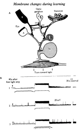
•mollusc Hermissenda crassicornis to localize sites of neuronal change which could play a causal role in associative learning, and which are accessible to quantitative analysis. We first found sites of convergence between three sensory pathways: the visual, vestibular and chemosensory (Alkon & Fuortes, 1972; Alkon, 1973, 1974a, 198(to, 1983; Alkon, Akaike & Harrigan, 1978). With simultaneous intracellular recordings from identified pre- and postsynaptic elements in thousands of adult ner-vous systems we constructed a working blueprint of the circumoesophageal nerner-vous system (Fig. 1). Within this blueprint, the flow of sensory information could be traced from the input stages through the integrative centres to the output motor cells of relevant muscle groups. The effects of sensory stimuli such as light (transduced by five photoreceptors in each eye) and rotation (a gravitational stimulus transduced by thirteen hair cells in each statocyst, a primitive vestibular organ) could be studied at every level of integration: sensory cells, interneurones and motor neurones (Fig. 2A). Based on the known convergences between the visual and statocyst pathways, and the specific responses of the network to light and rotational stimuli with specific temporal relationships, we hypothesized that Hermissenda could be conditioned by repetition of paired light and rotational stimuli.
In fact, it has been possible to demonstrate most of those features which charac-terize vertebrate classical conditioning. Rotation, which serves as an unconditioned stimulus, elicits a consistent and reliable 'clinging' response (Alkon, 19746) manifest by measurable contraction of the 'foot' (particularly the caudal half of the ventral surface). In addition, the animal moves towards a light source (Alkon, 19746). This is accompanied by lengthening of its foot, again particularly the caudal half. After light and rotation are repeatedly paired (Alkon, 19746) (i.e. maximal light precedes maximal rotation by l-0s), light elicits an entirely new response: contraction of the foot (Fig. 3), the response reflexly elicited by rotation. As a result of the conditioning procedure, light has taken on the meaning of rotation as measured by a new learned response (Lederhendler, Gart & Alkon, 1983). This learning behaviour is associative in that it is not produced by explicitly unpaired light and rotation, or light and rotation occurring with a randomly-varying interval during training (Crow & Alkon, 1978). It is stimulus specific, increases as a function of practice (i.e. it shows acquisition), can be retained for many weeks, extinguishes (Richards, Farley & Alkon, 1983) and shows savings. It also has a requirement for contingency (as well as contiguity) of the associated stimuli (Farley & Kern, 1983). This was shown by the degradation of the learning by interpolation of unpaired light or rotation stimuli. Finally, there is no long-term habituation or sensitization following training.
Intracellular recordings at each level of integration within the visual system of conditioned and control animals uncovered many learning-correlated changes of neuronal properties and response characteristics (Crow & Alkon, 1980; Farley & Alkon, 1982; Lederhendler, Goh& Alkon, 1982; West, Barnes & Alkon, 1982). Such recordings were made from progressively reduced preparations including living animals, isolated nervous systems and individual neurones isolated from all others, of conditioned and control animals. By this means it was possible to reconstruct a sequence of responses and changes during training.
RECEPTORS
COMMISSURE
u
t'
>
l?CENTRAL ?4 0 INTERNEURONES tMN 1 MOTOR NEURONES
MUSCLE GROUPS
Membrane changes during learning
99
Optic
ganglion Statocvst
I i I
Turn toward light40 s after first light step
V
30-s interval
20 mV
1
L .
Fig. 2. (A) Schematic diagram of visual pathway and its convergence with the statocyst pathway. The Type B photoreceptor (B) causes monosynaptic inhibition of the medial Type A photoreceptor (A). The medial Type A photoreceptor causes monosynaptic excitation of ipsilateral interneurones (I), which are also excited by ipsilateral hair cells (HC). Ipsiiateral hair cell impulse* and Type B impulses cause a transient inhibition (not shown here) and are followed by long-lasting effective excitation ( + ) of the S/E optic ganglion cell and thereby the Type B cell (Goh & Alkon, 1984). (B) Intracellular voltage recordings of Nermissenda neurones during and after light and rotation stimuli. Responses of a type B photoreceptor to the second of two succeeding 30-s light steps (with a 90-» interval intervening). The cell's initial resting potential, preceding the first of the two light steps in (1), (2), and (3), is indicated by the dashed lines. Depolarization above the resting level after the second of
the two light steps is indicated by shaded areas. (1) Light steps (~ 10* erg cm"2 »"') alternating with
[image:5.451.82.358.44.499.2]100
D. L. ALKON• P<00l
[image:6.451.78.377.49.306.2]Light Offset
Fig. 4. LLD (long-lasting depolanzation) responses of Type B photoreceptors. Values taken from actual voltage recordings at pre-chosen time points (0, 5, IS, 30, 60s) following the first light step. Note that the paired LLD values are significantly greater than random and control, using a two-tail Mann-Whitney U test. The significance levels indicated refer to comparison of paired with respect to random values (West, Barnes & Alkon, 1982). Inset: responses to first light step of Type B photoreceptors from paired, random and control groups. Shaded areas indicate LLD following the light step (monitored by top trace). Note that the paired LLD is clearly larger than random and control (West et al. 1982).
pairings cause progressively greater light-elicited depolarizations, an accumulation of membrane depolarization (Alkon, 1980ft) and an increased input resistance (Fig. 2B). After a training session (e.g. of 50 trials) the Type B cell remains depolarized for many minutes (Alkon, 19806; Crow & Alkon, 1980). On days following training (the reten-tion period) the Type B cell from condireten-tioned animals is no longer depolarized but remains more excitable. In response to injection of positive current or to light stimuli (Fig. 4), the conditioned Type B cell shows a greater depolarization (West et al. 1982). The increased impulse activity accompanying light-evoked depolarization in turn causes more inhibition of the medial Type A photoreceptor and, thereby,
Fig. 3. (A) Time lapse measurement of Hermissenda foot contraction. On the left are actual photographs (from below) of Hermissenda foot immediately before and 3 s after the onset of rotation. On the right are traces of the photographs on the left to highlight the contraction of the caudal half of the foot within 3 s of rotation onset. This contraction, part of a 'clinging response' is reliably elicited by the UCS (rotation) before and after training. Light, the CS, only elicits this response after associative training in which light and rotation are repetitively paired (I. Lederhendler & D. L. Alkon, in preparation). (B) Median response ratios for acquisition, retention and re-acquisition of a long-term behavioural change in response to a light stimulus in Hermissenda (random rotation, • ; random light, D; unpaired light and rotation, A ; random light and rotation, A ; nothing, • ; paired light and rotation, O). The response ratio [in the form of 1— A/(A+B)] compared the latency during the test (A) with the baseline response latency (B). Group data consist of two independent replications for all control groups and three independent replications for the experimental group (Crow & Alkon,
Journal of Experimental Biology, Vol. 112
Fig. 3
During
rotation
Before
rotation
ACQUISITION RETENTION RE-ACQUISITION
0-60
0-50
•B 0-40
c 8. 0-30
I
0-20
0 1 0
o
6 7 Days
Membrane changes during learning 101
Uecreases excitation of interneurones which receive monosynaptic EPSPs from it 'Farley & Alkon, 1982; Lederhendler et al. 1982; Goh & Alkon, 1984). Motor neurones which are monosynaptically excited by these interneurones are inhibited in this way by the Type B photoreceptor. In conditioned animals, increased light-evoked impulses in Type B cells decreases the excitation of motor neurones which in living animals cause turning toward a light source.
A number of other experiments taken together provide strong evidence that changes of Type B cells have a causal role in the production of associative learning. For exam-ple, the changes in Type B cells predict motor neurone changes (Lederhendler et al.
1982). The changes are present in Type B cells of conditioned but not control animals, physically isolated from all impulses and synaptic interactions with other neurones -i.e. these changes are intrinsic to the soma membrane (Westei al. 1982). When these same changes are produced in Type B cells of living animals by pairing current injec-tions (simulating the synaptic effects of stimulus pairing) with light, the learned behaviour measured on subsequent days is produced (Farley et al. 1983). This behavioural change does not follow unpaired light and current, or sham procedures. Given that the Type B cell is a site for storage of learned information, is it accessible to biophysical and biochemical analysis? Fortunately, substantial changes induced by the conditioning were found to be intrinsic to the soma membrane of the Type B cell. These changes were sufficiently large and the spike initiating zone and synaptic endings were electrically close enough, to account for many of the observed changes of impulse activity and thus for the modified response of the visual pathway to light.
-60.V *
- 2 0 mV
- l O m V
[image:9.451.146.304.346.588.2]SnA
Fig. 5. Light-induced inward Na+ current and voltage-dependent outward K+ currents across the soma membrane of the Type B photoreceptor. Voltage clamp recordings from Type B photoreceptor. Top current recording shows inward Na+ current during a light step whose onset is indicated by
arrow. Middle and bottom current recordings are of outward K+ currents (elicited in the dark by
102
D. L. ALKONSince the roughly spherical soma can be isolated from the small diameter (~l-2/imi and short axon, it is well suited for voltage-clamp studies (Alkon, 1979). Pharmaco-logical blockers and substitution of ions permitted the separation of six distinct ionic currents across the soma membrane of a Type B cell (Fig. 5).
ASW 3mmoir'4-AP
lOnA
3 mmol 1" 4-AP + 100 mmol 1"' TEA
J
L J JsOmV L J
I
10 mmol I"1 Ca2+ + K+ Mockers 2-5 nA
Is
Eh= - 6 0 m V
+20 + 20
Fig. 6. (A) Voltage-dependent outward currents across the membrane of the isolated Type B cell
soma. From left to right, ASW, 3 mmol I"1 4-aminopyridine (4-AP) added to ASW, 4-AP and
100 mmol I"1 tetraethylammonium (TEA) ion added to ASW. Note that addition of 4-AP and TEA
remove only a small portion of the late outward current elicited by command to 0 mV from a holding potential of — 60 mV. The dashed lines indicate the level of the non-voltage-dependent or 'leak' current. (B) Voltage-dependent outward current in presence of 4-AP and TEA. Voltage-dependent
activation of outward calcium-dependent K+ current (Ic) in Type B photoreceptor, and reduction by
pre-pulse depolarization. Current (top) and voltage (bottom) records from voltage-clamp experiment illustrate that a 3 8 depolarization to OmV elicits a small net outward current (top left), followed by a larger outward current when the membrane is stepped to +20mV (top middle). The pre-pulse depolarization reduced by ~ 4 0 % the outward current normally evoked by an 80 mV step from —60
to +20 mV (top right) and slowed the rise time as well. Bathing solutions included 10 mmol I"1 4-AP
and 100 mmol T1 TEA to block the fast (IA) and delayed (IK) K+ currents (Alkon, Farley & Hay,
Membrane changes during learning
103
(1) An early rapidly-activating and inactivating outward K+ current (Shoukimas & Alkon, 1980; Alkon, Lederhendler & Shoukimas, 1982a), blocked by l-SmmolT1 4-aminopyridine. It resembles IA currents previously identified (Connor & Stevens,
1971).
(2) A slowly-activating outward K+ current with much less inactivation, blocked by lOOmmoir1 tetraethylammonium ion. It is similar to delayed rectifying currents described for many neurones (Shoukimas & Alkon, 1980).
(3) A slowly-activating outward K+ current with substantial and prolonged in-activation (Fig. 6), which depends on elevation of intracellular Ca2+ and is therefore blocked by injection of Ca2+-chelators such as ethylene glycocol tetracetic acid. It can thus be termed a Caz+-dependent K+ current or Ic (Alkon, Farley, Hay & Shoukimas,
19836; Alkon, Farley, Sakakibara & Hay, 1984).
(4) A rapidly-activating voltage-dependent inward Ca2+ current which shows no inactivation and is blocked by 2-4mmol T1 Cd2+ (Fig. 7; Alkon et al. 19836, 1984). (5) A light-induced inward Na+ current which rapidly inactivates (Fig. 5; Alkon, 1979).
(6) A light-induced outward K+ current which substantially inactivates and results from a light-induced release of intracellular Ca2+ (Fig. 8). This current differs from the third one in that it arises from a light-induced rather than a voltage-dependent rise of intracellular Ca2+.
'Blind' experiments on Type B somata indicate that increase of excitability induced
- 1 4 0 - 1 2 0 - 1 0 0 - 8 0 40 mV
Fig. 7. Steady-state current-voltage plot of voltage-dependent inward current present in Type B
photoreceptor. Under conditions of high external Ba2+ (lOOmmoll"1) and blockade of the
voltage-dependent K+ currents (O O), the current is inward over the range of —40 to +40mV
(absolute), reaching its peak at ~ 0 mV. Removal of Ba2+ and Ca2+ from the bath, and addition of the
104
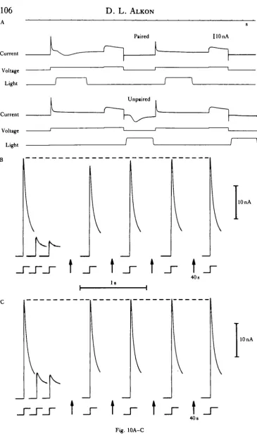
D. L. ALKONby conditioning arises from a persistent decrease of voltage-dependent K+ current/ (Fig. 9; Alkon et al. 1982a; Forman et al. 1984). These include the IA and the Ic*1+ - K+ outward K+ currents. A number of other studies were undertaken to deter-mine some of the biophysical steps leading to this long-term reduction of K+ currents. It was already known that progressively enhanced light-evoked depolarization and cumulative membrane depolarization of the Type B cell occurs during acquisition of associative learning (Alkon, 19806). During prolonged depolarization, prolonged elevation of intracellular Ca2+ was demonstrated by differential absorption spectrophotometry (Connor & Alkon, 1982, 1984). Elevation of intracellular Ca2+ by injection of Ca2+ causes prolonged reduction of both IA (Fig. 10; Alkon, Shoukimas & Heldman, 19826) and I O ^ - K * currents (Alkon et al. 1984; Alkon & Sakakibara,
1984).
The biophysical sequence (Fig. 11) underlying the transformation of the Type B membrane during learning is most probably as follows: repeated pairings of light and rotation elicit cumulative membrane depolarization. This arises from (1) the genetic-ally specified synaptic organization and (2) the response of this network during the
- 1 0 •
10 nA
50 mV
Fig. 8. Light-induced Ca2+-dependent K+ current. Current records (in response to command
depolarizations indicated on the left) obtained during voltage clamp of an isolated Type B photo-receptor soma in 0Na+, 3 0 0 m m o i r ' K+-artificial sea water. The light-induced current is inward below the equilibrium potential for potassium flux, approximately 0 mV. At 0 mV there is no appreci-able light-induced current. This current was reduced by intracellular injection of EGTA but not affected by 5 mmol I"1 Ccf+ in the external bathing medium, lowering of external Ca2+ or Na+, or substitution of Ba2+ for Ca2*. The trace beneath the current records indicates the duration of the light
Membrane changes during learning
105
fciteraction of the animal with its environment during the training stimuli. Cumulative depolarization is accompanied by prolonged elevation of the intracellular calcium
with-in the soma, and perhaps with-in the axon and termwith-inal branches. Elevated calcium causes inactivation of voltage-dependent K+ currents (IA. and most probably I c ^ - K+) which outlasts by many days the cumulative depolarization and elevation of intracellular
J 10 nA
Paired P<001
V
_r
Random
[image:13.451.44.404.114.590.2]l 5 0 m V
Fig. 9. Outward K+ currents of Type B cell (A and B). Outward currents elicited by command pulses
to OmV. Initial peaks are the early rapidly inactivating K+ currents (I*). Late outward K+ currents,
largely Ic»* - K*, attain a maximum value approximately 300 ms after the onset of the command pulse. Paired IA is smaller than random IA for both first and second command pulses. The ratio of paired
106
A
Current
Voltage
Light
Current
Voltage
Light
B
D. L. ALKON
Paired
Unpaired
UOnA
t j - t jr t _r t
40s Is
lOnA
K
t _r t j - t _r t
[image:14.451.42.408.35.671.2]40s
Fig. 10A-C
Membrane changes during learning
B
660-690 nm
107
SYNAPT1C EXCITATION
0-3-s flash
20 s
0001 AA
20mV
630-690 nm
• LIGHT-INDUCED DEPOLARIZATION
0-002 AA
Fig. 11. (A) Regenerative synaptic and light induced excitation of the Type B photoreceptor. Light-induced depolarization facilitates synaptic excitation and vice-versa in response to temporally associated light and rotation. Analysed in biophysical terms, synaptic depolarization causes
inactiva-tion of IA and Ica^-K* and enhancement of a voltage-dependent Ca2+ current. Increased
intracellular Ca2* causes further inactivation of IA and Ict^-K* and thus a further increase of
effective input resistance. These in turn cause more membrane depolarization. (B) Absorbance
changes at 660—690 (monitoring intracellular Ca2+) and 630—690 nm wavelength pairs (top and
bottom records) and membrane voltage response (middle) following a 0-3 s light flash (Connor & Alkon, 1984).
Ca2+. Reduction of voltage-dependent K+ currents increases the excitability thus permitting an enhanced depolarization to a light stimulus. The greater depolarization and the greater number of impulses that will be generated cause (1) greater inhibition of the medial Type A photoreceptor, (2) increased inhibition of ipsilateral inter-neurones, (3) decreased depolarization of ipsilateral motor neurones and finally (4) decreased turning toward a light source.
The change from transient (lasting minutes to hours) cumulative depolarization
Fig. 10. (A) Positive command pulses paired (upper records) and unpaired (lower records) with light steps. Each command step to 0 mV is followed after 30 ya by a brief step to +10 mV. The interstimulus interval illustrated here was used for paired vs unpaired comparisons (Alkon, Shoukimas & Heldman, 19826). (B) Rates of IA decrease and recovery during and following repetitive command depolariza-tions. Current responses to only three of the first five depolarizing steps used are shown. The steps following arrows were given at 40-8 intervals after the five depolarizing steps used for quantitation of differences. These five command depolarizations (2-2 a) to 0 mV occurred with a cycle time of 4-0 s. Each 2-2s step was followed by a second command (800ms) to — lOmV. (B) Command depolariza-tions paired with light. A light step (2-0 s) was presented 150 ms after the onset of each command
depolarization. Light intensity: 10 'Jcm~zs. (C) Command depolarizations alone. Arrows indicate
[image:15.451.47.416.48.284.2]108
D. L. ALKONControl P h K
Dark
Pair 2 (60 D)
10 nA
SOmVl
[image:16.451.37.412.47.226.2]I s
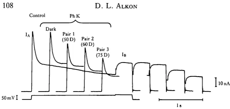
Fig. 12. Family of K+ currents (IA traces at left and matched IB traces at right) illustrate the effect of a single PhK (phosphorylase kinase) injection (60nC) under two conditions: in darkness (Dark) and after a 20-8 pairing (Pair 1 -3) of a light step with a variable command pulse to deliver a Ca2+ load. Control, control before injection. IA was elicited from a VH of — 60 mV, and decays within ~ 1 s to the steady state. l-9s later, a second superimposed command pulse ( + 10mV) elicits IB (delayed K+ current) uncontaminated by IA . (Full record only shown for control condition.) Note no effect
of PhK injection under dark conditions but a reduction of IA and a small (but not statistically significant) reduction of IB after the first pairing to a +50mV command (Pair 1, SOD). Further reduction of IA and also IB occurred after two additional pairings associated with increasing command steps (60 and 75 D, respectively) (Acosta-Urquidi, Alkon & Neary, 1984).
and elevated intracellular Ca2+ to a longer lasting reduction of open voltage-dependent K+ channels can most reasonably be thought to be mediated by biochemi-cal steps. Ca2+-calmodulin phosphorylation of proteins which regulate or are a part of the K+ channels themselves is implicated. Electrophoretic analysis revealed a difference in phosphorylation of low molecular weight proteins from the eyes of conditioned but not control Hermissenda (Neary, Crow&Alkon, 1981). Phosphory-lation of these proteins was in addition shown to be Ca2+-dependent (Alkon, 1983). Phosphorylation of one of these proteins (25 000 Mr) also changed under conditions
which inactivated the same voltage-dependent K+ current (IA) that was reduced during days on which the associative learning was retained (Neary & Alkon, 1983). Finally, iontophoresis of a Ca2+-calmodulin-dependent protein kinase (phosphory-lase kinase) caused a long-lasting reduction of the IA current (Fig. 12) which required some initial elevation of intracellular Ca2+ (Acosta-Urquidi, Alkon & Neary, 1982; Acosta-Urquidi, Neary & Alkon, 1984).
Membrane changes during learning 109
f crayfish (Jacobs & Atwood, 1981), and on neuromuscular junctions of the lobster fKravitz et al. 1980). Serotonin also produces effects on Aplysia similar to those observed after delivering a noxious stimulus such as shock or a 'pinch' (Camarado, Siegelbaum & Kandel, 1984).
These effects of pharmacological agents differ in many respects from the learning of an association between two specific stimuli as analysed for Hermissenda. First, a learned stimulus association is quite specific, while 'state' changes elicited by neurohumoral agents are generalized to many targets throughout the body. Second, the associative learning in Hermissenda persists for many weeks, while the pharmacological effects do not outlast the presence of the neurohumoral agent. Thus, there is no evidence that the neurohumorally-induced cellular changes involve storage of information, which persists beyond the initial experience of, for example, an arousal state. Nor mechanistically would the persistence of arousal effects be useful for the animal unless the arousing stimuli had been associated with other temporally-related events. Moreover, the effects of serotonin and octopamine are mediated by cyclic nucleotide phosphorylation (Batelle & Kravitz, 1978) rather than Ca2+ -calmodulin-dependent phosphorylations involved in the learned association in
Hermissenda (Acosta-Urquidi et al. 1982, 1984; AXkon etal. 1983a). The
serotonin-induced effects on Aplysia neurones are also independent of calcium (Siegelbaum, Camardo & Kandel, 1982). It may, however, be premature to compare directly calcium-dependent modification of Hermissenda membrane channels during associative learning with mechanisms of arousal and sensitization. Thus, although serotonin has been thought to be responsible for long-term reduction of potassium channels during sensitization, their prolonged reduction has never been correlated with long-lasting sensitization of intact animals. Furthermore, it has been recently shown that serotonin is not released or synthesized from presynaptic interneurones which have long been thought to convey the sensitizing effects on the synaptic endings of the sensory cell (Hawkins et al. 1981; Kistler et al. 1983).
Ultimately some intelligent and testable extrapolation from relatively simple to more complex nervous systems may become possible. Most promising to date are the properties of ionic channels within neural membranes. The voltage-dependent cal-cium channels of the Hermissenda Type B photoreceptors have been found in
Paramecium (Naitoh & Eckert, 1974), spinal cord motor neurones (Barrett & Barrett,
110 D. L. ALKON
dog and then to prove that these changes are intrinsic to particular neurones. It will b | more difficult still to reach any conclusion as to what portion of a discrete association learned by a cat or dog resides within a given set of neurones and is critical for its recall.
R E F E R E N C E S
ACOSTA-URQUIDI, J., ALKON, D. L. & NEAJLY, J. T . (1984). Intrasomatic injection of a Caz+-calmodulin
dependent protein kinase simulates biophysical effects of associative learning in aHermissenda photoreceptor.
Science, N.Y. (in press).
ACOSTA-URQUIDI, J., NEARY, J. T . & ALKON, D . L. (1982). Ca++calmodulin dependent protein kinase effects on voltage-dependent K+ currents. Soc. Neurosd. Abstr. 8, 825.
ALKON, D. L. (1973). Intersenaory interactions m Hermissenda. J. gen. Physiol. 62, 185-202.
ALKON, D. L. (1974a). Sensory interactions in the nudibranch mollusc Hermissenda crassicomis. Fedn Proc.
Fedn Am. Socs exp. Biol. 33, 1083-1090.
ALKON, D. L. (19746). Associative training of Hermissenda. J'. gen. Physiol. 64, 70-84.
ALKON, D. L. (1979). Voltage-dependent calcium and potassium ion conductances: A contingency mechanism for an associative learning model. Science, N.Y. 205, 810-816.
ALKON, D. L. (1980a). Cellular analysis of a gastropod (Hermissenda crassicomis) model of associative learn-ing. Biol. Bull. mar. biol. Lab., Woods Hole 159, 505-560.
ALKON, D. L. (19806). Membrane depolarization accumulates during acquisition of an associative behavioral change. Science, N.Y. 210, 1375-1376.
ALKON, D. L. (1982). A biophysical basis for molluscan associative learning. In Conditioning: Representation
of Involved Neural Functions, (ed. C. D. Woody) p. 147. New York: Plenum Press.
ALKON, D . L. (1983). Learning in a marine snail. Scient. Am. 249, 70-84.
ALKON, D. L., ACOSTA-URQUIDI, J., OLDS, J., KUZMA, G. & NEARY, J. T . (1983a). Protein kinase injection
reduces voltage dependent potassium currents. Science, N.Y. 219, 303—306.
ALKON, D. L., AKAIKE, T . & HARRIGAN, J. F. (1978). Interaction of chemosensory, visual and statocyst pathways in Hermissenda. J. gen. Physiol. 71, 177-194.
ALKON, D. L., FARLEY, J., SAKAKIBARA, M. & HAY, B. (1984). Voltage-dependent calcium and calcium-activated potassium currents of a molluscan photoreceptor. Btophys.J. (in press).
ALKON, D. L., FARLEY, J., HAY, B. & SHOUKIMAS, J. J. (19836). Inactivation of Ca++ dependent K+ current can occur without significant Ca++ current inactivation. Soc. Neurosd. Abstr. 9, 1188.
ALKON, D. L. & FUORTES, M. G. F. (1972). Responses of photoreceptors in Hermissenda. J. gen. Physiol. 60, 631-649.
ALKON, D. L., LEDERHENDLER, I. & SHOUKIMAS, J. J. (1982a). Primary changes of membrane currents during retention of associative learning. Science, N.Y. 215, 693-695.
ALKON, D. L. & SAKAKIBARA, M. (1984). Prolonged inactivation of a Caz+-dependent K+ current but not
Ca2+ current by light induced elevation of intracellular calcium. Soc. Neurosd. Abstr. (in press).
ALKON, D. L., SHOUKIMAS, J. J. & HELDMAN, E. (19826). Calcium-mediated decrease of a voltage-dependent potassium current. Biophys. J. 40, 245-250.
BARRETT, E. F. & BARRETT, J. N. (1976). Separation of two voltage-sensitive potassium currents, and demonstration of a tetrodotoxin-resistant calcium current in frog motorneurones. J. Physiol., Land. 255, 737-774.
BATELLE, B. A. &KRAVITZ, E. A. (1978). Targets of octopamine action in the lobster: cyclic nucleotide changes and physiological effects in haemotymph, heart and exoskeletal muscle. J. Pharmac. exp. Ther. 205, 438-448. BEBGER, T . W., ALGER, B. E. & THOMPSON, R. F. (1976). Neuronal substrate of classical conditioning in the
hippocampus. Science, N.Y. 192, 483-485.
BERGER, T . W. & THOMPSON, R. F. (1977). Limbic system interrelations: functional division among hippocampal-septal connections. Science, N.Y. 197, 587-589.
BEBGER, T . W. & THOMPSON, R. F. (1978). Identification of pyramidal cells as the critical elements in hippocampal neuronal plasticity during learning. Proc. natnAcad. Sd. U.SA, 75, 1572—1576.
BRONS, J. & WOODY, C. D . (1980). Long term changes in excitability of cortical neurons after Pavlovian conditioning and extinction. J. Neurophysiol. 44, 605-615.
CAMARADO, J. S., SIEGELBAUM, S. & KANDEL, E. R. (1984). Cellular and molecular correlates of serialization in Aplysia and their implications for associative learning. In Primary Neural Substrates of Learning and
Behavioral Change, (eds D. L. Alkon & J. Farley). New York: Cambridge University Press, (in press).
CAREW, T. J., HAWKINS, R. D. & KANDEL, E. R. (1983). Differential classical conditioning of a defensive withdrawal reflex in Aplysia califormca. Science, N.Y. 219, 397-400.
CAREW, T . J., WALTERS, E. T . & KANDEL, E. R. (1981). Classical conditioning in a simple withdrawal reflex in Aplysia caliform'ca.J. Neurosd. 1, 1426-1437.
CASTELLUCCI, V. F., PINSKER, H. M., KUPFERMAN, I. & KANDEL, E. R. (1970). Neuronal mechanisms of
Membrane changes during learning 111
KLARK, R. B. & WONG, R. K. S. (1983). Three components of outward current in isolated mammalian cortical W neurons. Soc. Neurosci. Abstr. 9, 601.
COHEN, D. H. & MACDONALD, R. L. (1976). Involvement of the avian hypothalamus in defensively con-ditioned heart rate change. J. camp. Neurol. 167, 465-480.
CONNOR, J. A. & ALKON, D . L. (1982). Voltage and light-dependence of intracellular Arsenazo signals. Soc. Neumsci. Abstr. 8, 944.
CONNOR, J. A. & ALKON, D. L. (1984). Light- and voltage-dependent increases of calcium ion concentration in molluscan photoreceptors. J. Newvpkyiiol. 51, 745-752.
CONNOR, J. A. & STEVENS, C. F. (1971). Voltage clamp studies of a transient outward current in gastropod neural somata. J. Physiol, Land. 213, 21-30.
CROW.T. J. & ALKON, D. L. (1978). Retention of an associative behavioral change inHermissenda crassicomis. Science, N.Y. 201, 1239-1241.
CROW, T. J. & ALKON, D. L. (1980). Associative behavioral modification inHermissenda: cellular correlates. Science, N.Y. 209, 412-414.
FARLEY, J. & ALKON, D. L. (1980). Neural organization predicts stimulus specificity for a retained associative behavioral change. Science, N.Y. 210, 1373-1375.
FARLEY, J. & ALKON, D. L. (1982). Associative neural and behavioral change in Hermissenda: Consequences of nervous system orientation for light- and pairing specificity. J. Neurophysiol. 48, 785-807.
FARLEY, J. & ALKON, D. L. (1984). Cellular analysis of gastropod learning. In Invertebrate Receptors, (ed. A. H. Greenberg). New York: Marcel Dekker, (in press).
FARLEY, J. & KE»N, G. (1983). Contingency-sensitive behavioral change in Hermissenda: temporally-specific attenuation of conditioning. Anim. Leant. Behav. (in press).
FARLEY, J., RICHARDS, W. G., LING, L. J., LIMAN, E. & ALKON, D. L. (1983). Membrane changes in a single
photoreceptor cause associative learning in Hermissenda. Science, N.Y. 221, 1201—1203.
FORMAN, R., ALKON, D . L., SAKAKIBARA, M., HARRIGAN, J., LEDERHENDLER, I. & FARLEY, J. (1984). Changes in IA and Ic but not INA accompany retention of conditioned behaviour in Hermissenda. Soc. Neurosci. Abstr. (in press).
GELPERIN, A., WIELAND, S. J. & BARRY, S. R. (1984). A strategy for cellular analysis of associative learning in a terrestrial mollusk. In Primary Neural Substrates ofLearning and Behavioral Change, (edsD. L. Alkon & ] . Farley). New York: Cambridge University Press, (in press).
GILLETTE, R. & DAVIS, W. J. (1977). The role of the metacerebral giant neuron in the feeding behavior of Pieurobranchaea.jf. amp. Pkysiol. 166, 129—159.
GILLETTE, R., GILLETTE, M. V. & DAVIS, W. J. (1980). Action-potential broadening and endogenously sus-tained bursting are substrates of command ability in a feeding neuron of Pleurobranchaea. J. Neurophysiol. 43, 669-685.
GOH, Y. & ALKON, D. L. (1984). Sensory, interneuronal and motor interactions within the Hermissenda visual pathway. J. Neurophysiol. 52, 156-169.
HAWKINS, R. D., CASTELLUCCI, V. F. & KANDEL, E. R. (1981). Interneurons involved in mediation and modulation of gill-withdrawal reflex in Aplysia. II. Identified neurons produce heterosynaptic facilitation contributing to behavioral sensitization. J. Neurophysiol. 45, 315.
HOYLE, G. (1982). Cellular basis of operant conditioning of leg position. In Conditioning: Representation of Involved Neural Functions, (ed. C. D. Woody), p. 197. New York: Plenum Press.
JACOBS, J. R. & ATWOOD, H. L. (1981). Long term facilitation of tension in crustacean muscle and its modula-tion by temperature, activity and circulating amines. J. comp. Physiol. 144, 335—343.
KANDEL, E. R. & SCHWARTZ, J. H. (1982). Molecular biology of learning: modulation of transmitter release. Science, N.Y. 218, 433-443.
KANZ, J. E., EBERLEY, L. B., COBBS, J. S. &PINSKER, H. M. (1979). Neuronal correlates of siphon withdrawal in freely behaving Aplysia. J. Neurophysiol. 42, 1538-1556.
KISTLER, H. B., HAWKINS, R. D., KUESTER, J., KANDEL, E. R. & SCHWARTZ, J. H. (1983). Imm-unocytochemical studies of neurons producing presynaptic facilitation in the abdominal ganglion of Aplysia califormca. Soc. Neumsci. Abstr. 9, 915.
KRAVITZ, E. A., GLU8MAN, S., HARRIS-WARRICK, R. M., LIVINGSTONE, M. S., SCHWARTZ, T. & GOY, M. F. (1980). Amines and a peptide as neurohormones in lobsters: action on neuromuscular preparations and preliminary behavioural studies. J. exp. Biol. 89, 159-175.
KRNJEVIC, K., MORRIS, M. E., REIFENSTEIN, R. J. & ROPERT, N. (1982). Depth distribution and mechanism
of changes in extracellular K+ and Ca2+ concentrations in the hippocampus. Can. J. Physiol. Pharmac. 60,
1658-1671.
LEDERHENDLER, I., GART, S. & ALKON, D. L. (1983). Associative learning in Hermissenda crassicornis (Gastropoda): evidence that light (the CS) takes on characteristics of rotation (the UCS). Biol. Bull. mar. biol. Lab., Woods Hole, Abstr. 165, 528.
LEDERHENDLER, I. I., GOH, Y. & ALKON, D. L. (1982). Type B photoreceptor changes predict modification of motorneuron responses to light during retention of Hermissenda associative conditioning. Soc. Neurosci. Abstr. 8, 824.
112 D. L. ALKON
LLINAS, R. & YAROM, Y. (19816). Properties and distribution of ionic conductances generating electrorespoii siveness of mammalian inferior olivary neurones in vitro. J. Physiol., bond. 315, 569-584.
MPITSOS, G. J. & COLLINS, S. D . (1975). Learning: rapid aversive conditioning in the gastropod mollusc
Pleurobranchaea. Science, NY. 188, 954-956.
NAITOH, Y. & ECKERT, R. (1974). The control of ciliary activity in protozoa. In Cilia and Flagella, (ed. M. Sleigh). New York: Academic Press.
NEARY, J. T . & ALKON, D. L. (1983). Protein phosphorylation/dephosphorylation and the transient, voltage-dependent potassium conductance in Hermissenda crassicornis.J. biol. Chem. 258, 8979—8983.
NEARY, J. T . , CROW, T . J. & ALKON, D . L. (1981). Change in a specific phosphoprotein band following associative learning in Hermissenda. Nature, Land. 293, 658-660.
RICHARDS, W., FARLEY, J. & ALKON, D . L. (1983). Extinction of associative learning in Hermissenda: Behavior and neural correlates. Soc. Neurosci. Abstr. 9, 916.
SAHLEY, C , RUDY, J. W. & GELPERIN, A. (1984). Associative learning in a mollusk: A comparative analysis. In Primary Neural Substrates ofLearning and Behavioral Change, (edsD. L. Alkon &J. Farley). New York: Cambridge University Press, (in press).
SHOUKIMAS, J. J. & ALKON, D . L. (1980). Voltage-dependent, early outward current in a photoreceptor of
Hermissenda crassicornis. Soc. Neurosci. Abstr. 6, 17.
SIEGELBAUM, S. A., CAMARADO, J. S. & KANDEL, E. R. (1982). Serotonin and cyclic AMP close single potassium channels in Aplysia sensory neurons. Nature, Land. 299, 413—417.
THOMPSON, R. F . , BERCER, T . W., BERRY, S. D . , CLARK, G. A., KETTNER, R. N., LAVOND, D . G., MAUK,
M. D., MCCORMICK, D. A., SOLOMON, P. R. & WEISZ, D. J. (1982). Neuronal substrates of learning and memory: hippocampus and other structures. In Conditioning: Representation ofInvolved Neural Functions, (ed. C. D. Woody), pp. 115-129. New York: Plenum Press.
WEST, A., BARNES, E. S. & ALKON, D . L. (1982). Primary changes of voltage responses during retention of associative learning. J. Neurophysiol. 48, 1243—1255.
WOODY, C. D . (1982). Memory, Learning and Higher Function. New York: Springer-Verlag.
WOODY, C. D. & BLACK-CLEWORTH, P. (1973). Differences in excitability of cortical neurons as a function of motor projection in conditioned cats. J . Neurophysiol. 36, 1104-1116.
WOODY, C. D . , VASSILEVSKY, N. N. & ENGEL, J. (1970). Conditioned eye blink: unit activity at coronal-precruciate cortex of the cat. J. Neurophysiol. 33, 851-864.
WOODY, C. D . , YAKOWSKY, P., OWBNS, J., BLACK-CLEWORTH, P. & CROW, T . (1974). Effect of lesions of